The closed circulatory system performs many functions in the body
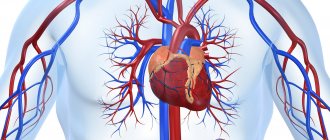
The heart is often called the human motor: this muscular organ begins beating in the embryo at the initial stage of intrauterine development and stops at the moment of death. Its anatomical structure is quite complex, and the functions performed are varied and have the main goal of maintaining a constant internal environment.
In our review and video in this article, we will try to understand how the cardiovascular system works: the structure and functions of this complex of organs, as well as common lesion syndromes and methods of functional assessment of its activity.
Circulation circles
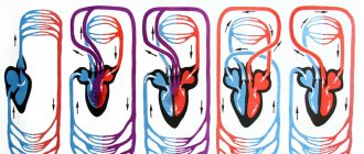
The blood flowing through the cardiovascular system can be compared to an athlete running different distances. When it passes through the pulmonary circulation, it is a sprint. And a big circle is already a marathon. The Englishman William Harvey described these circles back in 1628. During a large circle, blood is distributed throughout the body, not forgetting to provide it with oxygen and take away carbon dioxide. During this “race”, arterial blood becomes venous.
The pulmonary circulation is responsible for the flow of blood into the lungs, where the blood releases carbon dioxide and is enriched with oxygen. Blood from the pulmonary circulation returns to the left atrium. The systemic circulation, starting in the left ventricle, transports blood throughout the body. Oxygenated blood is pumped by the left ventricle into the aorta and its many branches - the various arteries. Then it enters the capillary vessels of organs and tissues, where oxygen from the blood is exchanged for carbon dioxide. The systemic circulation ends in small veins that merge into two large veins (vena cava) and return blood to the right atrium. The superior vena cava drains blood from the head, neck and upper extremities, and the inferior vena cava drains blood from the torso and lower extremities.
A little about physiology: how the cardiovascular system “works”
As we have seen from the anatomical review, the structure and functions of the cardiovascular system are quite complex.
Clear and coordinated operation of all its components allows us to ensure:
- transport of oxygen and substances necessary for the normal functioning of body cells;
- removal of metabolites from cells - processed substances and carbon dioxide;
- humoral (endocrine) connection between organs;
- adequate immune response in response to the introduction of infectious agents into the body;
- removal of toxins and harmful substances from the body;
- heat exchange.
Arteries
Not a single cell can survive without nutrients and oxygen. They are delivered by arteries. They carry oxygen-rich blood throughout the body. When you breathe, oxygen enters your lungs. where the delivery of oxygen throughout the body begins. First to the heart, then through the systemic circulation to all parts of the body. There, the blood exchanges oxygen for carbon dioxide and then returns to the heart. The heart pumps it back to the lungs, which take in carbon dioxide and give out oxygen, and so on endlessly. There are also pulmonary arteries of the pulmonary circulation, they are located in the lungs and through them blood, poor in oxygen and rich in carbon dioxide, enters the lungs, where gas exchange occurs. This blood then returns to the heart through the pulmonary veins.
Is my cardiovascular system healthy?
Are your heart and blood vessels healthy? To answer this question, the absence of complaints is not enough. It is important to regularly undergo a medical examination, during which the doctor will determine the main functional indicators of the cardiovascular system.
These include:
- Heart rate;
- arterial pressure;
- electrocardiogram;
- stroke volume of cardiac output;
- cardiac output;
- speed and other indicators of blood flow;
- breathing patterns during physical activity.
Heart rate
Determining the functional state of the cardiovascular system begins with calculating heart rate. The normal heart rate for adults is 60-80 beats per minute. A decrease in heart rate is called bradycardia, an increase is called tachycardia.
Note! In trained people, heart rate may be slightly lower than standard values - at the level of 50-60 beats/min. This is explained by the fact that the endurance heart of athletes “drives” a larger amount of blood in an equal period of time.
Heart rate norms change with age
Functional disorders of the cardiovascular system associated with changes in heart rate have various causes.
For example, bradycardia can be caused by:
- vegetative-vascular dystonia;
- stomach diseases (peptic ulcer, chronic erosive gastritis);
- hypothyroidism and some other endocrine disorders;
- previous myocardial infarction;
- cardiosclerosis;
- chronic heart failure.
Among the most common causes of tachycardia are:
- myocarditis;
- cardiomyopathy;
- pulmonary heart syndrome;
- acute myocardial infarction and left ventricular failure;
- hyperthyroidism and thyrotoxic crisis;
- acute infectious diseases;
- shock;
- massive blood loss;
- anemia;
- acute renal failure.
Note! Physiological (adaptive) tachycardia occurs with fever, increased ambient temperature, stress and psycho-emotional experiences, consumption of alcohol, energy drinks, and certain medications.
Arterial pressure
Blood pressure is one of the important indicators of the functioning of the circulatory system. The upper, or systolic, value reflects the pressure in the arteries at the peak of contraction of the walls of the ventricles of the heart - systole. The lower (diastolic) is measured at the moment of relaxation of the heart muscle.
The blood pressure of a healthy person is 120/80 mm Hg. Art. The difference between SBP and DBP is called pulse pressure. Normally it is 30-40 mmHg. Art.
The price of the simplest tonometers for measuring blood pressure starts from 450 rubles. Automatic models are more expensive.
Stroke and cardiac output
Stroke volume of blood is the amount of fluid that is ejected by the left ventricle of the heart in one contraction into the aorta. In a person with a low level of physical activity it is 50-70 ml, and in a trained person it is 90-110 ml.
Functional diagnostics of the cardiovascular system determines cardiac output by multiplying stroke volume by heart rate. On average, this figure is 5 l/min.
Heart and blood vessels
The human cardiovascular system is closed. This means that the blood moves only through the vessels and there are no cavities into which the blood flows. Thanks to the work of the heart and the branched system of blood vessels, every cell in our body receives oxygen and nutrients that are necessary for life.
Pay attention to the established name - cardiovascular system. The first place is taken by the heart muscle, which performs the most important function. We move on to studying this unique organ.
Heart
The branch of medicine that studies the heart is called cardiology (from the ancient Greek καρδία - heart and λόγος - study). The heart is a hollow muscular organ that contracts with a certain rhythm throughout a person’s life.
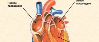
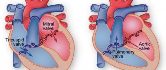
The outside of the heart is covered by a pericardial sac - the pericardium. Consists of 4 chambers: 2 ventricles - right and left, and 2 atria - right and left. Remember that between the ventricles and atria there are leaflet valves.
Between the right atrium and the right ventricle there is a tricuspid (tricuspid) valve, and between the left atrium and the left ventricle there is a bicuspid (mitral) valve.
In the heart, blood moves unidirectionally: from the atria to the ventricles, thanks to the presence of leaflet (atrioventricular) valves (from the Latin atrium - atrium and ventriculus - ventricle).
The largest human vessel, the aorta, with a diameter of 2.5 cm, departs from the left ventricle, in which blood flows at a speed of 50 cm per second. The pulmonary trunk departs from the right ventricle. Between the left ventricle and the aorta, as well as the right ventricle and the pulmonary trunk, there are semilunar valves.
The muscle tissue of the heart is represented by single cells - cardiomyocytes, which have transverse striations. The heart has a special property - automaticity: isolated from the body, the heart continues to contract without external influences. This is due to the presence of special cells in the thickness of muscle tissue - pacemaker cells (pacemaker cells, atypical cardiomyocytes), which themselves periodically generate nerve impulses.
The heart has a conduction system due to which the excitation that arises in one part of the heart gradually covers other parts. The conduction system includes the sinus node, atrioventricular node, His bundle and Purkinje fibers. It is thanks to the presence of these conducting structures that the heart is capable of automation.
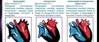
Cardiac cycle
The work of the heart consists of three phases successively replacing each other:
- Atrial systole (from the Greek systole - compression, contraction)
- Ventricular systole
- Total diastole (from the Greek diastole - expansion)
Lasts 0.1 sec. During this phase, the atria contract, their volume decreases, and blood flows from them into the ventricles. During this phase, the leaflet valves are open, the semilunar valves are closed.
Lasts 0.3 seconds. The leaflet (atrioventricular) valves close to prevent blood from flowing back into the atria. The muscle tissue of the ventricles begins to contract, their volume decreases: the semilunar valves open. Blood is expelled from the ventricles into the aorta (from the left ventricle) and the pulmonary trunk (from the right ventricle).
Lasts 0.4 seconds. In diastole, the cavities of the heart expand - the muscles relax, the semilunar valves close. Flap valves are open. During this phase, the atria fill with blood, which passively flows into the ventricles. Then the cycle repeats.
We have already discussed the cardiac cycle, but I want to focus your attention on some details. In total, one cycle lasts 0.8 seconds. The atria rest for 0.7 seconds during ventricular systole and total diastole, and the ventricles rest for 0.5 seconds during atrial systole and total diastole. Thanks to this energetically beneficial cycle, the heart muscle gets less tired during work.
Heart rate (HR) can be measured using the pulse - the jerky vibrations of the walls of blood vessels associated with the cardiac cycle. The normal average heart rate is 60-80 beats per minute. An athlete's heart rate is lower than that of an untrained person. With heavy physical exertion, heart rate can increase to 150 beats/min.
Changes in the heart rate are possible in the form of its excessive slowdown or increase, respectively, they are distinguished: bradycardia (from the Greek βραδυ - slow and καρδιά - heart) and tachycardia (from the ancient Greek ταχύς - fast and καρδία - heart). Bradycardia is characterized by a decrease in heart rate to 30-60 beats/min, tachycardia - above 90 beats/min.
The regulatory center of activity of the cardiovascular system lies in the medulla oblongata and spinal cord. The parasympathetic nervous system slows down and the sympathetic nervous system speeds up heart rate. Humoral factors also have an influence (from the Latin humor - moisture), mainly hormones: adrenal glands - adrenaline (strengthens the heart), thyroid gland - thyroxine (accelerates heart rate).
Vessels
Blood moves to tissues and organs inside the vessels. They are divided into arteries, veins and capillaries. We will discuss their structure and functions in general terms. I would like to note: if you think that venous blood flows through the veins and arterial blood flows through the arteries, you are mistaken. In the following article you will find specific examples that refute this misconception.
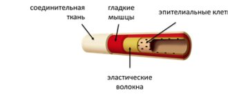
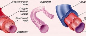
Arteries carry blood from the heart to internal organs and tissues. They have thick walls containing elastic and smooth muscle fibers. The blood pressure in them is the highest compared to the veins and capillaries, which is why they have the aforementioned thick wall.
The inside of the artery is lined with endothelium - epithelial cells that form a single layer of thin cells. Due to the presence of smooth muscle cells in the thickness of the wall, arteries can narrow and widen. The speed of blood flow in the arteries is approximately 20-40 cm per second.
For the most part, arteries carry arterial blood, but we must not forget about exceptions: venous blood flows from the right ventricle through the pulmonary arteries to the lungs.
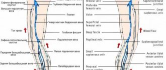
Veins carry blood to the heart. Compared to the arterial wall, veins have fewer elastic and muscle fibers. The blood pressure in them is low, so the wall of the veins is thinner than that of the arteries.
A characteristic sign of veins (which you will always notice on the diagram) is the presence of valves inside the vein. The valves prevent the reverse flow of blood in the veins - they ensure unidirectional blood flow. The speed of blood flow in the veins is about 20 cm per second.
Just imagine: veins lift blood from the legs to the heart, acting against gravity. The aforementioned valves and skeletal muscle contractions help them in this. That is why physical activity is very important, as opposed to physical inactivity, which is harmful to health by disrupting the movement of blood through the veins.
Mostly the veins contain venous blood, but we must not forget about exceptions: the pulmonary veins with arterial blood enriched with oxygen after passing through the lungs approach the left atrium.
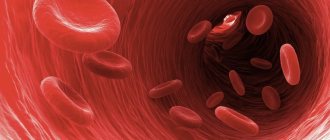
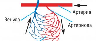
The smallest blood vessels are capillaries (from the Latin capillaris - hair). Their wall consists of a single layer of cells, which makes possible gas exchange and exchange processes of various substances (nutrients, by-products) between the cells surrounding the capillary and the blood in the capillary. The speed of blood movement through capillaries is the lowest (compared to arteries and veins) - 0.05 mm per second, which is necessary for metabolic processes.
The total lumen of capillaries is larger than that of arteries and veins. They are suitable for every cell of our body; they are the connecting link through which tissues receive oxygen and nutrients.
As blood passes through the capillaries, it loses oxygen and becomes saturated with carbon dioxide. Therefore, in the picture above you see that at first the blood in the capillaries is arterial, and then venous.
Hemodynamics
Hemodynamics is the process of blood circulation. An important indicator is blood pressure - the pressure exerted by blood on the walls of blood vessels. Its magnitude depends on the force of heart contraction and vascular resistance. There are systolic (on average 120 mm Hg) and diastolic (on average 80 mm Hg) blood pressure.
Systolic blood pressure refers to the pressure in the bloodstream at the moment of contraction of the heart, diastolic - at the moment of its relaxation.
With physical activity and stress, blood pressure rises and heart rate increases. During sleep, blood pressure decreases, as does your heart rate.
Blood pressure level is an important indicator for a doctor. Blood pressure may be elevated in a patient with kidney or adrenal disease, so it is extremely important to know and control its level.
Increased blood pressure, for example 220/120 mmHg. Art. doctors call arterial hypertension (from the Greek hyper - excessively; to say hypertension is not entirely correct, hypertension is increased muscle tone), and a decrease, for example, to 90/60 mm. rt. Art. will be called arterial hypotension (from the Greek hypo - under, below).
All of us have probably experienced orthostatic hypotension at least once in our lives - a decrease in blood pressure when suddenly rising from a sitting or lying position. It is accompanied by slight dizziness, but can also lead to fainting and loss of consciousness. Orthostatic hypotension can (within normal limits) occur in adolescents.
There is a nervous regulation of hemodynamics, which consists in the action on the vessels of fibers of the sympathetic nervous system, which constricts blood vessels (pressure increases), and the parasympathetic nervous system, which dilates blood vessels (pressure decreases accordingly).
The lumen of blood vessels is also affected by humoral factors that spread through the fluids of the body. A number of substances have a vasoconstrictor effect: vasopressin, norepinephrine, adrenaline, another part has a vasodilator effect - acetylcholine, histamine, nitric oxide (NO).
Diseases
Atherosclerosis (Greek athḗra - gruel + sklḗrōsis - hardening) is a chronic disease of the arteries that occurs as a result of a violation of the metabolism of fats and proteins in them. With atherosclerosis, a cholesterol plaque forms in the vessel, which gradually increases in size, ultimately leading to complete blockage of the vessel.
The plaque narrows the lumen of the vessel, reducing the amount of blood flowing through it to the organ. Atherosclerosis often affects the vessels that supply the heart - the coronary arteries. In this case, the disease may manifest itself as pain in the heart with minor physical exertion. If atherosclerosis affects the vessels of the brain, the patient’s memory, concentration, and cognitive (intellectual) functions deteriorate.
At some point, an atherosclerotic plaque may burst, in which case the incredible happens: the blood begins to clot right inside the vessel, because the cells react to the rupture of the plaque as if the vessel is damaged! A blood clot forms, which can block the lumen of the vessel, after which blood completely stops flowing to the organ that this vessel supplies.
This condition is called a heart attack (Latin infarcire - “to fill, stuff”) - a sudden cessation of blood flow due to artery spasm or blockage. A heart attack is expressed in the death of organ tissue due to an acute lack of blood supply. A cerebral infarction is called a stroke (Latin insultus - attack, blow).
© Bellevich Yuri Sergeevich 2018-2021
This article was written by Yuri Sergeevich Bellevich and is his intellectual property. Copying, distribution (including by copying to other sites and resources on the Internet) or any other use of information and objects without the prior consent of the copyright holder is punishable by law. To obtain article materials and permission to use them, please contact Yuri Bellevich
.