Chest pain is a symptom of many diseases, not necessarily heart diseases. Thus, diseases of the musculoskeletal system, respiratory and digestive organs, neurological disorders, and injuries may manifest themselves. However, you need to know how to determine if your heart is hurting, since this is where immediate help may be needed. It is especially important not to miss a dangerous condition, such as myocardial infarction.
Only a doctor can make a diagnosis, but some specific signs will help you understand that your heart is hurting.
How does the human heart work?
Heart (lat. сor) is a muscular-cavitary formation that ensures adequate blood supply to all cells and tissues.
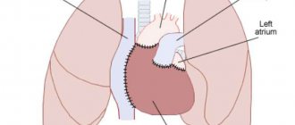
The peculiarity of the organ is its autonomy: individual innervation and regulation of contractile function. However, muscle, valve and conduction system structures are extremely sensitive to changes throughout the body. Topography of the organ: the heart is located in the chest cavity in a complex of mediastinal structures (a formation that is located between the two lungs), occupying the middle lower part. The organ “lies” on the diaphragm, enclosed in the pericardial sac. The lateral walls are adjacent to the roots of the lungs and the great vessels.
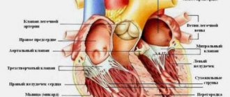
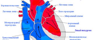
Schematic representation of the internal structure of the heart:
During a general clinical examination, relative and absolute cardiac dullness is determined by percussion (tapping) on the anterior wall of the chest. The predominant part of the organ is on the left side, the right border is along the outer edge of the sternum.
Listen to the activity of the heart and the functioning of the valves using a phonendoscope at the points of their projection.
Anatomy
The morphological structure of the heart is defined differently by experts. Anatomically, the organ is divided into right and left halves, which are connected through the vessels of the large and small circle of blood circulation.
During intrauterine development, the heart goes through different stages of chamber formation. In the case of an incomplete process at birth, pathological shunts remain between the left and right parts, which cause hemodynamic disturbances.
The chambers (cavities) of both halves are connected to each other using holes, where the direction of flow is regulated by the activity of the leaf valve structures.
The organ wall is represented by three main membranes:
- endocardium – lines the inner surface of the heart, forms tendon chords (threads) and valve apparatus;
- myocardium – a muscular layer that forms the organ wall, interventricular septum and papillary muscles;
- epicardium is the outer connective tissue membrane, which is considered the inner layer of the pericardial sac. Between the layers of the pericardium there is a small amount (up to 2 ml) of fluid, which ensures smooth sliding of the organ during different phases of the cardiac cycle.
Inflammatory pathologies of the pericardial sac or reactive changes against the background of other diseases (for example, pancreatitis or acute renal failure) lead to increased synthesis of fluid, which prevents the expansion of the cavities of the heart and adequate blood flow.
Cameras
The structure of the heart involves dividing the organ into halves, which are represented by four main and two additional chambers.
| Right part | Left departments |
| The atrium (atrium), which collects carbon dioxide-rich blood (venous) from throughout the body | The atrium, where the four pulmonary veins drain, carrying arterial blood with a high concentration of oxygen |
| The ventricle, which is connected to the superior chamber through the atrioventricular opening. The outflow tract carries blood through the pulmonary circulation for gas exchange | The ventricle is the largest chamber with a thick layer of muscle fibers, the contraction of which ensures adequate release of blood for delivery to the periphery |
| The auricle is a small cavity connected to the atrium (smaller than on the left) | Eyelet – additional chamber with entrance to the atrium |
The clinical significance of the ears is the additional volume with which the heart is filled under increased loads. However, stagnation of blood in the chambers increases the risk of developing blood clots (clots) with possible dissemination into the vessels of the brain or myocardium and subsequent stroke or heart attack.
Valve structures
Regulation of blood flow in a certain direction is determined by valve structures derived from the connective tissue inner membrane (endocardium). There are four main valves in the hemodynamic system of the organ:
- mitral (left atrioventricular) - represented by two valves that open into the ventricular cavity during atrial contraction;
- aortic (consists of three valves) - located at the exit from the left ventricle;
- tricuspid, which determines the movement of blood in the right sections;
- pulmonary valve (tricuspid), which regulates the flow of fluid from the ventricle into the pulmonary circulation.
The closing and opening of the valve flaps is ensured by the contraction of the papillary muscles and the length of the chordae tendineae (too short or long fibers of the latter lead to failure of the apparatus and reverse flow of blood).
Vascular system of the organ
The constant muscular work of the heart requires a large amount of energy, which is supplied through the coronary arteries with nutrients and oxygen. The coronary vessels of the organ are separated from the aorta directly at the base of the valve leaflets.
There are two main arteries supplying blood to the myocardium:
- The right one, emerging from the aorta onto the posterior surface of the heart, provides trophism to the right atrium and ventricle.
- The left one, which goes around the atrium and lies in the anterior groove, provides blood supply to the main muscle mass of the heart (the left sections, the interventricular septum and the anterior wall). Impaired blood flow in this vessel most often causes pain and a tingling sensation behind the sternum.
There are individual characteristics of the origin of the arteries, therefore, with contrast research methods, different types of blood supply to the heart are distinguished.
The outflow of venous blood occurs through the vessels of the same name, which open with small holes into the cavity of the right atrium.
Histology: what does the heart look like under a microscope?
The structure of the heart is organized by three main membranes, the cellular structure of which is determined by the functions performed. The microscopic location of tissues in the section (histology) is presented in the table:
| Layer | Picture under a microscope |
| Endocardium (tissue of valves, chordae tendineae and papillary muscles, inner lining) |
Cells feed on blood from the cavities of the heart |
| Myocardium | Muscle fibers built from mono- or binuclear cells. Contractile proteins are cross-striated, as in skeletal muscles. The individual fibers are connected to each other using insertion discs. The latter contribute to the rapid spread of contraction throughout the entire mass of the heart muscle. |
| Conduction system of the heart | Atypical cardiomyocytes (muscle) cells are of three types:
|
| Epicardium - the inner layer of the pericardium | A thin sheath of connective tissue containing elastic and collagen fibers. |
The photo shows the histological structure of the heart (muscle layer):
The nature of pain in cardiac diseases
Angina attack
The pain occurs behind the sternum, it can be squeezing, squeezing, sometimes cutting, but never sharp, but always dull. It arises exactly where the heart is. The person cannot pinpoint exactly where it hurts and puts his hands all over his chest. The pain radiates to the area between the shoulder blades, to the left arm, jaw, and neck. Usually appears during emotional stress, physical exertion, when leaving a warm room in the cold, while eating, at night. When your heart hurts, the discomfort lasts from a few seconds to twenty minutes. Usually the patient freezes in place, he develops shortness of breath, a feeling of lack of air, and a feeling of fear of death. Significant relief or complete relief of the attack occurs immediately after taking nitroglycerin. Pain in the heart does not depend on the position of the body, inhalation or exhalation.
Myocardial infarction
Sudden sharp pain behind the sternum of a pressing or burning nature, radiating to the left side of the chest and back. The patient feels as if there is a very heavy burden on his heart. A person experiences a feeling of fear of death. During a heart attack, breathing quickens, and the patient cannot lie down; he tries to sit up. Unlike angina, pain during a heart attack is very sharp and can be aggravated by movement. They cannot be removed with the usual medications for the core.
Inflammatory heart diseases
Heart pain occurs during inflammatory processes such as myocarditis and pericarditis.
With myocarditis, the sensations are almost the same as with angina pectoris. The main signs are aching or stabbing pain, radiating to the left shoulder and neck, a feeling of pressure behind the sternum, usually a little to the left. They are almost continuous and long-lasting, and can intensify with physical activity. After taking nitroglycerin, do not release it. Patients suffer from attacks of suffocation and shortness of breath during physical work and at night, swelling and pain in the joints are possible.
Signs of pericarditis are moderate, dull, monotonous pain and fever. Painful sensations can be localized in the left side of the chest, usually above the heart, as well as in the upper left part of the abdomen, the left shoulder blade. They get worse when coughing, when changing body position, when breathing deeply, or when lying down.
Aortic diseases
Aortic aneurysm is expressed by pain in the upper chest, which lasts several days and is associated with physical effort. It does not spread to other parts of the body and does not go away after nitroglycerin.
Dissecting aortic aneurysm is characterized by severe bursting pain behind the sternum, which may be followed by loss of consciousness. Emergency assistance required.
Pulmonary embolism
An early sign of this serious disease is severe chest pain that gets worse when you inhale. Resembles the pain of angina, but does not radiate to other parts of the body. Doesn't go away with painkillers. The patient experiences severe shortness of breath and palpitations. There is a bluish appearance of the skin and a rapid decrease in pressure. The condition requires immediate hospitalization.
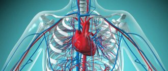
Circulation circles: where and from where does blood move through the vessels?
The main function of the heart is to ensure adequate delivery of blood to all structures of the body. This task is achieved through the coordinated work of the cardiovascular and respiratory systems.
Schematic representation of blood circulation in the body:
In functional anatomy, there are two circles through which the blood moves (large and small) and goes through the stages of providing the body with oxygen, nutrients and removing toxic metabolites (metabolic products).
Big circle
Arterial blood is transported through a large circle of circulation, starting from the cavity of the left ventricle. During contraction of the latter, fluid enters the aorta - the largest vessel of the human body, the individual branches of which deliver nutrients throughout the body:
- coronary vessels;
- the subclavian artery, the branches of which supply the organs of the head, neck, and structures of the upper limb;
- intercostal and bronchial, providing trophism to the mediastinal organs, lungs and structures of the chest wall;
- the celiac trunk, renal and mesenteric arteries supply all organs of the digestive tract, urinary system, and abdominal wall;
- bifurcation (bifurcation) of the aorta into common iliac arteries provides trophism to the structures of the pelvis and lower extremities.
Blood is transported through vessels with a gradual narrowing of diameter: from arteries and arterioles to capillaries. The cell wall of the latter has large pores through which oxygen and nutrients move to the tissues along a concentration gradient.
The waste blood is collected in the final section of the capillary, then through the venules and to the main vena cava, which flows into the cavity of the right atrium:
- lower - from the structures of the abdominal cavity, pelvis, soft tissues of the legs;
- upper - from the organs of the head and neck, part of the chest cavity.
Small circle
Venous blood entering the right side of the heart is enriched with carbon dioxide, high concentrations of which have a depressing effect on the respiratory and vasomotor centers of the brain. Gas is removed using the pulmonary circulation, starting from the right ventricle:
The pulmonary trunk, which divides into the right and left arteries.- Lobar and segmental arteries.
- Pulmonary capillaries, which are part of the airborne barrier. The thin walls of the alveoli and blood vessels facilitate the movement of oxygen and carbon dioxide along a diffusion mechanism (concentration gradient).
- Venules that flow into the main veins (two from each lung) and carry blood to the left atrium.
The name of the vessels is determined not by the composition of the blood, but by the direction in relation to the heart: fluid moves through the veins towards the organ, through the arteries - away from it.
Evolutionary development of organisms
The evolution of vertebrates began with the lancelet.
This organism already has a notochord and a neural tube. And also the most primitive heart for vertebrates: a pulsating abdominal vessel.
Further complexity of the organization led to the formation of fish. Organisms that breathe with gills have a two-chambered heart and one circulation.
Amphibians and most reptiles have a three-chambered heart. This also increases their vital energy.
Birds and mammals are at the pinnacle of evolution. The heart is formed by four chambers. There are no openings between the atria, nor between the ventricles. The two circles of blood circulation are completely separated. Therefore, birds and mammals have warm blood, which sharply distinguishes them from other animals. Humans, of course, also belong to this group.
Cardiac cycle
Adequate blood supply to the body is ensured by the coordinated contraction of the muscle fibers of the heart wall, which determine the organ’s work cycle.
There are two main phases:
- systole – contraction;
- diastole – relaxation.
The different speeds of impulse conduction through atypical cardiomyocytes with the presence of a delay in the atrioventricular node ensure the coordinated functioning of the organ: during atrial systole, blood penetrates into the ventricles. The latter are in the relaxation phase, which forms a sufficient volume to be filled with liquid (up to 100 ml on the left).
During contraction of the ventricles, the valves of the aorta and pulmonary artery open, the valves of the atrioventricular connections are closed - blood flows into the circulation. The pulse is determined in the peripheral vessels, and the heartbeat is determined in the chest area.
At this time, the atria are in the diastole phase and are filled with blood from the hollow (right sections) and pulmonary veins (left).
There is a statement that the heart works half its life and rests half of its life, since the duration of systole and diastole is the same (0.4 seconds each).
Pain of non-cardiac origin
Intercostal neuralgia
Intercostal neuralgia is often mistaken for heart pain. It does resemble angina, but there are significant differences. Neuralgia is characterized by a sharp shooting pain, which intensifies with movements, turns of the body, coughing, laughter, inhalation and exhalation. The pain may go away quickly, but can last for hours or days, intensifying with every sudden movement. Neuralgia is localized locally on the left or right between the ribs; the pain can radiate directly to the heart, lower back, back or spine. Usually the patient can pinpoint the exact location of the pain.
Osteochondrosis
With thoracic osteochondrosis, a person experiences pain in the heart, which radiates to the back, upper abdomen, shoulder blade and intensifies during movement and breathing. There may be a feeling of numbness in the interscapular area and left arm. Many people mistake their condition for angina, especially if the pain occurs at night and there is a feeling of fear. You can distinguish heart pain from osteochondrosis by the fact that in the latter case nitroglycerin will not help.
Digestive diseases
Pain in the chest usually occurs due to muscle spasms in the walls of the stomach. Symptoms such as nausea, heartburn, and vomiting will help you find out their true origin. These pains last longer than heart pains and have a number of features. They depend on food intake: for example, they appear on an empty stomach and disappear after eating. Nitroglycerin does not help with such conditions, but antispasmodics are effective.
Symptoms of acute pancreatitis are very severe pain that can be mistaken for heart pain. The condition is similar to a heart attack, and in both cases nausea and vomiting are possible. It is almost impossible to remove them at home.
With spasm of the gallbladder and bile ducts, it seems that the heart hurts. Although the liver and gall bladder are located on the right, severe pain radiates to the left side of the chest. In this case, antispasmodics help.
Severe pain due to a hernia of the esophagus (the opening of the diaphragm) is similar to angina. It appears at night when a person is in a horizontal position. Once you take a vertical position, your condition improves.
central nervous system
With disorders of the central nervous system, frequent and prolonged pain is observed in the chest area, namely in the apex of the heart, that is, in the chest from the bottom left. Patients describe the symptoms differently, but, as a rule, these are constant aching pains, which are sometimes acute and short-lived. Pain due to neuroses is always accompanied by sleep disturbances, irritability, anxiety and other manifestations of autonomic disorders. In this case, sedatives and sleeping pills help. A similar picture can be observed during menopause.
In some cases, cardioneuroses are difficult to distinguish from ischemic heart disease, since there may be no changes on the ECG in both cases.
Heart functions
The heart is rightfully considered the main organ of the human body, because disruption of its functions causes total disorders, and stopping activity leads to the death of the patient.
The main functions of the human heart:
automatism - independent synthesis of nerve impulses for myocardial contraction;- conductivity – atypical cells ensure the smooth functioning of different parts of the organ’s muscles;
- pumping function - pumping blood throughout the body with sufficient pressure for delivery to the periphery;
- gas exchange is ensured by the work of a small circle on the principle of an oxygen concentration gradient;
- endocrine role - natriuretic hormone is produced in the wall of the left atrium, which affects the functioning of the kidneys and the excretion of salts from the body.