heart transplantation
Until recently, decompensated heart failure was considered a death sentence. However, today, when medicine has stepped forward, heart transplant techniques have appeared, patients have a chance to live. But, despite all the progress, heart transplantation is still considered a complex, high-tech operation that requires careful preparation.
Indications for heart transplantation
For transplantologists, careful selection of the recipient – the patient who will receive a heart transplant – is important. First of all, we consider those people who are no longer helped by medications, but who can return to an active life if they have a healthy organ. Indications for surgery are:
- left ventricular ejection fraction less than 20%;
- Na in blood serum is less than 135 mEq/l;
- wedge pressure in the pulmonary artery more than 25 mmHg;
- the level of norepinephrine in the blood plasma is more than 600 pkg/ml;
- cardiothoracic index more than 0.6;
- decrease in maximum VO2 less than 10 ml\kg\min.
It is important that these indicators remain intact with the maximum possible drug support. With these indicators, the prognosis for one-year survival without a heart transplant is less than 50%.
ASK ANY QUESTION ABOUT HEART TRANSPLANTATION TO SPECIALISTS OF LEADING CLINICS
Who is a transplant indicated for?
Candidates for a heart transplant are patients with pathology that does not predict more than a year of life using other treatment methods. These include patients with:
- pronounced signs of heart failure with the slightest movements, at rest, if the ejection fraction during ultrasound examination is below 20%;
- dilated and ischemic cardiomyopathy;
- malignant arrhythmias;
- congenital heart defects.
Previously existing age restrictions (up to 65 years) are currently not considered decisive. For a child, the timing of the operation is determined by the most optimal preparation and the ability to provide complete immune protection.
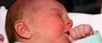
This baby needs urgent surgery to save his life.
Preparing the patient for surgery
Heart transplantation is a complex procedure that requires careful preparation. The patient undergoes a multi-stage examination before a decision is made about the possibility of organ transplantation. Necessary:
- carefully collect anamnesis of the disease;
- perform a chest x-ray;
- take a maximal oxygen uptake (VO2) stress test;
- undergo a series of tests for infections to exclude hepatitis, HIV and a number of other diseases that may be a contraindication;
- undergo routine urine and blood tests to assess the general condition of the body;
- undergo cardiac catheterization with tonometry of the right side (necessary to exclude pulmonary hypertension, which will be a contraindication to transplantation);
- take a test to evaluate human lymphocyte antigen (HLA);
- undergo an ECG, EchoCG.
The examination may reveal both absolute and relative contraindications that exclude the patient from being placed on the waiting list for a donor heart.
Late stage complications
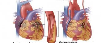
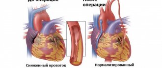
After several years, the likelihood of infections and rejection decreases significantly. But a person faces other complications, the most common being a decrease in the lumen of the capillaries. This is the leading cause of death in late stages after transplantation.
The problem of capillary narrowing can only be determined when all complications can be overcome in the early stages. Today, doctors are successfully coping with this pathology. Saving a life after replacing a person’s heart is possible only if the narrowing of the arteries is diagnosed in a timely manner.
Today, heart transplantation is the method of choice for many patients with cardiovascular diseases. The number of people who have already had this operation increases every year. Despite the fact that transplantation carries many complications, this operation is in great demand.
Transplantation technique
Due to decompensation of the disease, not all patients have the opportunity to immediately undergo transplantation. If the patient's condition is unsatisfactory, they can use:
- Pharmacological bridge to transplantation is a technique in which the patient’s condition is brought to satisfactory with the help of medications (mainly infusion ionotropic agents).
- A mechanical bridge to transplantation is a technique based on mono- or biventricular bypass of the ventricles or the use of an artificial heart in the preoperative period.
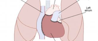
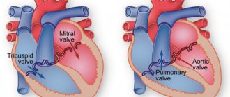
The operation takes place under conditions of established artificial circulation. Transplantation can be performed heterotopically or orthotopically. In the first case, the donor heart is placed under the patient's heart without removing the latter. This technique is used for severe hypertension in the pulmonary circulation. In other cases, a donor heart replaces the patient's own organ.
Complications at an early stage
Most often, the new organ is rejected. To prolong life after a heart transplant, patients are required to take medications that inhibit the synthesis of T-lymphocytes. Moreover, the use of these drugs is lifelong.
Clinical symptoms of rejection may vary. First signs:
- weakness;
- elevated temperature;
- dyspnea;
- migraine.
Treatment of rejection consists of administering increased dosages of glucocorticosteroids, performing plasmaphoresis and other measures aimed at removing toxic substances.
Another complication is infectious diseases, which are the result of a decrease in human immunity.
Postoperative period
After the operation, the patient is under medical supervision for a long period of time. During this period, immunosuppressive therapy with the help of hormones and cytostatics is selected, thanks to which it is possible to avoid rejection processes, the condition of the postoperative wound is assessed, and complications are corrected if they arise.
In the first month after surgery, a myocardial biopsy is performed every 1.5-2 weeks. This is necessary to assess the condition of the donor heart. Over time, this procedure is performed less and less often.
Demikhov's early years
Vladimir Petrovich Demikhov was born on July 31, 1916 on the Kuliki farm, Volgograd province, into a peasant family. The father, Pyotr Yakovlevich, died three years after the birth of his son, during the Civil War. His mother, Domnika Aleksandrovna, raised three children alone: the eldest Slava was only 6 years old at the time of her father’s death, Vladimir was 3 years old, and her daughter Yulia was still a baby. Already in childhood, the boy was drawn to the secrets of the heart - his mother later recalled that once little Vladimir received a scolding for trying to cut a dog.
While still a young man, Demikhov attended a factory apprenticeship school to become a repairman, and in 1934 he entered Voronezh University. There, at the conference, having made a report, he so impressed the physiologist Sergei Bryukhonenko that he recommended young Demikhov to transfer to the Faculty of Biology of Moscow State University, to the Department of Human and Animal Physiology.
The urge to construct models of internal organs was already there - immediately after moving, Demikhov sold his only suit and used the money to buy silver plates to create a model of an artificial heart.
Already in his junior year, Demikhov was actively involved in scientific work and in 1937 he independently designed and transplanted an artificial heart into a dog. It was driven by an electric motor. The dog lived for another two hours after the operation - a huge success at that time.
In 1940, Demikhov graduated from the university, and a year later the war began, which Demikhov went through as a senior laboratory assistant in a pathological-anatomical laboratory - since he was a biologist by profession, not a physician - and finished his service with the rank of senior lieutenant in Manchuria. During the war, life confronted him with the extremely talented surgeon Boris Petrovsky. Sincerely caring for the truth, Demikhov several times pointed out to Petrovsky certain errors in the procedures. The ambitious Petrovsky harbored a grudge. Many years later, he will be one of those who will prevent Demikhov from receiving his well-deserved fame.
In 1945, almost immediately after the war, he came to the Institute of Experimental and Clinical Surgery. Then he meets his future wife, Liya Nikolaevna, and the marriage gives birth to a daughter, Olga, now a professor and doctor.
In 1946, Demikhov successfully transplanted a second heart into a dog, and soon completely replaced both the heart and the lung - this became a sensation in the world of science and medicine, but in the USSR the operation was treated extremely coolly.
From 1946 to 1959, Demikhov performed a total of 250 second heart transplant operations, with one of the dogs surviving for 32 days.
Source
In 1948, Demikhov began experiments on liver transplantation, and a few years later - heart transplantation. All this is being done in order to finally move on to the most difficult and seemingly impossible operation - a human heart transplant. Demikhov is sure that it is only a matter of time. But a heart transplant from a deceased donor, in addition to medical issues, also raised an ethical question: if the heart is still beating, can the donor be considered officially dead? And if the donor is not dead, then how ethical is it to transplant his organs into the recipient? In fact, it was precisely because of this that a ban on organ transplants was imposed in the USSR, and the desire of doctors to develop was considered “contrary to the principles of communist morality.” This issue was finally resolved only in 1992, when the law on organ transplantation was passed, defining irreversible death as brain death.
World experience and prices
As practice shows, a transplanted heart, if all medical recommendations are followed, can work without interruption for 5-7 years. However, the aging process in the donor organ proceeds faster than in its own, and therefore sooner or later the patient will note the return of all symptoms of heart failure.
The actual practice of heart transplantation differs significantly from country to country. In some countries, these operations are not carried out at all because they are not regulated at the legislative level. In other countries, such operations are clearly regulated and both residents of the country itself and foreign citizens can resort to it. For example, in the Republic of Belarus, organ transplantation is carried out in accordance with the law, which is based on the “presumption of consent”: organ collection from a donor can be carried out after declaring brain death, unless wishes to the contrary are expressed by the patient before death. The organ can be used for further patients in need.
SELECT A FREE CLINIC FOR HEART TRANSPLANTATION
Among the factors leading to deterioration of the condition of the transplanted heart in the long term, specific graft vasculopathy is especially distinguished. However, this is only one of the significant factors, the final result of which is the development of post-transplant restrictive cardiomyopathy [1, 3, 5].
Understanding the reasons for failure in the long-term period after heart transplantation is the main opportunity to overcome the stagnation of results. This publication provides an analysis of the clinical results and histological and ultrastructural organization of the myocardium of the donor heart, based on the study of heart biopsies of patients who survived 10-22 years after transplantation.
Material and methods
By July 2012, at the Russian Scientific Center for Surgery named after. acad. B.V. Petrovsky performed 29 orthotopic heart transplantation operations. 23 patients survived the hospital period (1-3 months p/o) - hospital mortality was 20.7%. A detailed analysis of the results and causes of failure during the hospital period and the first 10 years after transplantation is presented in our earlier publication [1].
9 (45%) patients survived the ten-year period, 5 - 15 years, of which 2 people have already lived more than 20 years after surgery.
Ultrastructural studies of the myocardium and microvasculature were performed regularly from the 10th to the 22nd year after transplantation in 6 recipients (5 men and 1 woman) aged from 33 to 66 years (average age - 47.6 years). Coronary angiography followed by biopsy of the right ventricle was performed annually (or twice a year).
Biopsy samples obtained from each recipient were studied: 3-4 samples from the wall of the right ventricle and the interventricular septum. One of the biopsy specimens, after routine histological processing, was embedded in paraffin and the prepared sections were stained with hematoxylin and eosin and Massan. Semi-thin sections were prepared from other samples embedded in epoxy resin and, after removing the resin, stained with histological dyes: hematoxylin and eosin, toluidine blue, and hyacinth violet.
Ultrathin sections with a thickness of 50-70 nm were prepared from the same blocks, contrasted with uranyl acetate, lead citrate, viewed and photographed in a JEM-100CX electron microscope at an accelerating voltage of 80 kV with a magnification from 5000 to 32,000.
Results and discussion
The morphological picture in the myocardium of the donor heart had similar qualitative changes in the long-term period after surgery (more than 10 years), despite clinical differences between recipients (gender, age, initial diagnosis, features of the postoperative period and treatment). This allows us to assume certain patterns in the process of morphological adaptation of donor hearts in general (see table).
The state of the microvasculature had an uneven mosaic character.
Patient K. There was a pronounced reduction of the microvasculature, which progressed over time. But even these few capillaries were often surrounded by a connective tissue “muff”. Electron microscopy revealed a sharp expansion of the basement membrane of the capillaries. Over the years, the decrease in the density of the microvasculature became even more pronounced.
Patient P. The walls of the capillaries are tortuous, thinned, unevenly expanded; signs of “sludge” of blood cells in the lumen of the capillaries could often be seen. Small intramural arteries and arterioles had thickened, homogenized walls, sometimes their lumen was practically not defined (Fig. 1).
Figure 1. Micrograph. Myocardium of patient P. Thickening of the wall of a small artery, the lumen is practically not defined. Semi-thin section, stained with hematoxylin and eosin. About. 60, approx. 10.
Electron microscopy revealed that the endothelial cells of the capillaries were thinned, and the level of micropinocytosis in them was reduced. Over time after transplantation, the number of capillaries per unit area visually decreased.
Patient Sh. The number of capillaries is noticeably reduced. The endothelium of the capillaries is thickened and thinned in some places. The lumen of the vessels was expanded, the “sludge” phenomenon was observed, as well as the adhesion of blood cells to the capillary wall. Diapedetic hemorrhages occurred. Over time, this structure of the microvasculature did not change significantly.
Patient G. By the 10th year of research, the capillary bed was practically unchanged, however, thickening and homogenization of the wall were noted in some arterioles. Over time, a gradual decrease in the density of the capillary bed, perivascular sclerosis, and a decrease in the level of micropinocytosis in the endothelial cells of the capillaries were revealed; in some capillaries, tortuosity of the luminal surface was noted, sometimes with the separation of protruding parts of the endothelial cells into the lumen of the vessel (Fig. 2).
Figure 2. Micrograph. Ultrastructure of the myocardium of patient G., tortuosity of the luminal surface of the capillary with separation of parts of endothelial cells into the lumen of the capillary. Ultra-thin section, double contrast. Uv. 12,000. Adhesion of blood cells to the surface of endothelial cells was often observed. The walls of the microvessels were sharply osmiophilic—impregnated with plasma; diapedetic hemorrhages were observed in the myocardial interstitium near the vessels.
Patient R. Entire myocardial fields were observed, where individual hibernating cardiomyocytes were immured among masses of fibrous tissue (Fig. 3).
Figure 3. Micrograph. Myocardium of patient R. Fields of interstitial sclerosis, single cardiomyocytes with signs of hibernation are “immured” in the thickness of the sclerotic interstitium. Semi-thin section, stained with hematoxylin and eosin. About. 40, approx. 12. Other areas of the biopsy specimens were sharply hypertrophied cardiomyocytes, among which single capillaries were found in the layers of connective tissue.
Patient Kr. There was a sharp expansion of the capillary bed, plethora of microvessels; there was no visible decrease in the density of the capillary bed during the entire observation period (Fig. 4).
Figure 4. Micrograph. Myocardium of patient Kr. Expansion of the capillary bed, signs of “sludge” of formed elements in microvessels. There is no visible decrease in the density of the capillary bed. Semi-thin section, stained with hematoxylin and eosin. About. 20, approx. 12.
There was an increase in the electron density of the cytoplasm of endothelial cells, apparently associated with their plasma impregnation. Over time after transplantation, the state of the microvasculature did not change significantly.
Changes in cardiomyocytes and the interstitial substance of the myocardium of the donor heart also had similar features with certain quantitative differences.
In the long-term period after transplantation
There were no episodes of pronounced rejection reactions.
Some cardiomyocytes are in a state of dedifferentiation, characteristic of the hibernation state: atrophy and loss of striation of myofibrils, accumulation of glycogen and small mitochondria in the central part of the cardiomyocyte (Fig. 5, a).
Figure 5. Micrograph. Ultrastructure of the myocardium of the right ventricle of a transplanted heart 20 years after surgery. a — chromatin margination in the nucleus; accumulation of small enlightened mitochondria, devoid of intermitochondrial contacts, uv. 11,000.
Another group of cardiomyocytes has oddly shaped nuclei containing large nucleoli, often with accumulation of chromatin at the periphery. In this case, mitochondria have different electron densities, are reduced in size, and the specific contours between them are disrupted. Taken together, this indicates both the initial signs of apoptotic degeneration and an extensive form of apoptosis of cardiomyocytes with the formation of apoptotic bodies (see Fig. 5, b).
Figure 5. Micrograph. Ultrastructure of the myocardium of the right ventricle of a transplanted heart 20 years after surgery. b — advanced phase of apoptosis of cardiomyocytes, condensation of nuclear remnants and organelles, beginning of the formation of apoptotic bodies, uv. 12,000.
And finally, as the duration of the post-transplantation period increases, in the myocardium of the donor heart, cells with large nuclei and a narrow rim of cytoplasm are more often detected in the immediate vicinity of cardiomyocytes, which, by their morphology, can be classified as pluripotent (stem?) cells (see Fig. 5 , V).
Figure 5. Micrograph. Ultrastructure of the myocardium of the right ventricle of a transplanted heart 20 years after surgery. c - a cell with a large nucleus and a narrow rim of cytoplasm (near the modified cardiomyocytes), resembling a stem cell in ultrastructure, uv. 12,000.
It is important that in the long-term period after surgery the number of capillary vessels per unit area of myocardial section decreases. This process is natural, but uneven and is expressed to varying degrees in different patients.
An electron microscopic study showed that over time, in biopsy samples of a transplanted heart, the capillary becomes an almost rare structure. The preserved capillaries are often surrounded by a connective tissue “muff”.
It can be assumed that progressive microcirculation disorders may underlie chronic ischemia of the donor myocardium, resulting in hibernation and apoptosis of cardiomyocytes [2].
We have previously reported that from the first months after transplantation, all recipients experience hyperlipidemia, which is more pronounced with the initial diagnosis of ischemic cardiomyopathy. It was this fact that served as the basis for the fact that since the mid-90s we have included exchange plasmapheresis in the mandatory protocol for postoperative therapy [4]. Planned plasmapheresis sessions, starting from the first month after transplantation, were performed in only one patient, Kr., who underwent anatomical heart transplantation 16 years ago. Then they were repeated at least 3-4 sessions annually. Despite the constant tendency towards severe hyperlipidemia and initial severe ischemic heart disease, it was in this recipient that minimal microcirculation disorders were noted in the donor heart. In our opinion, this observation requires careful further analysis.
An interesting and significant fact is that after 10 years, not a single recipient showed morphological signs of a rejection reaction. Despite this, coronary angiography is absolutely necessary for these patients (sometimes 2 times a year!) for the timely detection of local stenoses in the epicardial coronary arteries and the performance of angioplasty in order to prevent myocardial infarction.
Data obtained from ultrastructural study of the donor heart correlate with the results of clinical and functional monitoring of recipients in the long-term period after transplantation. Transplant dysfunction begins to develop according to the diastolic type of heart failure, which is based on increasing myocardial sclerosis (subendocardial, interstitial). Compensatory hypertrophy of functioning cardiomyocytes (in the absence of a rejection reaction) allows maintaining normal ejection fraction values for a long time. According to M. Bilingham and C. Berry [3], the myocardial mass of a transplanted heart increases significantly with time from the moment of transplantation.
Thus, the condition of the donor heart in the long-term period after transplantation (dystrophy-hypertrophy of cardiomyocytes, impaired microcirculation, interstitial sclerosis) can, in our opinion, be considered as a special form of post-transplantation cardiomyopathy, the causative factors of which cover all periods of possible myocardial damage, starting from the donor stage .
Sources
- Toker E., Aktaş S. The childbirth experiences of Syrian refugee mothers living in Turkey: a qualitative study. // J Reprod Infant Psychol - 2021 - Vol - NNULL - p.1-17; PMID:33896296
- Wang B., Zhao D., Lu T., Liu S., Rong C. Quantifications and Applications of Relative Fisher Information in Density Functional Theory. // J Phys Chem A - 2021 - Vol - NNULL - p.; PMID:33891419
- Dimentberg E., Cardaillac C., Richard E., Plante AS., Maheux-Lacroix S. Translation and Cultural Validation of the WERF EPHect Endometriosis Patient Questionnaire into Canadian French. // J Obstet Gynaecol Can - 2021 - Vol - NNULL - p.; PMID:33887447
- Bhandari D., Kotera Y., Ozaki A., Abeysinghe S., Kosaka M., Tanimoto T. COVID-19: challenges faced by Nepalese migrants living in Japan. // BMC Public Health - 2021 - Vol21 - N1 - p.752; PMID:33874937
- Samkange-Zeeb F., Samerski S., Doos L., Humphris R., Padilla B., Bradby H. “It's the First Barrier” - Lack of Common Language a Major Obstacle When Accessing/Providing Healthcare Services Across Europe. // Front Sociol - 2021 - Vol5 - NNULL - p.557563; PMID:33869495
- Elshahat S., Newbold K.B. Physical activity participation among Arab immigrants and refugees in Western societies: A scoping review. // Prev Med Rep - 2021 - Vol22 - NNULL - p.101365; PMID:33868904
- Ono K., Yoshioka N., Hage D., Ibaragi S., Tubbs RS., Iwanaga J. Correction to: Duplication of the external jugular vein: a language barrier of database search in classic anatomical studies. // Surg Radiol Anat - 2021 - Vol - NNULL - p.; PMID:33856506
- Noack E.M., Schulze J., Müller F. Designing an App to Overcome Language Barriers in the Delivery of Emergency Medical Services: Participatory Development Process. // JMIR Mhealth Uhealth - 2021 - Vol9 - N4 - p.e21586; PMID:33851933
- Schrot-Sanyan S., Kolanska K., Haimeur Y., Varlas V., Parisot-Liance L., Daraï E., Bornes M. Language barrier as a risk factor for obstetric anal sphincter injury—A case-control study. // J Gynecol Obstet Hum Reprod - 2021 - Vol50 - N8 - p.102138; PMID:33831603
- Kyaw PP., Geater AF. Healthcare seeking preferences of Myanmar migrant seafarers in the deep south of Thailand. // Int Marit Health - 2021 - Vol72 - N1 - p.1-9; PMID:33829467
Things are getting worse
Then Kovanov and academician Boris Vasilyevich Petrovsky, leaders of the delegation of Soviet doctors in Munich, organized a party meeting. Everyone was present except Demikhov (he was a non-party member). To prevent Demikhov from finding out anything, the meeting was disguised as a walk. Lined up in rows, the doctors conveyed to each other the thoughts of the speakers in a chain. Kovanov said that Demikhov was supposedly going to stay, and the task of the communists was to prevent this. It is necessary to inform the authorities, and a trusted comrade must lure the vile defector out of the room. This was entrusted to Gleb Mikhailovich Solovyov, Petrovsky’s most talented and beloved student.
Demikhov was generally trusting, and especially towards gifted people. From such a bright young surgeon as Gleb Mikhailovich, he did not expect a trick. Solovyov lied that another magazine, Der Spiegel, was also asking to do the operation live, and now a car would arrive from them. And the car turned out to be an embassy car: under the escort of KGB officers, the imaginary defector went to Moscow. From around the corner, Petrovsky and Kovanov personally observed the progress of the operation. And then the three of them with Solovyov went to celebrate their luck in the very beer hall where Hitler once proclaimed the creation of his party.
Go to action
Arriving from the front in Moscow, he got a job teaching physiology at the Fur Institute, where he received premises for experiments. Then I found my classmate Aron Gurvich, with whom I had once argued a lot about immunity. Wounded in the war, Gurvich worked at the Institute of Biological and Medical Chemistry. He helped a friend - he brought morphine for anesthesia from work and caught two stray puppies in Neskuchny Garden. On February 23, 1946, the heart of one dog was transplanted into another, and the recipient lived with both hearts for 15 minutes. This was done without hypothermia, a heart-lung machine, or immune suppression drugs.
In the same year, Demikhov managed to transplant both a heart and lungs into a dog, and the animal lived for 6 days. This was the end of the researcher’s work at the Fur Institute - the authorities considered that he was doing nonsense.
Main works
In 1951, at a session of the USSR Academy of Medical Sciences, Demikhov transplanted a donor heart and lungs into the dog Damka. She lived another whole week. The dog died not from heart problems, but from damage to the larynx. It was a worldwide sensation, but the achievement went unnoticed in the USSR.
Demikhov did not stop at the cardiopulmonary device and in the same year he transplanted a liver, and a little later, already in 1952, he came up with a mammary-coronary bypass scheme: a healthy vessel was connected to the heart, and it began to work in place of the damaged one. The scheme was immediately tested - of course, on a dog: plastic cannulas and tantalum clips were used to fasten the arteries. The work was carried out quickly - 2 minutes for all manipulations. For this work, Demikhov received the first prize named after Burdenko.
One of the dogs operated on by Demikhov lived for more than 7 years without experiencing any particular health problems. Ultimately, Demikhov learned to connect up to 4 cardiopulmonary complexes to one animal, and most importantly, to keep them in a functioning state for a whole week.
In 1955, Demikhov took aim at an almost fantastic idea - and transplanted a second head into the dog. This becomes one of his most famous works - he will create about 20 such dogs over all the years of work. Demikhov then worked at the 1st Moscow Medical Institute named after Sechenov. The laboratory was located in a former church and was very small; instead of an operating lamp, the surgeons had an ordinary pre-war incandescent light bulb.
Dog head transplant. Source
The first two-headed dog seemed like a fantastic alien from outer space - the heads had united blood vessels. For the experiment, Demikhov selected a large adult dog and a large puppy. Next, the puppy's torso was cut through the middle part of the chest, and the front part, with the heart and lungs removed, was transplanted onto the neck of the whole dog. In the process of stitching, a general circle of blood flow was formed, and the puppy's head began to live due to the breathing and blood circulation of the large dog. Both heads stuck out their tongues in the heat, eating and drinking; one of the heads systematically tried to bite the second on the ears - unlike the experiment of Alexis Carrel, whose dog only demonstrated a number of reflexes, Demikhov’s creation seemed quite alive and adequately perceives the world around him. The longest-lived head lived for a whole month and even managed to grow. So Demikhov proved that even the head - this incredibly complex organ - can be successfully transplanted.
Despite the success, most of the animals still died quickly, and Demikhov sincerely believed that infections and technical limitations were responsible for this, being unable to prove the need for tissue compatibility.
In those years, compatibility was checked only by the similarity of blood groups - Demikhov checked them in both dogs and even checked blood smears after surgery, finding nothing other than a slight inflammatory process. There was no scientific proof of tissue compatibility, nor was there a way to test for tissue compatibility before surgery. Demikhov himself wrote:
“It is necessary to carefully study the antigenic composition of donor and recipient cells, as well as to find ways to eliminate their immunobiological differences... Immunologists need to find more sensitive methods that allow them to detect differences during transplantations in animals within a species.”
Diagram of a two-headed dog with united circulation circles.
Source In 1958, Demikhov was invited to Germany. He performed 12 operations there: 10 heart transplants and 2 head transplants for dogs. But, being a keen scientist, at some point, while making a report, he entered into a debate too hotly and told more than the KGB required. Very quickly after this, Demikhov was deported back to the Union, accused of revealing state secrets and wanting to stay in the West. Then his colleagues and cousin General S.M. stood up for Demikhov. Shtemenko - and the pressure on him was eased. But since then Demikhov has been restricted from traveling abroad.
In 1960, his laboratory was visited by a then unknown surgeon from South Africa, Christian Barnard, who would later call Demikhov “the father of heart and lung transplantation.” He came one more time - in 1963, like many other young transplantologists from all over the world.
All this time, Vladimir Petrovich remained only a junior researcher, having neither degrees nor titles. In 1960, Demikhov, due to a quarrel with Vladimir Kovanov, his immediate director, was forced to move to the Sklifosovsky Institute of Emergency Medicine - Kovanov did not allow Demikhov’s dissertation “Experimental Vital Organ Transplantation” to be defended.
At the Institute of Emergency Medicine he was given a laboratory, but it was located in a damp basement: the floor was covered with boards under which water squelched, most of the equipment was built on the knee, and instead of a compressor they used a Buran vacuum cleaner.
Here, in 1962, Demikhov transplanted a second heart into the dog Grishka. Grishka lived a record 142 days after the operation - and would have lived longer if he had not fallen victim to a drunken robbery: the dog was trying to protect his possessions. Demikhov had a hard time with the death of his experimental subject and since then he always took all the operated dogs home.
Demikhov was still trying to defend his dissertation - he had long accumulated material not only for a candidate’s thesis, but also for a doctorate. Finally, in 1963, at the Faculty of Biology and Soil Sciences of Moscow State University, he defended both at once, receiving a Doctor of Science degree. The defense was difficult - many opponents of the brilliant surgeon were present, and there were cries from the audience about the unscientific nature of the experiment and quackery.
Heritage
In 1996, Michael DeBakey, a renowned surgeon who was to become a consultant on President Yeltsin’s upcoming operation, came to Moscow, and the first thing he asked was: “Can I bow to Academician Demikhov?” Demikhov was found with difficulty. The state remembered the outstanding surgeon and then awarded the first and only order - “For Services to the Fatherland”, III degree.
Demikhov’s works are known and remembered all over the world - Christian Barnard was not lying when he called Vladimir Petrovich the father of transplantology.
Crisis and later years
By 1965, Demikhov, already a Doctor of Science, a world-famous transplant surgeon, was aiming for new heights: at one of the meetings of the Society of Transplantologists he was going to present a project for organ conservation. Demikhov wants to create a bank of animals that would house the organs of deceased donors. But not everyone likes the idea.
Petrovsky, who once met Demikhov by chance during the war, did not forget the arrogant, from his point of view, laboratory assistant. By 1965, he became Minister of Health and tried to destroy his rival. He writes about Demikhov :
“Demikhov is certainly an interesting person, a fanatic, and spoiled by the lack of leadership. From the surgeon's point of view, it has very large defects. From a physiological point of view, there is a complete lack of experimental control. He performs the operation and then does not monitor the dog. Dogs often die from bleeding. He has no surgical training. It works on a pure idea.”
Source
When Vladimir Petrovich talks about his idea at the transplantology section, those present accuse him of quackery. The chairman of the section, Professor Ostroverkhov, reproaches Demikhov for the low level of his experiments and calls Demikhov’s experiments “nonsense.” Everyone is silent. To defend Demikhov means to directly go against Petrovsky. But one person still stands up - Tatyana Andreevna Grigorieva, head of the department of histology at the 2nd Moscow Medical Institute.
“What is happening is not a meeting of a scientific society. This is a vile trial of an outstanding scientist of the world,” says Grigorieva.
Others stand behind her, and most importantly, they give Demikhov himself the opportunity to speak. In the end, the scientist is acquitted, but both he and his supporters know that this is only the first attack. Demikhov is not broken yet, but he is already close to it. His health begins to deteriorate due to the nervous tension he has experienced.
A few years later, the state decides to take away Demikhov’s new apartment, justifying this by the fact that it was given to him by accident, by mistake. Demikhov is getting worse. His wife Liya Nikolaevna said that Demikhov was on the verge: he decided that if they came to evict him, he would throw himself out the window - but he would not complain.
Then the director of the Sklifosovsky Institute stands up for him, winning Demikhova a few more years.
In 1968, Demikhov suffered a stroke. He never recovered from it, but continued to work - his laboratory existed until 1986, and in 1988, Demikhov, as part of a group of surgeons, was awarded the State Prize for introducing bypass surgery into practice.
Gradually, the great scientist began to lose his memory and towards the end of his life he did not even leave the house alone. By the 1990s, he was almost completely forgotten, but most of his ideas not only came true, but also became absolutely routine - both coronary bypass surgery and organ transplants are now found everywhere.
On November 22, 1998, Vladimir Petrovich died from a ruptured aneurysm.
Animals can also change body parts
The driver explained to him that any part in the car can be replaced – it’s always like new. And then he uttered a phrase that decided the fate of world transplantology: “Animals can also change body parts.” And he said that the lizard grows a new one instead of a severed tail. Volodya immediately asked questions: “will the dog also grow a new tail?”, “and why does blood come from a cut off tail?” The teacher explained that a dog has a heart that pumps blood. Soon the mother caught Volodya with a knife in his hand, when he was about to cut the puppy’s side and understand how the heart works. The puppy was saved, but the son was flogged for being a knacker. It didn’t help: young Demikhov fell ill with a thirst for anatomical knowledge. Realizing that he would not make a gardener, his family sent Volodya to study.