What is total peripheral resistance?
Total peripheral resistance (TPR) is the resistance to blood flow present in the body's vascular system.
It can be understood as the amount of force opposing the heart as it pumps blood into the vascular system. Although total peripheral resistance plays a critical role in determining blood pressure, it is solely an indicator of cardiovascular health and should not be confused with the pressure exerted on arterial walls, which is an indicator of blood pressure.
Components of the vascular system
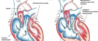
The vascular system, which is responsible for the flow of blood from and to the heart, can be divided into two components: the systemic circulation (systemic circulation) and the pulmonary vascular system (pulmonary circulation).
The pulmonary vascular system delivers blood to and from the lungs, where it is oxygenated, and the systemic circulation is responsible for transporting this blood to the body's cells through the arteries, and returning the blood back to the heart after being supplied.
Coronary
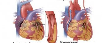
Regulation of coronary artery tone is a complex subject. There are a number of mechanisms to regulate coronary vascular tone, including metabolic demands (ie, hypoxia), neurological control, and endothelial factors (ie, EDRF, endothelin).
Local metabolic control (based on metabolic demand) is the most important mechanism for controlling coronary blood flow. A decrease in tissue oxygen content and an increase in tissue CO2 contents act as vasodilators. Acidosis acts as a direct coronary vasodilator and also enhances the effects of adenosine on the coronary vasculature.
What is opps in cardiology
Total peripheral resistance influences the functioning of this system and can ultimately significantly affect the blood supply to organs.
The total peripheral resistance is described by the partial equation:
OPS = change in pressure/cardiac output
The change in pressure is the difference between mean arterial pressure and venous pressure.
Mean arterial pressure equals diastolic pressure plus one-third of the difference between systolic and diastolic pressure. Venous blood pressure can be measured using an invasive procedure using special instruments that physically detects the pressure inside the vein.
Cardiac output is the amount of blood pumped by the heart in one minute.
Factors influencing the components of the OPS equation
There are a number of factors that can significantly influence the components of the OPS equation, thereby changing the values of the total peripheral resistance itself.
These factors include vessel diameter and the dynamics of blood properties. The diameter of blood vessels is inversely proportional to blood pressure, so smaller blood vessels increase resistance, thus increasing OPS. Conversely, larger blood vessels correspond to a less concentrated volume of blood particles exerting pressure on the vessel walls, meaning lower pressure.
Hydrodynamics of blood
Blood hydrodynamics can also significantly contribute to an increase or decrease in total peripheral resistance.
Behind this is a change in the levels of coagulation factors and blood components that can change its viscosity. As one might expect, more viscous blood causes greater resistance to blood flow.
Less viscous blood moves more easily through the vascular system, resulting in lower resistance.
An analogy is the difference in force required to move water and molasses.
References
- Fuster, V.; Alexander, R.W.; O'Rourke, R.A. (2004) Hearst's Heart, Book 1
. 11th edition, McGraw-Hill Professional, Medical Pub. Division. Page 513. ISBN 978-0-07-143224-5. - ^ a b
Table 30-1 c: Trudie A Goers;
Washington University School of Medicine, Department of Surgery; Klingensmith, Mary E; Lee Ern Chen; Sean S. Glasgow (2008). Washington Manual of Surgery
. Philadelphia: Wolters Kluwer Health/Lippincott Williams & Wilkins. ISBN 978-0-7817-7447-5 .CS1 maint: several names: list of authors (link to site) - ^ a b c d
Derived from values in dyn s/cm5 - University of Virginia Health System. "Physiology: Pulmonary Artery Catheters"
- ^ a b c d e
J. B. Thurston, Viscosity and viscoelasticity of blood in small diameter tubes, Microvascular Research 11, 133 146, 1976 - "Cardiac output and blood pressure." biosbcc. Retrieved April 7, 2011.
- Measuring Real Pulsatile Blood Flow Using X-Ray PIV Method with CO2 Microbubbles, Hanook Park, Eunseop Yeom, Seung-Joon Seo, Jae-Hong Lim and Sang-Joon Lee, NATURE, Scientific Reports 5
, Article Number: 8840 (2015), Doi: 10.1038/srep08840. - Masugata, H., Peters, B., Lafitte, S. et al. (2003). "Evaluation of adenosine-induced coronary steal in coronary occlusion based on the degree of opacification defects on myocardial contrast-enhanced echocardiography." Angiology
.
54
(4): 443–8. Doi:10.1177/000331970305400408. PMID 12934764.
Table 30-1 describing normal values for hemodynamic parameters is found in the fifth edition of the Washington Manual of Surgery.
Hemodynamic parameters
At the same time, depending on the greater or lesser severity of changes in regional vascular resistance, they will accordingly receive a smaller or larger volume of blood ejected by the heart.
This mechanism is the basis for the effect of “centralization” of blood circulation in warm-blooded animals, which ensures redistribution of blood, primarily to the brain and myocardium, in difficult or life-threatening conditions (shock, blood loss, etc.).
Resistance, pressure difference and flow are related by the basic equation of hydrodynamics: Q=AP/R.
Since the flow (Q) must be identical in each of the successive sections of the vascular system, the drop in pressure that occurs throughout each of these sections is a direct reflection of the resistance that exists in that section.
Thus, a significant drop in blood pressure as blood passes through the arterioles indicates that the arterioles have significant resistance to blood flow. The average pressure decreases slightly in the arteries, as they have little resistance.
Likewise, the moderate pressure drop that occurs in the capillaries is a reflection of the fact that capillaries have moderate resistance compared to arterioles.
The flow of blood flowing through individual organs can change tenfold or more.
Since mean arterial pressure is a relatively stable indicator of the activity of the cardiovascular system, significant changes in the blood flow of an organ are a consequence of changes in its general vascular resistance to blood flow. Consistently located vascular sections are combined into certain groups within the organ, and the total vascular resistance of the organ must be equal to the sum of the resistances of its sequentially connected vascular sections.
Since arterioles have significantly greater vascular resistance compared to other parts of the vascular bed, the total vascular resistance of any organ is determined to a large extent by the resistance of the arterioles.
Arteriolar resistance is, of course, largely determined by arteriolar radius. Therefore, blood flow through the organ is primarily regulated by changes in the internal diameter of the arterioles through contraction or relaxation of the muscular wall of the arterioles.
When the arterioles of an organ change their diameter, not only does the blood flow through the organ change, but the drop in blood pressure that occurs in that organ also undergoes changes.
Arteriolar constriction causes a greater drop in arteriolar pressure, resulting in an increase in blood pressure and a concomitant decrease in changes in arteriolar resistance to vascular pressure.
(The function of arterioles is somewhat similar to that of a dam: closing the dam gates reduces the flow and raises the dam level in the reservoir behind the dam and lowers the level downstream.)
On the contrary, an increase in organ blood flow caused by the dilation of arterioles is accompanied by a decrease in blood pressure and an increase in capillary pressure.
Due to changes in hydrostatic pressure in the capillaries, arteriolar constriction leads to transcapillary fluid reabsorption, while arteriolar dilation promotes transcapillary fluid filtration.
Peripheral vascular resistance refers to the resistance to blood flow created by blood vessels. The heart, as a pumping organ, must overcome this resistance in order to pump blood into the capillaries and return it back to the heart.
Peripheral resistance determines the so-called subsequent cardiac load. It is calculated by the difference in blood pressure and CVP and by MOS. The difference between mean arterial pressure and CVP is designated by the letter P and corresponds to a decrease in pressure within the systemic circulation.
To convert the total peripheral resistance to the DSS system (length•s•cm-5), it is necessary to multiply the obtained values by 80. The final formula for calculating peripheral resistance (Pk) looks like this:
To determine P, it is necessary to recalculate the CVP values in centimeters of water column into millimeters of mercury.
For such a recalculation there is the following relationship:
1 cm water. Art. = 0.74 mm Hg. Art.
In accordance with this ratio, it is necessary to multiply the values in centimeters of water column by 0.74. So, the central venous pressure is 8 cm of water. Art. corresponds to a pressure of 5.9 mmHg. Art. To convert millimeters of mercury to centimeters of water, use the following ratio:
1 mmHg Art. = 1.36 cm water. Art.
CVP 6 cm Hg.
Art. corresponds to a pressure of 8.1 cm water. Art. The value of peripheral resistance, calculated using the above formulas, reflects the total resistance of all vascular sections and part of the resistance of the systemic circle.
Peripheral vascular resistance is therefore often referred to in the same way as total peripheral resistance.
Pulmonary
The main factor determining vascular resistance is the small arteriolar
(known as arteriole resistance) tone. These vessels range from 450 microns in diameter to 100 microns. (For comparison, the diameter of the capillary is from 5 to 10 microns.)
Another factor determining vascular resistance is precapillary arterioles
. These arterioles have a diameter of less than 100 µm. They are sometimes called autoregulatory vessels because they can dynamically change in diameter to increase or decrease blood flow.
Any changes in blood viscosity (eg due to changes in hematocrit) will also affect the measured vascular resistance.
Pulmonary vascular resistance (PVR) also depends on lung volume, and PVR is lowest in functional residual capacity (FRC). The high compliance of the pulmonary circulation means that the degree of lung expansion has a major influence on PVR. This is primarily due to effects on the alveolar and extra-alveolar vessels. During inspiration, an increase in lung volume causes expansion of the alveoli and longitudinal distension of the interstitial alveolar vessels. This increases their length and decreases their diameter, thereby increasing the resistance of the alveolar vessels. On the other hand, a decrease in lung volume during exhalation leads to a narrowing of the extra-alveolar arteries and veins due to a decrease in the radial tract from adjacent tissues. This leads to an increase in extra-alveolar vascular resistance. PVR is calculated as the sum of the alveolar and extra-alveolar resistance, since these vessels are located in series with each other. Because alveolar and extraalveolar resistance increase at high and low lung volumes, respectively, the overall PVR takes the shape of a U-shaped curve. The point at which the PVR is lowest is near the FRC.