Published: 03/30/2021 17:00:00 Updated: 03/30/2021
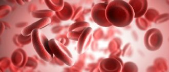
Erythrocytes are red blood cells, the most numerous blood cells. Formally, they are not cells, since during the process of maturation they lose many of the structures necessary for cells. For example, they lack nuclei and do not synthesize any protein molecules, unlike other cells in the body. So the name “cell” in this case is used for convenience. Red blood cells are formed in the bone marrow and constantly circulate in the body, performing the most important function of maintaining life - they carry oxygen from the lungs to tissues and organs and remove carbon dioxide.
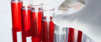
In addition to red blood cells, blood contains plasma, platelets, and leukocytes. However, the number of red blood cells is so large that just a couple of drops of blood contains about one billion of these cells. They make up about 40% of the total blood volume. Actually, it is red blood cells that give our blood its characteristic red color due to its hemoglobin content.
Red blood cells do not last forever, they wear out over time and eventually die. The average life cycle of a red blood cell is approximately 120 days—a total of four months. However, do not worry, the bone marrow is constantly producing new cells and maintaining the required level of red blood cells. Various unfavorable circumstances can reduce or, conversely, increase their reproduction rate and affect their life expectancy - thus, the balance of blood composition is disrupted. An increase or decrease in red blood cells is associated with various pathological conditions. Let's consider this issue in more detail.
Where are the cells grown?
Red blood cells are produced in the bones of the skull, bone marrow, spine and ribs. In childhood, there is another place of synthesis - the ends of the long tubular bones of the legs and arms.
The destruction of aged red blood cells occurs in the liver and spleen. They live on average 3 months. Any processes that disrupt the “production” or increase the destruction of red blood cells lead to disease.
Reticulocytes have fibrous structures inside
There are always about 3% reticulocytes in the blood. These are the precursor cells of maturing red blood cells. The presence of “earlier” progenitors means pathology.
Platelets
It is very difficult for a person to tolerate massive blood loss. However, our body has a mechanism that protects it from blood loss, and platelets play a major role in this mechanism.
Platelets are colorless, irregularly shaped bodies that circulate in the blood. They have the ability to form clots (thrombi) that stop bleeding.
If bleeding begins, platelets gather at the wound and try to block the bleeding. Calcium, vitamin K and the protein fibrinogen help platelets form a clot to close a bleeding vessel. As the curd dries, it hardens, forming the well-known “crust”.
“Portrait” of a middle-aged red blood cell
Cell size is determined by diameter, which is 7.5 microns (micrometers). This is 6 times smaller than the thinnest human hair. The total surface of all red blood cells is 1.5 thousand times greater than the covering of the human body. The change in size is called anisocytosis.
The shape of the cells is flat, with thickenings along the edges, forming a disk concave on both sides. The “design” of the cell is determined by the optimal distance of each surface point to the center, this increases the possibilities of contact with transported gas molecules. There is no nucleus inside the cell (fish, birds and amphibians have one), which is associated with an adaptation to binding more hemoglobin.
Disturbance in the shape of blood cells is called poikilocytosis. Up to 15% of altered cells are allowed.
Red blood cells do not synthesize their own protein; 71% of the cell mass is water, 10% is the membrane-covered membrane. Cells are economically powered by energy obtained without oxygen.
Reticulocytes are larger in size; inside there is a mesh formation containing amino acids and fats.
The plasma membrane is half composed of glycoproteins; it is capable of transmitting oxygen, carbon dioxide, electrolytes sodium and potassium, and water. This suggests that a violation of the protein-lipid composition of the blood (cholesterol levels) leads to premature wrinkling and destruction.
By weight, up to 90% is occupied by hemoglobin (a chemical compound of iron and protein).
Tasks and functions
The main functions of red blood cells are related to:
- with the transfer of oxygen from the pulmonary lobes to the tissues, and carbon dioxide in the opposite direction;
- representation of the species antigenic specificity of human blood (the AB0 blood group determination system is based specifically on the properties of erythrocyte agglutinogens);
- supporting the acid-base ratio (balance) and osmotic pressure necessary for the course of biological processes in the body;
- simultaneous transfer of fatty organic acids into tissues.
Red blood cells carry oxygen molecules
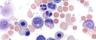
White blood cells - leukocytes
Leukocytes perform protective functions; as soon as an infection enters the body, white blood cells - leukocytes - come into play. Leukocytes are constantly on guard. Some white blood cells (lymphocytes) produce protective antibodies - proteins that neutralize or destroy viruses and pathogenic bacteria.
The life cycle of leukocytes is relatively short - from several days to several weeks. One cube of blood from a healthy person contains from 4 to 8 thousand leukocytes. If the body is fighting an infection, this number may increase. A persistently too high or too low number of white blood cells in the blood may indicate the presence of serious illnesses.
What is considered normal
The total number of these cells in the body is determined by the figure 25 × 1012. Laboratory calculations are based on the cell content in one cubic mm of blood.
According to the rules, the analysis is taken from capillary or venous blood in the morning after a quiet rest and before meals. The level of red blood cells is influenced by external conditions and the nature of nutrition.
The norm changes throughout life. There is a dependence on a person’s age, gender and climate zone where people live.
In a child during the neonatal period, the maximum number of erythrocyte cells is observed (within the range of 4.3 - 7.6 x 10¹²/l). The destruction of the mother's red blood cells immediately after birth and their replacement with her own causes yellowing of the skin. By the year the amount decreases to 3.6 - 4.9 x 10¹² / l, and in adolescence it increases slightly to “adult” levels (3.6 - 5.1 x 10¹² / l).
The level in women (3.7 - 4.7 x 10¹² /l) is lower than in men (4.0 - 5.1 x 10¹² /l). This is due to physiological blood loss during menstruation. During pregnancy, a woman’s body begins to increase the consumption of iron, and with it red blood cells. Mild anemia (anemia) indicates this feature.
A decrease in red blood cell levels is called anemia. The degree and form of the disease are influenced by various reasons.
An increase in the number of red blood cells (erythrocytosis) is possible with significant dehydration or with blood pathology associated with increased synthesis of red blood cells and impaired utilization.
The structure of frog red blood cells
Scientists have long established that human red blood cells have structural features that ensure the most efficient gas exchange. This applies to form, quantity, and internal content. This is especially obvious when the structure of human and frog red blood cells is compared. In the latter, red blood cells are oval in shape and contain a nucleus. This significantly reduces the content of respiratory pigments. Frog red blood cells are much larger than human ones, and therefore their concentration is not so high. For comparison: if a person has more than 5 million of them per cubic mm, then in amphibians this figure reaches 0.38.
How does agglutination occur?
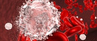
Agglutination of erythrocytes is the reaction of interaction of agglutinogens (antigens) located on the surface of the cell membrane with specific plasma agglutinins. The result of the interaction can be seen when determining the blood group on a white plate - the formation of small sticky lumps.
In a healthy person, this process is reversible and is possible when the cells lose their electrical charge. In pathological conditions, agglutination promotes thrombus formation. At the same time, the number of free red blood cells in the blood decreases.
Below shows agglutination of red blood cells with slow blood flow
Cell shapes
A healthy red blood cell has a classic appearance: a smooth and rounded surface, elastic walls, and a flattened central part. Deviations in the structure are rare; the main varieties are presented:
- Spherocytosis - abnormal elements have a spherical shape associated with pathology of the cell membrane. Red blood cells are characterized by increased fragility; their destruction is fixed in the spleen, which leads to the formation of anemia. When an organ is removed, the processes of destruction of red blood cells stop.
- Elliptocytosis is a similar pathology to the previous one. The cells have a non-standard cake-like appearance. Both diseases are associated with genetic predisposition.
- Acanthocytosis - the elements develop a star structure. The deviation is associated with hereditary factors and liver diseases.
- Sickle-shaped erythrocytosis - formed after malaria, formed when the erythrocyte membrane ruptures. In such cells, hemoglobin e performs its main functions due to crystallization.
How does a red blood cell participate in respiration?
Red blood cells are responsible for saturating the blood with oxygen and removing unnecessary carbon dioxide accumulations. To achieve this, most of the cell mass is occupied by hemoglobin (globin protein + 4 heme/iron molecules). It is called "blood pigment" because heme is what gives blood its color. Depending on the sequence of amino acids, different types of pigment are distinguished in globin.
The oxyhemoglobin complex is formed by combining with oxygen. It is created in the pulmonary capillaries, and in the tissues it breaks down again and releases free oxygen to the cells.
The appearance of methemoglobin or carboxyhemoglobin in the blood during poisoning and intoxication disrupts the process of oxygen transfer and leads to tissue hypoxia.
Elevated red blood cells
Red blood cells can be elevated due to many reasons, ranging from banal dehydration to erythremia - chronic leukemia.
Therefore, if there are any deviations in test results, you should consult a specialist to determine the cause. An increase in the number of red blood cells is called erythrocytosis, which can be: 1. Primary. A rare hereditary disease characterized by loss of energy, dizziness and darker color of the mucous membranes. 2. Secondary. Caused by other diseases or conditions (for example, smoking or staying in high mountains) and is associated with oxygen starvation of cells.
Thus, the following reasons for the increase in red blood cells can be identified:
- Dehydration. When the volume of fluid in the body is reduced, the percentage of red blood cells (and other blood cells) artificially increases.
- A lack of oxygen, which the body tries to compensate by producing more red blood cells.
- Congenital heart defect. If the heart cannot pump blood effectively, the amount of oxygen reaching the tissues is reduced. The body creates more red blood cells to compensate for oxygen deprivation.
- Genetic causes (changes in sensitivity to oxygen, impaired release of oxygen by hemoglobin).
- Polycythemia vera is a rare disease in which the body produces too many red blood cells.
Increased production of red blood cells can cause blood to thicken, slow blood flow, and related problems (eg, headaches, dizziness, vision problems, excessive blood clotting).
Often, elevated red blood cell levels are due to dehydration, hot weather, extreme stress, or excessive exercise. A pathological increase in red blood cells is a fairly rare pathology. Much more often, patients encounter reduced levels.
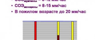
Erythrocyte sedimentation rate
Since red blood cells have their own mass, the blood, when drawn into a graduated tube, is stratified due to cell sedimentation. To prevent cell elements from sticking together, a special solution is added.
The result of the reaction is assessed after an hour by the height of the transparent column.
The reaction in men is considered normal - from 12 to 32 mm/hour, in women - from 18 to 23. In pregnant women, ESR increases to 60 - 70 mm/hour. The reaction is widely used in the diagnosis of diseases along with other tests.
Red blood cell stability
The ability to maintain its shape and work steadily in the blood is called resistance. It is important to consider that for this to happen, an isotonic concentration of sodium chloride in the blood must be maintained.
- As the concentration increases (hypertonic solution), red blood cells lose water, shrink, and are unable to carry oxygen.
- In the case of blood thinning and hypotonic concentration, water tends to enter the blood cells, they swell, rupture, and hemoglobin passes into plasma. Such blood is called “lacquered”, and the process is called hemolysis.
In severe conditions, doctors monitor the need to add saline solutions or water to prevent disruption of tissue respiration.
The properties of red blood cells provide the body with resistance to environmental conditions and compatibility with external influences. The test for red blood cells is part of the blood formula and must be checked in case of any disturbances in the patient’s well-being.
General principles of the existence of red blood cells
An erythrocyte is a cell derived from the red germ of hematopoiesis. About 2.4 million of these cells are produced per day, they enter the bloodstream and begin to perform their functions. During the experiments, it was determined that in an adult, red blood cells, the structure of which is significantly simplified compared to other cells of the body, live 100-120 days.
In all vertebrates (with rare exceptions), oxygen is transferred from the respiratory organs to the tissues through hemoglobin in erythrocytes. There are exceptions: all representatives of the family of “white-blooded” fish exist without hemoglobin, although they can synthesize it. Since at the temperature of their habitat, oxygen dissolves well in water and blood plasma, these fish do not require more massive oxygen carriers, which are erythrocytes.
- Indicators of red blood cells and how they are indicated in a blood test, red blood cell mass and treatment of anemia