Angiodysplasias are organ embryopathies from embryonic capillaries, veins, arteries, arteriovenous anastomoses, causing characteristic functional and morphological changes in regional circulation in childhood and young age.
It is believed that vascular malformations occur in the early phases of the formation of the vascular system of the embryo in the period from 4 to 8 weeks. intrauterine development.
The etiology of vascular dysplasia has not yet been fully elucidated, but it should be assumed that teratogenic factors affect the fetus and disrupt the formation of blood vessels precisely in the early phases of embryogenesis [1].
Depending on which pathological vessels dominate, the following clinical forms of angiodysplasia are distinguished in the defect: capillary, venous, arterial, mixed, arteriovenous anastomosis [2, 3, 4]. There are also mixed variants of the defect, for example capillary-venous dysplasia.
The incidence of angiodysplasia, according to various authors, ranges from 3 to 7% of the population [1, 4]. Angiodysplasia in the face and neck area in children and adolescents leads to the formation of neurotic conditions in patients and their parents, and as a result, the problem develops from a medical one into a social one. Appearing at birth, areas with dilated pathological vessels not only do not disappear (unlike hemangiomas), but also slowly progress, disfiguring the patient, bringing severe emotional suffering, and worsening the quality of life.
In 1981, R. Anderson and D. Parrish of Harvard University first proposed the concept of selective photothermolysis (SP), based on the ability of skin chromophores such as hemoglobin, oxyhemoglobin and water to selectively absorb a specific wavelength. The maximum energy absorption by hemoglobin and oxyhemoglobin is observed in the green (500–560 nm) and yellow (560–590 nm) spectra. The peak of interaction of laser radiation with water occurs in the mid- and far-infrared region of the spectrum (more than 1000 nm) [5].
Over 24 years, this theory has been confirmed many times and is currently the theoretical basis for the treatment of vascular pathology [6]. The gold standard of treatment is the use of laser and IPL systems that generate radiation in these spectra of visible and invisible light. The purpose of this study: to develop treatment tactics for pediatric patients with capillary and capillary-venous and venous dysplasia in the face and neck.
Materials and methods
The study was conducted at the Department of Laser Surgery of the Federal State Budgetary Institution Russian Children's Clinical Hospital of the Ministry of Health and Social Development of the Russian Federation for the period from 2008 to 2015. The results of examination and treatment of 4192 patients with capillary and capillary-venous and venous dysplasias in the head and neck area aged from the first days of life to 17 years are summarized.
Among the diagnostic methods, in addition to a general clinical examination and description of the local status, the most important were ultrasound examination of soft tissues in the projection of areas of capillary dysplasia and Doppler ultrasound to identify the feeding and drainage vessels of the pathological area.
In patients with a predominance of the venous component in the vascular defect, an MRI study was performed in vascular mode or with contrast enhancement of the vessels. A neurological and ophthalmological examination was also carried out to identify possible damage to the meninges and organ of vision. For retrospective analysis, follow-up data of treated patients were used.
To treat patients, a multifunctional device for photo and laser therapy was used with the ability to connect laser and non-laser interchangeable radiation sources with different spectral characteristics. In our study, an IPL nozzle with a radiation spectrum in the range of 515–1200 nm, with a 560 nm filter to cut off the unwanted part of the spectrum that causes tissue overheating, was used to coagulate pathological capillaries; for coagulation of pathological venous vessels - a replaceable Nd:Yag laser attachment generating a wavelength of 1064 nm. The device was equipped with a skin contact cooling system integrated with the emitter to -100 C on the skin surface. The operation of the laser used, which generates wavelengths of the “vascular spectrum”, is based on the principle of selective photothermolysis (SP), that is, the destruction of the main target (pathological vessel) when exposed to light, which transforms when absorbed into heat, does not damage the surrounding tissues.
Laser removal of wine stains
At the First Children's Medical Center, a medical device based on a copper vapor laser is used for children. Laser skin treatment allows the patient to get rid of the defect in several sessions without pain and without damaging surrounding tissues. Selective laser photothermolysis for angiodysplasia is a procedure that has the maximum cosmetic effect! Removing wine stains with a laser is the best method today. The procedure is performed painlessly, quickly, without cosmetic defects, on an outpatient basis. After exposure to the laser, the spot gradually begins to fade and disappears completely. In the vast majority of cases, anesthesia is not required.
Diagnosis of capillary, capillary-venous and venous dysplasias
To diagnose and treat capillary and capillary-venous dysplasia, we applied the gradation of “port-wine stains” proposed by American experts.
Grade 1 - a light pink spot consisting of small, sparsely located vessels, which, at six times magnification, look like discrete tips of vessels like grains of sand. Pathological capillaries are located in the deep layers of the dermis. This form of pathology was diagnosed in 458 children.
2nd degree - a spot from intense pink to light red in color, at six times magnification it appears as separate large vessels with significantly smaller areas of normal skin between them. Pathological vessels are located in the deep and middle layers of the dermis. This form of pathology was diagnosed in 1092 patients.
3rd degree - separately dilated vessels, almost touching each other and lying in the middle layers of the dermis and on the border with the epidermis. 1804 children had it.
4th degree - a spot from red to purple, observed as a homogeneous, slightly raised mass of blood vessels. Normal skin is not visible between them. Pathological vessels can be located in layers in all layers of the dermis and under it. This degree was observed in 501 patients.
5th degree - the damage becomes nodular and lumpy. Pathological vessels can be located in layers in all layers of the dermis and under it. This degree was observed in 34 children.
In this sample of children, type 3 capillary dysplasia was most common. For the diagnosis of venous dysplasia in 303 patients, the reference point was the results of Dopplerometry of the pathological area with an assessment of the intensity of blood flow and the caliber of pathological vessels.
Angiodysplasia in the facial area was observed in 3479 children (83%), and in the neck area - in 712 patients (17%). By type of pathology, the distribution of patients with angiodysplasia in the face and neck was as follows: capillary dysplasia was noted in 3005 cases (71.7%); capillary venous dysplasia – in 1042 cases (24.8%); venous dysplasia – in 145 children (3.5%). It should be noted that the majority of patients were children with lesions in two or more anatomical areas of the face. Angiodysplasia with damage to the meninges was diagnosed in 154 children, with damage to the organ of vision - in 84 patients, a combination of angiodysplasia with damage to the central nervous system and visual apparatus - in 123 patients.
Preparing for treatment
Modern laser therapy capabilities make it possible to perform SF of pathological vessels in young patients, starting from two weeks of age and on an outpatient basis. Before starting therapy, the possibility of treating vascular pathology using laser and IPL, the advantages and disadvantages of each, and the possibility of application in each specific case are discussed. It is also necessary to warn patients and their parents that the treatment of vascular pathology is long-term and the number of procedures is not limited to one manipulation.
4 weeks before and after the 4th week of treatment, it is recommended to avoid exposure to the sun, and when going outside in sunny weather, apply sunscreen with a protection factor of at least 50 units to all exposed areas. 2 weeks before the procedure, it is recommended to avoid taking any anticoagulants and non-steroidal anti-inflammatory drugs.
Contraindications to SF are:
- the presence of an infectious process at the site of intended exposure, especially herpes infection;
- oncological diseases;
- predisposition to the formation of keloid scars;
- intense tan;
- use of isotretinoin within the last year. Before the start of treatment, all patients signed informed consent for the manipulation and were photographed in two projections.
Possibilities for treating vascular skin pathologies with laser
In the practice of pediatricians, dermatologists and plastic surgeons, patients sometimes encounter skin lesions of the so-called “port-wine stain” type and some similar vascular formations. Unfortunately, not all doctors are familiar with the characteristics of these diseases and methods of treating them.
The laser department of the Russian Children's Clinical Hospital has been treating vascular skin disorders since 1992; per year, 300-400 sick children are successfully treated, who manage not only to restore normal skin, but also to provide psychological comfort.
| Figure 1. Capillary angiodysplasias |
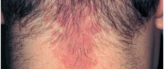
In Fig. 1 shows a patient with capillary angiodysplasia. In the English-language literature, this pathology is called port wine stains (wine stains), and the term nevus flammeus (flaming nevus) is occasionally used. Sometimes this pathology is mistakenly called hemangioma. Capillary angiodysplasias are congenital and are sometimes combined with Sturge-Weber syndrome. They occur in 0.3% of the population (in Moscow and the Moscow region - more than 30 thousand patients), never go away on their own, with the exception of a rare form - nevus of Unna. Localization most often is the face, less often - the chest, neck, arms, and very rarely - legs. Capillary angiodysplasia (CAD) appears in infants in the form of light pink or pink spots, which upon closer examination look like the tips of individual vessels (grades I-II). As the child grows, the CAD becomes light red or red and looks like a continuous surface of the vessels without light spaces between them (II-III degree).
Further, the vessels of the CAD can expand more and more, the CAD thickens, rises above the surface of the skin and looks like a solid mass of highly dilated vessels (IV-V degree). At this stage, the skin bleeds easily, becomes infected, CAD can cause asymmetry and disruption of facial proportions, and malignant degeneration is also possible. CAD of all degrees is accompanied by severe psychological discomfort.
Before the advent of medical lasers emitting yellow light, skin transplantation offered some hope of success, otherwise the pathology was practically incurable.
| Figure 2. Stellate hemangioma (telangiectaxia) |
In Fig. Figure 2 shows stellate hemangioma, which is also called stellate or spider-shaped telangiectasia. Like other types of telangiectasia, it is treated with yellow light lasers. Telangiectasia (TAE) are dilated capillaries of the skin that are visible to the naked eye. Usual localization: on the face - cheeks, nose, on the legs - hips, under the knees, ankles. TAE, unlike the pathologies described above, do not pose a threat to health and are rather a cosmetic defect. However, TAE of the legs is recognized as a chronic pathology of the lower extremities and, therefore, requires medical correction. TAE is the mildest, but also the most widespread pathology of skin vessels, affecting about 30% of the adult population. TAEs occur in both women and men, and they cause no less trouble for the latter, because people mistakenly associate TAEs on the nose with alcohol abuse. There are quite a few reasons that cause TAE. This is primarily a genetic predisposition and abuse of sun tanning, as well as sometimes frostbite of the skin. TAE on the legs is often accompanied by varicose veins.
Sclerotherapy and laser are used in combination and separately to treat TAE on the legs. The only way to treat TAE on the face is to use a yellow light laser.
A huge number of publications, mostly foreign, are devoted to the treatment of vascular skin pathologies described in our article. The best results with minimal side effects, according to numerous studies, are provided by pulsed yellow light lasers - a dye laser pumped by a flash lamp and a copper vapor laser, emitting at wavelengths of 585 and 578 nm, respectively. Recently, a number of works have appeared on successful treatment with pulsed green light laser - Nd:YAG with double frequency, emitting at a wavelength of 532 nm. Several articles have also been published on the experience of using non-laser pulsed light sources emitting in the yellow range, an example of which is Photoderm. At the same time, information appeared about unsatisfactory results of treatment using the Photoderm installation - low efficiency and significant side effects in the form of necrosis, hyperpigmentation, etc.
Since 1992, at the children's laser center of the Russian Children's Clinical Hospital, we have been treating vascular disorders using a copper vapor laser, and since 1994 we have been working on the Yakhroma-M copper vapor laser system, created by the Physics Institute named after. P. N. Lebedev RAS together with the State Scientific and Production Enterprise “Istok”. Light from the laser is transmitted to the patient's skin through a flexible light guide, at the end of which there is a laser pen. The laser beam is blocked by a shutter that opens for a split second. The exposure time (0.1 s, 0.2 s, 0.3 s, etc.) is selected by the doctor depending on the disease and the patient’s skin type. Using a thin aiming beam, the laser beam is directed precisely into the vessel. The shutter opening is controlled using a foot pedal.
The thin laser beam of yellow light is strongly absorbed by the blood, namely by the hemoglobin contained in the blood. The tissue surrounding the vessels, which does not have red pigment, absorbs yellow light much less. Therefore, it is possible to select the parameters of laser radiation in such a way that the dilated vessels are heated to a certain temperature sufficient for their coagulation, and at the same time the surrounding tissue is not damaged. Selective destruction of abnormal blood vessels provides a unique opportunity to remove vascular pathologies without damaging the skin itself. The excellent cosmetic result obtained with a copper vapor laser is especially important, since vascular pathologies are very often localized on the face.
An individual vessel is treated by tracing along the vessel from the thinner to the thicker part until the vessel visibly disappears or, if the vessel is thick enough, until the color changes visibly to purple. 7-10 days after treatment of telangiectasias, healthy skin remains in their place. Continuous pathologies such as CAD are treated using the point-by-point method, leaving small untreated areas around each point. After the operation, the treated areas are lightened. On the third day, the treated area becomes covered with a crust, and healing occurs under the crust. After a week, the crusts come off, a pink or red spot remains on the treated area, which lasts for one and a half to two weeks, then the spot turns brownish and remains that way for another one and a half to two weeks. After two to three months, the treated area acquires the color characteristic of the patient's skin.
For the treatment of telangiectasia, 1-3 sessions are required, for the treatment of CAD - 2-8 sessions, depending on the degree and size of the pathology.
To achieve good treatment results, proper preparation and especially careful care after treatment are important. The main components of postoperative care are the daily use of bactericidal ointment and protecting the crusts from accidental removal. This is especially true for children who like to pick at scabs. Peeling off the crusts can lead to the formation of scars. The attending physician must explain postoperative care to the patient during each visit.
Anesthesia
In pediatric practice, the use of high-energy laser and non-laser systems for coagulation of pathological vessels of various diameters in the face and neck is associated with intense pain. In this regard, we used inhalation anesthesia when the pathological process was localized on the face in the eye area and when the area of influence was more than 10 cm2. In other cases, manipulations were performed without anesthesia. The use of local anesthesia for coagulation of pathological vessels is impractical, since under the influence of the anesthetic the pathological vessel narrows, reducing the formed blood elements in its lumen and, accordingly, hemoglobin - the main chromophore that absorbs laser radiation. As a result, the procedure is insufficiently effective.
An area of no more than 4% of the body surface was treated at a time, since in two cases when treating 5% of the body surface area, a temperature reaction was observed over the next 24 hours.
How does laser treatment for port-wine stains work?
The procedure for laser removal of port-wine stains is quite simple and does not require the use of anesthesia. In some cases, the procedure is performed using a local anesthetic cream. During the procedure itself, the child may feel a slight tingling sensation. In general, the operation is very easy to tolerate for children of different ages and does not require long-term rehabilitation. In most cases, a series of several procedures is performed. As a result of each session, as a rule, the stain changes color (becomes less noticeable) and ultimately disappears completely.
Treatment of capillary, capillary-venous and venous dysplasias
Treatment began with a test procedure, the purpose of which was to select optimal treatment parameters individually for each patient, taking into account the clinical form of angiodysplasia and skin type. According to the types of procedures performed, all patients can be divided into two large groups - SF and transdermal laser coagulation with a wavelength of 1064 nm. The distribution of patients by age and type of manipulation performed is presented in Table 1.
Table 1. Distribution of patients by age and type of laser manipulation
| Types of manipulations | Age | Total | ||||
| 0–1 years | 1–4 years | 4–7 years | 7–15 years | Over 15 years old | ||
| Selective photothermolysis face, neck | 841 | 1552 | 604 | 834 | 58 | 3889 |
| Transdermal photocoagulation | 78 | 93 | 59 | 69 | 4 | 303 |
| Total: | 919 | 1645 | 663 | 903 | 62 | 4192 |
As can be seen from Table 1, the main number of patients occurs in the first 4 years of life (1645 people), with a gradual decrease by the age of 15 (62 patients). Accordingly, the main age period in which we carry out treatment is the first 4 years of life.
The treatment method itself consists of a test procedure to select the radiation dose and evaluate the results after 6 weeks. All subsequent procedures were carried out with complete treatment of pathological vascular tissue, provided that it did not occupy more than 4% of the body area. Otherwise, plots were treated at two-week intervals. When carrying out procedures, in addition to protective glasses, the doctor is required to use eye protection for the patient. Gauze pads, folded in 8 layers and moistened with saline, are placed on the eye area, on top of which metal shields are placed. When working in the periorbital area, after the procedure, 1–2 drops of a solution of oftan catachrom are instilled into the conjunctival sac. Immediately after the procedure, we applied Bepanten Plus ointment to the treatment area, and to relieve pain, we applied cooled gel thermal packs for 10–15 minutes.
5–10 minutes after the procedure, hyperemia, slight swelling and hemorrhages appear in the area of treatment. In the next 24 hours, with large treatment areas (more than 10 cm2), moderate swelling develops, which is reduced over the next 24–48 hours.
The effectiveness of treatment was assessed by the number of procedures required to achieve lightening of the pathological area to the color of normal skin or to a barely noticeable light pink spot with blurred boundaries.
Removal of capillary angiodysplasia (“port-wine stain”): advantages of laser treatment
The following advantages of using laser equipment in removing port-wine stains can be highlighted:
- laser radiation with certain parameters is able to penetrate the skin without damaging it.
- The wavelength used in the laser coincides with the absorption peak of hemoglobin, therefore it is maximally absorbed by hemoglobin and noticeably less by other tissues. Due to this, the deep layers of the skin are not damaged.
- Copper vapor laser radiation is almost not absorbed by water, which prevents overheating of surrounding tissues.
- Focusing the laser beam into a small spot allows you to limit the area of influence to the pathological area, avoiding irradiation of normal tissue.
- The pulsed nature of the radiation and the laser radiation dosage system allow you to control the absorbed energy and avoid overheating.
Thus, high-tech laser equipment allows procedures to be performed without damaging the surrounding tissue with maximum cosmetic effect, which is extremely important, since most operations are performed on cosmetically significant areas of the face or body.
For more detailed information regarding the treatment of skin pathologies or to schedule a consultation, please call 8 (8452) 244-000.
Children's health means parents' peace of mind!
Make an appointment with a surgeon
Choose a doctor
Treatment results
When studying the anamnesis of 160 patients who received laser treatment before contacting our clinic, a high frequency (54% of patients) of the formation of intradermal fibrosis, hyper- and atrophic scars in the projection of the port-wine stain was noted against the background of frequently repeated SF procedures (on average 1 time per month ) regardless of whether a “vascular” laser or an IPL device was used (Fig. 1). It should be noted that in conditions of frequently repeated procedures, even with an adequately selected dose of exposure, a positive result is not achieved, since scar tissue keeps the walls of pathological capillaries in an expanded state, preventing them from collapsing during subsequent laser exposure, and the number of procedures completed by the patient is equal to on average 10–15.
Rice. 1. Patient, 17 years old, with capillary facial dysplasia, linear hypertrophic scars in the chin and upper lip, which appeared as a result of frequently repeated (every 4 weeks) IPL procedures. In total, the patient underwent 12 IPL procedures (A). Patient, 16 years old, with capillary dysplasia of the nasal clivus, eyelids and upper lip on the left. Hypertrophic scars on the nasal slope and left cheek. Intradermal fibrosis in the projection of capillary dysplasia. Condition after 10 procedures with a “vascular” laser (578 nm) (B)
In patients with scarring skin changes due to aggressive SF procedures in terms of power and frequency, the use of IPL therapy turned out to be highly effective (Fig. 2). The average number of procedures in this group of children was 6.
Rice. 2. Patient A., 16 years old, with capillary dysplasia of the right cheek and upper lip on the right. Intradermal fibrosis of the skin. The patient has a history of 14 SF procedures with a “vascular” laser (595 nm) at intervals every 3 weeks (A). Condition after 2 IPL procedures (B). A consistent positive result was obtained
Capillary malformation - symptoms and treatment
Laser therapy is the main treatment method for capillary malformation. It is carried out only with selective lasers, since their waves act exclusively on blood hemoglobin [2][3][4][10].
Laser radiation affects the upper layer of capillaries, which causes sharp heating of red blood cells. As a result, the capillary either ruptures or sharply narrows.
Laser therapy can be started at any age, but optimally from the 1st month of life. If treatment is carried out under anesthesia, laser treatment is performed on average three times a year, but no more than four. It is believed that for successful socialization in society it is necessary to achieve the maximum result of treatment before starting school.
Laser treatment has its complications : temporary (reversible) and permanent (irreversible). All of them arise due to the thermal effect of laser radiation on tissue.
Swelling, redness and bruising are temporary complications. They indicate the effectiveness of the procedure and usually disappear 7-10 days after treatment.
Irreversible changes occur when the skin is overexposed to radiation. The resulting burn can lead to the appearance of a scar. These complications can be avoided by choosing a competent specialist and the right laser.
Another hardware treatment method is IPL - intensive pulselight, i.e. intense pulsed light . Unlike laser, IPL represents broadband light. Essentially, this is a powerful lamp, to cut off unnecessary ranges of which special filters are used. This technology is often used in cosmetology to treat skin diseases, including the treatment of capillary malformation.
Before the advent of laser technology, the main method of treating port-wine stains was surgical removal of the altered skin with plastic correction of the resulting defect. Hours of traumatic surgery were performed, often leaving disfiguring scars that caused more discomfort than the spot itself.
Sclerosation - administration of a drug (sclerosant) through an injection - is used in the treatment of combined malformations, for example capillary-venous. This method is not used in the treatment of simple port-wine stains due to the lack of a cavity into which the drug must be injected.
The use of beta-blockers in the form of systemic (propranolol, atenolol) or local therapy (timolol, arutimol) in the treatment of capillary malformation is ineffective, since they do not act on capillary angiodysplasia.
Among other things, experimental work is currently being carried out on the use of the drug sirolimus as systemic and local therapy, but this method has not yet received widespread use.
Treatment of cutaneous capillary dysplasia
For skin forms of capillary dysplasia in the face and neck area or types 1-3 of capillary dysplasia, the use of laser devices that generate a wavelength in the spectrum with maximum absorption of radiation by hemoglobin and oxyhemoglobin (532, 578, 595 nm) and IPL installations will be highly effective.
When carrying out SF using IPL, the selection of exposure parameters was carried out depending on the skin phototype according to Fitzpatrick. A more aggressive program was used for skin phototypes I and II, with subsequent softening of the parameters when moving to the darker V phototype. The flash generated by the IPL attachment consists of two pulses with a duration of 2.4 ms and 4.0–6.0 ms (depending on the program) with an interval of 15 ms. The radiation power depended on the type of capillary dysplasia and amounted to 26–31 J/cm2.
The average number of procedures for IPL therapy for capillary dysplasia in the face and neck was 4.2.
Recognizing venous dysplasia
There are frequent cases of venous dysplasia developing into phleboangiotic dysplasia. In this case, disability cannot be avoided. In the case of sepsis or rupture of an aneurysm due to venous disease, death is also possible.
For this reason, diagnosis of dysplasia should be carried out as early as possible - at the first suspicion of venous insufficiency. In modern phlebology practice, various tactics for recognizing deep vein dysplasia are used. Since the external signs of the disease make it possible to accurately determine its location, the following is carried out in the problem area:
- CT/MRI/ultrasound;
- angiography and other phlebological manipulations.
Brain imaging (MRI) is often done to confirm.
Treatment of capillary venous and venous dysplasias
When treating capillary-venous and venous dysplasias with the presence, in addition to pathological capillaries, of pathological vessels of larger diameter, it is necessary first of all to remove large-diameter vessels located under the skin. For this purpose, the optimal treatment method is Nd:YAG transdermal laser coagulation.
The operating principle of the Nd:YAG laser module is to generate a series of single pulses with a power of 90 to 120 mJ/cm2 through a sapphire tip with a diameter of 6 mm. In this case, radiation, penetrating through the skin, does not damage it, but causes the liquid part of the blood to boil, leading to thrombosis of coagulating pathological venous vessels. At the moment of action of the pulses, there was contact cooling of the surface of the treated skin area to -100 C. The average number of Nd:YAG transdermal laser coagulation procedures was 2.5.
Discussion and conclusions
The most optimal timing for repeated manipulation in pediatric patients when using an IPL installation is after 6 weeks, Nd:YAG after 4–6 months. It is necessary to adhere to these time intervals to avoid the development of complications.
IPL therapy, according to some experts, is considered more aggressive than laser therapy with a strictly defined wavelength from the vascular spectrum. When analyzing the results of treatment of patients with capillary dysplasia in the face and neck, it turned out that the number of complications with IPL therapy is significantly lower compared to laser coagulation.
A single IPL flash is evenly distributed in tissues, accumulating in target skin chromophores (hemoglobin and oxyhemoglobin). Since we used a 560 nm filter, which cuts off wavelengths from 515 nm to 560 nm, there is practically no energy accumulation in melanin and, as a result, there is no heating of the dermis. In addition, simultaneously with the flash, the skin surface is contact cooled to -100 C, which ensures both the safety of the procedure and its comfort. Complications after IPL therapy and transdermal laser coagulation 1064 nm are presented in Table 2.
As can be seen from Table 2, the most common complication was the formation of a scab in the affected area in 1% of cases (39 patients). Other types of complications that arise during the treatment of angiodysplasia and are characteristic of the use of “vascular” lasers are practically equal to 0.
Rice. 3. Patient V., 8 years old, with capillary venous dysplasia of the face before treatment (A); after 2 Nd:YAG laser coagulations of pathological vessels in the area of the left and right cheek and 4 IPL procedures (B). A consistent positive result was obtained. Treatment will continue
It should be noted that in the case of capillary dysplasia that is resistant to traditional treatment, it is necessary to remember the possibility of the presence of larger pathological vessels and carry out their coagulation first. This will reduce the total number of procedures and obtain maximum results in a short time (Fig. 3 and 4).
Rice. 4. Patient P., 1.5 years old, capillary-venous dysplasia of the left cheek, clivus and eyelids OS. State before treatment with a “vascular” laser 578 nm (A); condition after 9 SF procedures with a “vascular” laser (578 nm). Lack of positive effect from the therapy (B); 7 years – condition after 1 procedure of Nd:YAG coagulation of a pathological venous vessel in the area of the outer corner of the eye, followed by treatment of the CD area using IPL (4 procedures). A persistent positive result was obtained (B). Treatment will continue
Nd:YAG transdermal laser coagulation of pathological venous vessels located in and under the dermis is by far the safest and most effective treatment method. Moreover, during the entire observation period, with the high effectiveness of treatment for venous and capillary-venous dysplasia in the face and neck in children and adolescents, not a single complication was obtained (Fig. 5).
Rice. 5. Patient O., 7 years old, venous dysplasia of the forehead, nose and upper eyelid on the left before treatment (A); immediately after Nd:YAG coagulation (1064 nm) in the forehead area (B); after 1 month after Nd:YAG coagulation (1064 nm) in the forehead area (B); after Nd:YAG coagulation (1064 nm) in the area of the nasal slope and upper eyelid (after 4 months) (D). The treatment was completed due to the patient’s complete satisfaction with the result obtained
The use in clinical practice of universal platforms that allow the connection of several laser and non-laser interchangeable radiation sources with different spectral characteristics is the most clinically and economically justified, as it allows one device to effectively solve problems in the treatment of a wide range of vascular pathologies in children and adolescents.
[1] Geraskin A.V., Shafranov V.V. National guidelines for pediatric surgery. – M.: Geotar Media, 2009. – P. 1042–1080. [2] Dan V.N. Angiodysplasia (congenital malformations of blood vessels) / comp. V.N. Dan, S.V. Sapelkin. – M.: Verdana, 2008. – 200 p. [3] Roginsky V.V., Nadtochiy A.G., Grigoryan A.S. and others. Classification of formations from blood vessels of the maxillofacial region and neck in children. Dentistry No. 4, 2011, pp. 71-76. [4] Dan V.N. Modern classifications of congenital vascular malformations (angiodysplasias) / V.N. Dan, A.I. Shchegolev, S.V. Sapelkin // Angiology and vascular surgery. – 2006. – T. 12. – No. 4. – P. 28–33. [5] Potekaev N.N., Kruglova L.S. Laser in dermatology and cosmetology. – M., 2012. – P. 24. [6] Anderson RR, Parris JA Microvasculature can be selectively damaged using dye lasers. A basic theory and experimental evidence in human skin. Lasers Surg Med 1981; 1:263.
At Premium Aesthetics you can purchase the following types of cosmetology equipment:
|
|


