Pelvic phleboliths as a sign of varicose veins of the pelvic organs in men
A.A. Kapto1, 2 1 Multidisciplinary medical holding “SM-Clinic” (Moscow); 2 Federal State Autonomous Educational Institution of Higher Education "Russian Peoples' Friendship University" of the Ministry of Education and Science of the Russian Federation (Moscow)
Introduction. Pelvic phleboliths were first described by CV Rokitansky (1856) in the periprostatic venous plexus. JH Orton (1908) was the first to conduct a differential diagnosis of pelvic phleboliths and ureteral stones. G. Dillon and H. Cody (1928) were the first to associate pelvic phlebolithiasis with pain in the pelvic area. Phlebolith, or vein stone, is formed due to the germination of a thrombus (blood clot) by connective tissue and the deposition of lime salts in it during thrombophlebitis (usually against the background of varicose veins). Pelvic phleboliths are commonly seen on radiography and computed tomography. Carriage of phleboliths is most often asymptomatic and does not require treatment. However, their presence indicates venous congestion of the pelvic organs.
Materials and methods of research. From July 20, 2015 to February 15, 2019, 192 patients with bilateral varicocele, varicose veins of the pelvic organs, and May-Thurner syndrome were examined. The diagnosis of varicocele was verified by examination and confirmed by scrotal Doppler ultrasound and thermography (since April 2021). The diagnosis of varicose veins of the pelvic organs was verified using TRUS using the criteria - dilatation of the veins of the paraprostatic plexus of more than 5 mm and/or the presence of blood flow reflux during the Valsalva test with TRUS. May-Thurner syndrome, Nutcracker syndrome and Posterior nutcracker syndrome were verified using MRI data of the inferior vena cava and pelvic vessels and in some patients were confirmed by the results of phlebography and phlebotonometry. X-ray visualization of the pelvis during dynamic pharmacocavernosography (n = 37) and venography (n = 54) was performed in 73 patients.
Research results. The age of the patients ranged from 17 to 69 years and averaged 34 years. The combination of May-Thurner syndrome and Nutcracker syndrome was identified in 56 patients (29.2% of cases). The combination of May-Thurner syndrome and Posterior nutcracker syndrome was found in 5 (2.6% of cases) out of 192 patients. Pelvic phleboliths were identified in 56 patients (76.7% of cases). In 17 (23.3%) patients, pelvic phleboliths were on the right side, with a diameter ranging from 1.5 to 8.6 mm (average 4 mm). In 39 (53.4%) patients, pelvic phleboliths were on the left side, with a diameter from 1.6 to 11.9 mm (average 4.4 mm). Pelvic phleboliths on both sides were recorded in 8 patients (11% of cases).
Discussion. Pelvic phleboliths are considered an incidental finding and are of interest only in the differential diagnosis of ureterolithiasis. Understanding the causes of pelvic phlebolithiasis leads to a diagnosis of varicose veins of the pelvic organs. The predominantly left-sided localization of pelvic phleboliths and their larger size compared to the right side are apparently due to the left-sided localization of both compression of the left common iliac vein (May-Thurner syndrome) and compression of the left renal vein (Nutcracker syndrome), which are the cause of venous congestion pelvic organs.
Conclusions. The presence of pelvic phleboliths indicates previous thrombosis of the pelvic veins due to varicose veins of the pelvic organs. Clinically, in retrospect, this could manifest itself as symptoms of exacerbation of chronic prostatitis. When pelvic phleboliths are detected, it is advisable to diagnose varicose veins of the pelvic organs, varicoceles and arteriovenous conflicts.
Topics and tags
Lower Urinary Tract Symptoms
Magazine
Urological bulletins 2021 No. 1 - Special issue
Comments
To post comments you must log in or register
Diseases that develop against the background of blood stagnation
As a result of prolonged stagnation, diseases develop that have to be treated with medication and, in some cases, surgery.
The following pathologies are observed in women:
- Adnexitis, inflammation of the appendages;
- Menstrual irregularities;
- Miscarriage;
- Infertility.
In men, the diseases are as follows:
- Prostatitis;
- Varicocele;
- Impotence.
One of the most common ailments is hemorrhoids, and almost everyone notices a deterioration in their emotional state.
Treatment
Treatment of diseases caused by blood stagnation is only a temporary measure. In any case, if there is a reason why the outflow is disrupted, then it must be eliminated; in order to receive recommendations, you need to schedule a visit to a therapist. It is very important to deal with the root problem; in most cases, diet and increased physical activity help, but this will have to be approached under the supervision of a doctor so as not to stimulate deterioration.
At the Heratsi Medical Center you can visit a therapist for free. It is also possible to invite a doctor to your home.
The cost of all services of the medical center can be viewed in the “Price” section or by calling the 24-hour hotline.
Watch a video with a simple exercise that will help get rid of blood stagnation in the pelvis:
Predisposing factors
Today, the issue of the formation of phleboliths does not cause controversy among specialists. Vein stone is the result of calcification of a blood clot, which means that the causes of its occurrence are similar to those of thrombosis.
Predisposing factors to the appearance of phleboliths are considered:
- Varicose veins with slow blood flow, venous regurgitation, and a tendency to thrombosis;
- Thrombophlebitis and thrombosis, when blood clots become a matrix for lime deposits;
- Heredity;
- Heavy lifting and sedentary lifestyle;
- Childbirth.
As you can see, the formation of stones in the veins is also caused by reasons that contribute to venous stagnation and varicose veins.
The most common localization of phleboliths are the vessels of the lower extremities, pelvic cavity, spleen , much less often these formations occur in the veins of the arms, lungs, and liver. Phleboliths can also become a peculiar finding in vascular tumors (hemangiomas).
Prevention
It is easier to prevent pelvic diseases than to treat pathological processes and their consequences.
You can protect the female pelvic organs from the inflammatory process by following certain rules:
- Compliance with intimate hygiene standards;
- Elimination of hypothermia;
- Minimization of intrauterine manipulations;
- Use of barrier methods of contraception (condom);
- Regular examination of both the woman by a gynecologist and the sexual partner by a urologist-andrologist.
- When the first symptoms of the disease appear, seek medical help early.
Mechanism of disease development and localization
Calcified thrombi have an unclear mechanism of development. Doctors suggest that their formation is associated with hemodynamics, changes in blood volume and flow, and a violation of venous pressure.
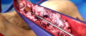
The developmental process occurs at the onset of intravascular thrombosis. In this case, the epithelium is damaged, coagulation and fibrinolysis are disrupted.
Initially, the phlebolith core is formed due to the sediment of inactive crystals. Then the connective tissue fibers undergo calcification.
Stones inside the pelvic vessels occur when there is an excessive concentration of phosphatase. Due to this, calcium begins to accumulate on the blood clot. The formations have an oval, round shape. The diameter depends on the course of the vein.
The surface is white or brown. The shell is multi-layered; inside there is a core of dense consistency.
The disorder is localized in the kidneys, pelvis, and head.
Symptoms
Phleboliths in the pelvis by themselves do not bother patients in any way and can exist in their body for years, being detected only on an X-ray or ultrasound examination.
The symptoms of varicose veins and venous stasis in the pelvic veins are quite obvious:
- Chronic pelvic pain syndrome. Patients are bothered by dull, aching pain in the lower abdomen and lower back with unclear localization. In the vast majority of cases, this syndrome worries women.
- Long and heavy menstruation can also be combined with venous congestion and pelvic varicose veins.
- Infertility is quite often combined with pelvic varicose veins, but is not always its consequence.
- Swelling of the genital organs.
- Varicose veins of the legs and external genitalia with a high degree of probability suggest the presence of venous stagnation in the veins of the pelvis - a trigger point for the formation of blood clots and phleboliths in the future.
Possible complications
Phlebostasis is the formation of multiple stones. In medical practice, phleboliths can cause a danger comparable to thrombosis. The risk of recurrence of occlusion increases.
With varicose veins, the likelihood of recurrent blood clots increases. If stones migrate and enter the portal vein, this is a dangerous complication. This can lead to pulmonary embolization, which has serious consequences.
Types of stones according to different criteria
Different patients may develop one or more stones (multiple). They are small (microlites) and large (macrolites). If stones are formed in the bladder, then they are called primary. Stones that enter the organ from the kidneys are considered secondary. They are characterized by an increase over time.
According to their chemical composition, stones are divided into:
- to oxalate,
- phosphate,
- cystine,
- urate,
- struvite,
- mixed.
Diagnostics
The absence of characteristic signs complicates diagnosis. If the presence of phleboliths is suspected, the following examination methods are performed:
- Ultrasound ultrasound of veins The study makes it possible to evaluate anatomical features.
- X-ray. The image allows you to determine the exact location of the stones. They have an oval shape and a characteristic concentration.
- . Tomography with contrast allows you to clearly see the location of the phlebolith, its structure, shape and size.
- MRI. Soft tissue examination is carried out to accurately diagnose the surrounding soft tissues and the extent of vascular damage.
The phlebologist additionally prescribes a biopsy of the stones. This is necessary to establish a reliable analysis.


