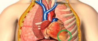
Every modern person must know the minimum practical medical actions that can be applied in an emergency situation. It is especially important to understand the location of the main organs, bones, and arteries.
In medical practice, cases are often described when people of any age lose consciousness or simply lie on the ground, without any signs of a normal condition. In this case, the person needs to feel for a pulse in the carotid artery. If it is absent, it is necessary to perform artificial respiration.
What is the carotid artery? It is important to understand the process that it carries out. The carotid arteries direct blood to the brain, and its amount accounts for 70% of the total volume. The name of these vessels is directly related to their ability to put the human body into a state of sleep, when compressed with a certain force for a period of 10 seconds.
What happens if you press on an artery: instructions
When you squeeze it lightly, you feel discomfort and sometimes mild pain. Strong compression easily puts a person to sleep. Receptors perceive pressure as a stimulus to increase pressure, so they do the opposite - they sharply lower it. A person’s heartbeat calms down on the neck, and due to the minimal supply of oxygen, the body experiences drowsiness. Long-term and severe compression of the carotid artery can be fatal.
Typically, a person's pulse is checked on their arm. But in complex traumatic cases this is not enough. Therefore, the pulse is felt in the neck. It should be noted that the pressing force must be controlled. On a thin body, the pulse will be clearly felt even with weak pressure, but if you have developed muscles, you will have to try.
Structure of the carotid artery
The common carotid artery and its branches are divided into two halves: on the right side of the body it passes in the neck from the brachiocephalic trunk, and on the left, it comes from the aortic arch in the thoracic region. To understand where the carotid artery is located in a person, you need to feel the pulse in the neck, you need to press the vertical depression in the neck on the right or left side. Everything is clearly shown in the photo.
Medical students often wonder where the carotid artery is located in the neck? To feel the pulse of the carotid artery in the neck, you need to lay the person down or sit on a chair. Place several fingers, usually the middle and index fingers, in the cavity between the Adam's apple and the lateral neck muscle.
Normal heart rate values are about 60-80 beats per minute. Each of the arteries runs diagonally upward; in the lower part they are connected by the trachea.
External carotid artery
The paired organ provides brain tissue with 55 ml of blood per 100 g. tissue, and also saturates with oxygen molecules 3.7 ml per minute. Such provision is considered normal. The external artery in humans is located slightly higher from the larynx, closer to the face, and is part of the front of the head.
In the area of the Adam's apple, the artery diverges to the front and back of the head. The first directs blood flow to the eyeballs, the second to the brain cells. The external artery consists of:
- front end;
- medial part;
- rear end;
- terminal branches - a system of capillaries directed to the eyes and into the oral cavity.
Proof of the presence of a capillary network is the redness of the face. In stressful situations, excessive physical strain, and hot weather, blood rushes to the skin. For some, this effect is practically unnoticeable, for others it is clearly pronounced. This is influenced by genetic predisposition, skin properties, etc.
The external artery directs blood to the following organs:
- thyroid;
- auricles, tympanic cavity;
- eyes;
- facial muscular system, ligaments;
- skin of the face and head;
- some parts of the meninges, the occipital vessel;
- roots of teeth, soft palate, lingual vessels.
The branches of the external artery do not send blood directly to the brain. However, pathologies in the functioning of blood vessels can lead to negative consequences. All that can be done if the external artery is malfunctioning is to contact a neurosurgeon.
In some cases, plastic surgery is required. What diseases can appear if the artery malfunctions:
- Benign tumors in the facial and cervical regions.
- Arteriovenous pathology is the connection between an artery and a vein (blood does not flow into many capillaries).
- Congenital malformation of blood vessels. Signs are external facial defects, migraines and regular pulsation in the head at night, bleeding from large arteries that cannot be stopped.
As a rule, such diseases arise due to dangerous facial injuries, plastic surgery on the nose, ears, throat, etc. Also, there is a high probability of pathologies occurring due to improper implementation of basic procedures.
For example, aesthetic punctures, rinsing the sinuses, tooth extraction, cosmetic injections in the eye area. Rare cases of the disease can cause hypertensive complications.
Internal carotid artery
The branch of the internal artery is directed through the temporal bone. The strength of blood flow increases due to external and internal factors. The number of oxygen molecules entering the brain with the blood exceeds the norm and a person feels a surge of strength, energy for work and other vigorous activities.
But a long process can lead to the opposite - a person feels a loss of strength, drowsiness and fatigue. Oxygen must be supplied normally - excess or deficiency harms the body. Components of the internal artery:
- cavernous - in the dura mater;
- cervical - located deep under the muscle layer;
- part in a torn hole;
- brain;
- stony - laid in the bone canal.
The artery runs laterally, along the edge of the pharynx, parallel to the ear gland. It is divided into small vessels. The complex system supplies brain cells well with oxygen. In the cranium, the branch of the clivus and the pituitary vessel depart from the internal artery. Small arteries are directed to:
- gray matter;
- nuclei of the medulla oblongata;
- white matter;
- cortical parts.
Pathological deformations of the carotid artery Lack of treatment for diseases such as atherosclerosis, syphilis, tuberculosis can lead to deformation of the artery. Against the background of inflammatory processes, internal tissues begin to grow, and the lining of the artery ruptures, letting blood into the area between the walls.
As a result, medical intervention can lead to narrowing of the artery, as a result, a lack of oxygen molecules provokes an ischemic stroke. The narrowing can also cause other health problems - an internal artery that twists incorrectly, or splits into three, a blood clot, or an aneurysm.
Features of blood supply in the facial area.
Features of blood supply in the facial area.
The maxillofacial area is supplied with blood by the branches of the external carotid artery, which form a group of anterior, middle and posterior branches.
The anterior group includes the thyroid, lingual and facial arteries.
The middle group consists of the ascending pharyngeal artery, superficial temporal and maxillary arteries.
The posterior group is formed by the sternocleidomastoid branch, the occipital artery and the posterior auricular artery.
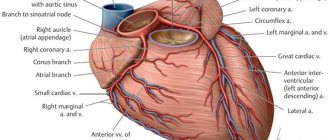
Branches of the external carotid artery.
1 – superficial temporal artery; 2 – occipital artery;
3 – maxillary artery; 4 – external carotid artery;
5 – ascending pharyngeal artery; 6 – internal carotid artery;
7 – muscle that lifts the scapula; 8 – trapezius muscle;
9 – suprascapular artery; 10 – brachial plexus;
11 – thyrocervical trunk; 12 - common carotid artery;
13 – superior thyroid artery; 14 – lingual artery; 15 – facial artery;
16 – anterior belly of the digastric muscle; 17 – buccal muscle; 18 – middle meningeal artery.
The outflow of venous blood from the organs and tissues of the maxillofacial region is carried out into the internal jugular vein, which receives blood from the facial vein, pterygoid venous plexus, lingual, thyroid and mandibular veins.
Lymph from the head and neck area collects in the jugular lymphatic trunks, which also receives lymph from the mastoid, parotid, submandibular, peripharyngeal and mental regional nodes.
Regulation of blood circulation in the vascular system of the maxillofacial region is carried out by the nervous and humoral pathways. In addition, the vessels have their own basal tone (myogenic regulatory mechanisms).
The vasomotor center of the medulla oblongata sends impulses along nerve fibers through the cervical sympathetic nodes to the vessels of the maxillofacial region.
The vessels of the face form an abundant network with well-developed anastomoses, so wounds on the face heal quickly.
When carrying out all procedures , you should be as careful as possible to avoid intra-arterial and intravenous administration of the drug.
It is safe to inject the drug into the periosteum using cannulas, which are less dangerous than needles.
DANGEROUS AREAS FOR INSTRUCTION OF FILLERS.
Projection of the bony openings of the facial part of the skull.
- supraorbitalis (supraorbital foramen) - the place of exit of the supraorbital SNP - the place of intersection of the upper bony edge of the orbit with a vertical line drawn through the medial edge of the iris. SNP is covered by m. orbicularis oculi, direction of movement - up under m. corrugator and m. frontalis.
- infraorbitalis (infraorbital foramen) – the exit point of the infraorbital SNP – the intersection of a point 1 cm below the lower bony edge of the orbit with a vertical line drawn through the medial edge of the iris. SNP is covered by m. orbicularis oculi and m. levator labii superioris direction of movement is downward and medial.
- mentalis (mental foramen) – the place of exit of the mental SNP – the place of intersection of the mid-height of the lower jaw at the intersection with a vertical line drawn through the medial edge of the iris. SNP is covered by m. depressor labii inferioris, the direction of travel is upward and medial.
Deep injections into the projection of the neurovascular bundles can lead to compression of blood vessels and disruption of blood supply, can be painful and cause changes in skin sensitivity.
The nasal region contains many terminal arteries.
When correcting this area, it is important to take special care, because the terminal branches of the arteries pass there, and the injection of Hyaluronic acid can have catastrophic consequences.
With increasing scientific evidence regarding embolization of small facial arteries following filler injections, nasal procedures should only be performed using cannulas.
From the ophthalmic artery Dorsal nasal artery Vessels of the angle of the nose Lateral nasal artery
From the external carotid artery to the columella artery.
Interbrow area.
When injecting fillers into the glabella area, local necrosis may develop due to the small number of vessels in this area.
In the area delimited by the points of fixation to the bone m. orbicularis oculi from the sides, m. corrugator supercilii above and m. procerus from below, the distribution of the filler is difficult (especially drugs with high viscosity HA), creating a high local pressure of the drug on the tissues and vessels.
Temporal and periorbital regions.
The superficial temporal (sentinel) vein is located in the temporal region behind the artery of the same name and follows its course. Crossing the temporal region 1-1.5 cm above the zygomatic arch, the vein in the layer of subcutaneous fatty tissue is directed to the auricle.
At the medial edge of the orbit, the angular vein is located superficially ; it communicates through the veins of the orbit with the cavernous sinus of the dura mater.
Careless injection of filler into the lumen of the vein or its excessive amount can lead to thrombosis, hematoma or later complications of an infectious nature.
temporales (temporal branch) of the facial nerve in the temporal region lies under the SMAS and goes to the tail of the eyebrow.
The place of its surface occurrence is located in the projection of a triangle, the apex of which is located 2 cm above the end of the eyebrow, and the base is along the lower zygomatic arch.
The parotid salivary gland is in the shape of an inverted triangle with its base on the zygomatic arch and its apex in the area of the angle of the mandible.
The duct of the parotid salivary gland lies below and parallel to the zygomatic arch under the SMAS layer, it horizontally crosses m. masseter and, entering the buccal muscle, appears in the vestibule of the oral cavity.
Damage to the duct leads to the development of chronic local inflammation of the adjacent soft tissues.
transversa facies (transverse artery of the face) is located in the zygomatic region. Parallel and superior to the parotid duct. The vessel supplies blood to the soft tissues of the area, including the skin and subcutaneous tissue through perforator vessels; the permanent perforator is located in the middle of the distance between the wing of the nose and the ear canal or 3 cm medially and 3.5 below the orbital edge.
When performing procedures with a cannula in the zygomatic region, damage to the permanent perforator should be avoided. transversa facies.
marginalis mandibulae (marginal branch of the mandible) of the facial nerve is located under the SMAS and descends first behind the branch and angle of the mandible and, not reaching the posterior edge of the m. depressor anguli oris, extends onto the face, located at this point on the bone.
Deep bone injections in this area should be carried out with caution, because this branch innervates the muscles of the lower lip and part of the subcutaneous muscle of the neck.
Educational phone numbers : 8-812-248-99-34, 8-812-248-99-38, 8-812-243-91-63, 8-929-105-68-44
Application for ordering products here
Seminar schedule here
Concept of carotid artery aneurysm
An aneurysm is an increase in the size of a small area of an artery with an accompanying thinning of its lining. This is a very dangerous disease that can be fatal if the walls of the vessel rupture. Aneurysm can be congenital or acquired, due to muscle atrophy, prolonged inflammatory processes, etc.
What can cause a wall rupture:
- stress, prolonged nervous and physical tension;
- sudden surges in pressure;
- injury to the cervical spine or head.
Blood gradually builds up after the rupture, putting pressure on the surrounding brain tissue. Very often death occurs, but in the case of a small hematoma, the situation can be corrected with surgery.
Pain relief during stenting
The intervention is performed under local anesthesia using light sedatives, since it is necessary to assess the patient’s neurological status during stenting. When the balloon is inflated, the patient may experience a slow heart rate (bradycardia) and a decrease in blood pressure (hypotension), so continuous monitoring of cardiac activity during surgery is necessary.
The patient lies on his back, with his arm abducted, on which a blood pressure cuff is attached. The electrodes of the device for taking a cardiogram are glued to the chest. The surgical field is processed and the patient is covered with a sterile sheet.
Artery disease - thrombosis: concept and characteristics
Thrombosis is a disruption of normal blood circulation in the brain. As a rule, a thrombus forms at the site where the artery branches into external and internal. In this area, blood does not move as intensely, which allows platelets to form deposits on the walls of blood vessels. What can affect platelet formation:
- congenital or acquired heart disease;
- brain damage;
- increased blood clotting;
- an autoimmune hypercoagulable state associated with the production of antiphospholipid antibodies;
- a disease associated with heart rhythm disturbances - sudden excitation and slowdown.
How does thrombosis manifest itself, symptoms:
- without any symptoms;
- sharp pain;
- subacute;
- chronic.
The patient experiences tinnitus, migraines in the head, constant pain in the cervical spine, blurred vision, the inability to chew normally due to muscle problems, and possible loss of consciousness.
Thrombosis of the internal carotid artery is characterized by a lack of sensation in the limbs, nervousness, hallucinations, inability to speak normally, and skin pain on the head. If a section of the intracranial artery is damaged, vomiting, lack of sensation in the limbs, and sleep disturbances are possible.
How to identify problems with the carotid artery When visiting a clinic, an accurate diagnosis can only be made based on research:
- Magnetic resonance angiography.
- Rheoencephalography.
- Electroencephalography.
- Computer tomography.
- Ultrasound examination of blood vessels.
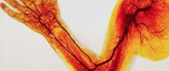
A fascinating “walk” through the veins and arteries
I bring to your attention an article about a medical diagnostic procedure - duplex scanning of blood vessels.
Duplex scanning of blood vessels, otherwise known as vascular Doppler ultrasound (USDG), is essentially an ordinary, familiar, well-known ultrasound scan.
Many patients undergoing medical examination are prescribed an ultrasound examination of the brachiocephalic vessels. I will try to talk about the essence of this study, what information it gives to your attending physician, and why this study was included in the list of diagnostic measures for medical examination of the adult population.
So, as a “tour guide,” I invite you to a short and, I hope, educational journey through the veins and arteries. The study is called duplex because it combines two modes - a two-dimensional image and a Doppler mode - a study based on measuring blood flow velocity. The 2D image looks like this.
This is a familiar black and white image, or rather gray, in which the manufacturers of modern ultrasound scanners promise not just 50, but as many as 256 shades! Doppler is a mode for studying blood flows, their speed and direction. It is the inclusion of the Doppler that is accompanied by the appearance of somewhat unusual sounds during the examination, sometimes slightly frightening to patients. These sounds are a kind of interpretation of the characteristics of blood flow. Each artery has its own “melody”. The external sleepy one is harsh and rough, the internal sleepy one is softer and more pleasant. And the veins generally “sound” like a light breeze.
This is what duplex scanning looks like on the monitor.
There is a color doppler that adds bright colors to a boring black and white picture. Patients, having seen the image, are usually interested in what is “red” and “blue” there?
Both “red” and “blue” are blood. The color depends only on the direction of blood flow relative to the sensor. What moves towards the sensor is colored red, what moves away from the sensor is colored blue.
This is no longer a duplex, but a triplex study. Actually, any modern ultrasound examination is triplex, but the name “duplex” has been preserved as more familiar.
Vascular examination is harmless to the patient, non-invasive (no penetration into the body) and does not require prior preparation. And now a little about the study of the brachiocephalic arteries (BCA), the same study that is included in the list of studies under the clinical examination program.
Brachiocephalic arteries is a definition that includes the carotid and vertebral arteries. The carotid arteries have nothing to do with the quality of sleep. They owe their unusual name to a small anatomical formation on the internal carotid artery. This formation, or rather the expansion of the artery, is called the carotid or carotid sinus. If you squeeze the neck in the area of this sinus, the number of heart contractions decreases, the person seems to fall asleep and may even lose consciousness. This is where the name carotid arteries comes from.
The examination is performed with the patient lying on his back, sometimes with a cushion under the neck for better vision of the arteries with the sensor.
This is what the common carotid artery looks like on an ultrasound scanner.
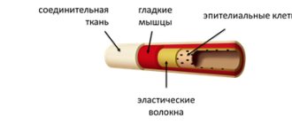
The carotid arteries can tell a lot of interesting things about a person. First of all, we are interested in the diameter and shape of the arteries, as well as the presence of atherosclerotic plaques that are dangerous to human health. The presence of atherosclerotic plaques or the predisposition of blood vessels to their appearance is indicated by such an indicator as the thickness of the “intima-media” complex or, in other words, the thickness of the inner layer of the vascular wall.
IMM thickness is an indicator of the severity of the atherosclerotic process in the body. Normally, it should be no more than 1.0 mm. The intima is a thin white stripe, the media is a thin black stripe following the intima, and the dark inside is the lumen of the artery.
The following image shows a vessel that is not affected by atherosclerosis. The thickness of the intima-media complex is 0.6 mm. The blue line is drawn along the inner layer of the vascular wall - the intima.
But in the next image, the intima-media complex, in other words, the vessel wall, is already thickened (1.4 mm) - these are already manifestations of atherosclerosis.
As atherosclerosis progresses, the intima-media complex not only thickens, but atherosclerotic plaques form, which cause narrowing (stenosis) of the vessel. The degree of stenosis is expressed as a percentage. Atherosclerotic plaques look something like this. This is the site of bifurcation (branching) of the common carotid artery (CCA) into its two branches - the internal carotid artery (ICA) and the external carotid artery (ECA). Arterial branching sites are favorite sites for the appearance of atherosclerotic plaques. I outlined the contours of the vessel itself in blue, and the atherosclerotic plaque in red. The degree of vasoconstriction in this patient is about 30%.
And in the next two pictures the process is already more advanced, the degree of narrowing of the vessel is 68%. The blue outline shows the vessel itself, and the red outline shows what the lumen of the vessel has become, changed due to atherosclerosis.
Atherosclerotic plaques can be extremely dangerous. Increasing in size, the plaque impairs blood flow in the vessel, up to its complete blockage. In addition, sometimes a fragment of atherosclerotic plaque can break off and block a smaller blood vessel, thus causing a stroke or heart attack.
If complicated plaques are detected and with a pronounced degree of narrowing of the vessel, a consultation with a vascular surgeon is indicated to decide on surgical treatment. If the process is moderate, the doctor prescribes treatment with statins - drugs that strengthen the surface of the plaque and prevent its destruction. It is also necessary to control your blood cholesterol levels, correct your diet and get rid of bad habits.
It is generally accepted that if atherosclerotic plaques are detected in the carotid arteries, then in the coronary arteries - those that feed the heart, there is almost one hundred percent probability that they are also present. We cannot easily and simply look into the arteries of the heart. Coronary angiography, the study of the blood vessels of the heart, is a complex study performed in a hospital setting. Therefore, ultrasound examination of brachiocephalic vessels (neck vessels) is a simple and informative method for diagnosing atherosclerosis. It is believed that atherosclerosis develops if a person has elevated levels of “bad” cholesterol - low-density lipoproteins.
To paraphrase popular wisdom, we can say that by the age of 50 every man should plant a tree, raise a son, build a house and... check his cholesterol level. It is advisable for a man who smokes to pay attention to his cholesterol by the age of forty. The same recommendations apply to representatives of the fair sex. In addition to the carotid arteries, vertebral arteries are also available for study. At the level of the sixth cervical vertebra, the vertebral arteries enter the bony canal formed by the transverse processes of the cervical vertebrae. Sometimes the artery enters the bone canal higher, at the level of the fourth or even third cervical vertebra.
The vertebral artery with a diameter of 3 mm is shown in the following image. Its walls are highlighted with blue lines. Dark vertical formations that interrupt the course of the artery are transverse processes of the cervical vertebrae. Ultrasound does not penetrate bone tissue, so the artery wall is not visible in these areas.
The vertebral arteries (VA) are smaller in diameter than the carotid arteries. The normal size is considered to be a diameter of 3 mm, if less than 3 but more than 2, this is called a “small” diameter, and less than 2 mm is already hypoplasia (underdevelopment) of the artery. This is a congenital condition, and it is impossible to influence the diameter of the arteries. When hypoplasia of the vertebral artery is detected on one side, the vertebral artery on the other side is usually increased in diameter. This ensures balance and ensures adequate blood supply to the brain. In addition to the diameter, we evaluate the shape of the arteries and the presence of tortuosity. The photo below shows an artery with a deformed course. It can be seen that the artery is curved, and this arc is painted in different colors.
Sometimes there are simply arteries curled into a ring! How can you avoid getting sick or dizzy with these? The vertebral vein runs parallel to the vertebral artery and above it in the bone canal. Blood in veins and arteries moves in different directions, so the vessels are painted in different colors.
Typically, the vertebral vein does not appear so bright and deep blue on an ultrasound. As a rule, it is slightly stained. Such brightness and expressiveness is already a bit of a pathology, which in conclusion is reflected in the phrase “difficulty of venous outflow.” This is not a disease yet, but rather a feature of the patient’s venous tone. But this condition already requires treatment, since it often manifests itself as tedious headaches and heaviness in the head. Remember what the doctor said from the movie “Formula of Love”? “The head is a dark object and cannot be examined.” A hundred years ago, maybe it was like that. Well, now it is very possible, and ultrasound examination of the brachiocephalic vessels (neck vessels) is an informative and safe diagnostic method that allows one to obtain important information about the blood supply to the brain.
It is enough just to do the research during medical examination. An ultrasound examination of the carotid and vertebral arteries at the second stage of clinical examination is prescribed to everyone who has elevated cholesterol levels or has complaints as prescribed by a neurologist.
Davidenko Yulia Gennadievna, head of the functional diagnostics office of the polyclinic of OKB No. 3.