Reception is conducted by:
Krivonosov Denis Sergeevich -
cardiologist, candidate of medical sciences
Make an appointment
Svitova Elena Olegovna —
Obstetrician-gynecologist. Functional diagnostics doctor. Ultrasound diagnostics doctor. Doctor of prenatal ultrasound diagnostics.
Make an appointment
Pelvic varicose veins (PVVD) is a chronic vascular pathology characterized by dilation of the pelvic veins. With the disease, the return flow of blood worsens, causing stagnation and poor circulation. Symptoms of pelvic varicose veins in women are nonspecific and are inherent in many gynecological pathologies, so literally a few decades ago the disease could not be diagnosed in time and only the symptoms were stopped, and their root causes were not eliminated.
According to a study conducted in 1999, in approximately 15-20% of cases in patients with vein disease, the reason for visiting a gynecologist was pain in the lower abdomen. However, the diagnosis was correctly made only in 2% of cases. It’s scary to imagine that about 15% of women had their uterus removed as a result of an incorrect diagnosis.
The Profimedica clinic employs professionals with extensive experience. Modern equipment from the world's best manufacturers allows specialists to diagnose and eliminate pathology at the earliest stages.
Varicose veins of the pelvis in women
Varicose veins, or ectasia (expansion of the lumen of a vessel in a limited area), is a disease that affects the venous network of the pelvic region. Varicose veins of the pelvis lead to disruption of normal blood flow from the internal and external female genital organs. In the vast majority of cases (80%), varicose veins are dilated by the ovarian veins, and extremely rarely (1%) with pelvic varicose veins, the veins of the broad ligament of the uterus are affected. Most often, varicose veins of the pelvis occur in women of fertile age - from 25 to 45 years.
Varicose veins of the small pelvis, associated with the expansion of its vessels, have recently become a fairly common disease in women. Varicose veins of the small pelvis occur in 15-20% of cases. Unfortunately, this pathology in most cases is accompanied by a number of other gynecological diseases, in which their clinical symptoms often hide the manifestations of pelvic varicose veins.
It is precisely because of the difficult diagnosis of pelvic varicose veins that doctors can choose incorrect and ineffective ways to treat varicose veins of the small pelvis, and therefore many patients are not satisfied with the results of standard treatment of veins and intrapelvic organs.
Request a call back Get a free consultation
Prevention of varicose veins and its complications during pregnancy
Basic recommendations:
- Early contact with a gynecologist, registration for pregnancy. Following the recommendations of a specialist. Regular visits to the antenatal clinic.
- Healthy, active lifestyle : walks in the fresh air, Pilates for pregnant women, yoga, swimming, aqua aerobics. Avoidance of saunas, steam baths, hot tubs, and exposure to the open sun.
- Proper nutrition with enough vitamins, microelements, fiber. Exclusion of sweet, fatty, fried, smoked, flour, starchy foods. Body weight monitoring .
- Drinking enough water.
- Rejection of bad habits.
- Loose clothing made from natural fabrics. Refusal of high-heeled shoes and tight ballet shoes. Wearing comfortable, soft shoes made of natural materials with a comfortable last and instep supports.
- Breaks during work to warm up and walk.
- Sleeping on the left side , which reduces the pressure of the uterus on the inferior vena cava and right ovarian vein.
This will prevent further development of varicose veins and its complications.
Diagnostics
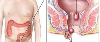
If the superficial veins of the lower extremities, vulva, or vaginal opening are affected, or external hemorrhoids appear, there are no problems with making a diagnosis. The doctor assigns it during examination, taking into account the patient’s complaints and general information from the medical history.
It is more difficult to detect varicose veins of the internal organs of the pelvis : deep hemorrhoids, vagina, uterus and bladder. Signs:
- dilation of the veins of the uterus, visualized on ultrasound.
- internal hemorrhoids can be palpated during rectal digital examination.
- Vaginal varicose veins are determined during a vaginal examination.
- damage to the bladder when blood appears in the urine - if there are no other reasons and there are changes in the veins in neighboring organs.
The results of routine examinations of the pregnant woman, which are carried out in accordance with the standard protocol for pregnancy management, are important. For example, a urine test containing blood.
A decrease in hemoglobin levels is often associated with internal chronic bleeding from damaged vessels. Increased sugar increases the vulnerability of vascular walls to infection.
During pregnancy, it is safe to perform Doppler ultrasound ( USDG ) of the venous vessels .
The method defines:
- degree of development of varicose veins;
- localization and degree of deformation of venous valves;
- the severity of the disturbance in the direction of blood movement through the veins (in the opposite direction from the heart down and towards emergency collaterals);
- the presence of changes in surrounding tissues.
In severe cases (if spontaneous rupture of internal dilated venous vessels and bleeding is suspected), magnetic resonance imaging is performed .
Symptoms of pelvic varicose veins
Dilated veins and impaired venous blood flow in the pelvis can lead to quite serious health consequences and also negatively affect women’s self-esteem.
With varicose veins, the venous vessels (perineal and vulvar) dilate in such a way that swollen, enlarged veins become visible and protrude on the patient’s skin; in addition, this process is often accompanied by:
- local edema;
- a feeling of heaviness and bursting pain in the pelvis;
- bleeding;
- mucous discharge.
The undoubted signs of varicose veins of the pelvic veins include:
- Regular long-term pain in the lower abdomen, which manifests itself primarily after physical exertion.
- Increasing discomfort in the second half of menstruation.
- Dysmenorrhea.
- Sharp painful sensations due to hypothermia of the lower extremities, stress;
- Dyspareunia.
If a patient experiences several of the listed symptoms at once, we can with a high degree of probability speak of pelvic varicose veins.
Rice. 2. Cosmetic defects of the external genitalia with pelvic varicose veins (PVVD)
Causes of varicose veins
The venous system of the pelvis in women is very complex; varicose veins of the small pelvis are partly provoked by the anatomical and physiological characteristics of the blood supply to this area. In the female body, the uterovaginal, vesical, and rectal plexuses anastomose with each other and carry blood to the internal iliac vein, and the veins of the round and broad ligaments of the uterus, merging with the branches of the uterine vessels, to the hilum of the ovary. The female genital organs and their veins also experience increased stress during pregnancy, which affects the elasticity of the walls of blood vessels.
Typically, pelvic vein pathology results from:
- constant physical overload;
- pregnancy and childbirth;
- age-related changes;
- hormonal imbalances.
Often, patients notice the first manifestations of a disease associated with dilated veins during pregnancy. Please note: with pelvic varicose veins, poorly functioning veins lead, in addition to internal discomfort, to external unaesthetic manifestations on the skin. Since a vein affected by varicose veins makes a depressing impression, it can cause the patient to develop psychological complexes.
Request a call back Get a free consultation
In what cases is CT of the pelvic vessels prescribed?
Computed tomographic angiography of the pelvic vessels is indicated for suspected development of vascular diseases and to determine the etiology of dysfunction of internal organs. The examination helps in the differential diagnosis of the following pathological conditions:
- Leriche syndrome;
- thrombosis, thromboembolism of the iliac vessels;
- atherosclerosis of the pelvic arteries;
- neoplasms developing in nearby organs and tissues and compressing the vessel;
- phlebitis, vasculitis;
- vascular tumors;
- abdominal injuries;
- aneurysm;
- disturbances in the functioning of the pelvic organs of unknown etiology;
- congenital vascular anomalies;
- impotence in men;
- infertility.
CTA is prescribed in case of low effectiveness of other examination methods, if necessary, to clarify the diagnosis and select effective therapy.
In the process of preparing for surgical treatment, computed tomography is recommended to determine the location and extent of the upcoming intervention. After surgery, a study is prescribed to monitor the restoration of circulatory system function and tissue regeneration.
Peripheral arteries of the pelvis, three-dimensional image (A)
If a tumor is suspected in the pelvic area, vascular CT helps determine the direction of further treatment, predict the possible consequences of surgical intervention, and adjust the scope of radiation and chemotherapy.
Diagnosis of varicose veins of the small pelvis
Specialists who suspect varicose veins in a woman use the following modern methods for diagnosis:
- transabdominal scanning;
- transvaginal scanning;
- Ultrasound angioscanning of the venous system.
It is these methods that make it possible to give an accurate assessment of the condition of the ovarian, inferior vena cava, iliac, and renal veins. During diagnostics in women with possible varicose veins, specialists immediately determine the main parameters of the venous system:
- Vessel diameter.
- Characteristics of blood flow.
- The presence or absence of pathological reflux (reverse flow) of blood in any vein.
- The presence of other venous pathologies.
The second stage of examination of venous vessels involves radionuclide venography and computed tomography with in vivo labeled red blood cells. These procedures allow you to objectively assess the condition of the vessels, detect varicose veins and the presence of signs of pelvic venous congestion. Also an important factor in the successful treatment of venous vessels of the pelvic region is the correct and objective assessment of pain syndrome, which is carried out using the modified McGil visual analogue scale.
| Stage | Characteristic features of affected vessels, diameter in mm | Localization |
| first | less than 5 | any pelvic venous plexus |
| second | from 6 to 10 | ovaries or uterus |
| third | more than 10 | total vascular damage |
Absolutely correct and accurate information about the presence of the disease, the condition of the blood vessels and pelvic organs at the final stage of the examination can be obtained by performing an invasive X-ray contrast study. During selective ovariography and pelvic venography, examining literally every vein, doctors as clearly as possible:
- clarify the anatomical features of the position and structure of the ovarian vessels;
- measure the diameter of the ovarian vessels;
- identify the number and nature of the tributaries of the gonadal vessels;
- identify the presence of reflux characteristic of the internal and ovarian iliac veins;
- detect the connection of subcutaneous and intrapelvic vessels.
Note! If a comprehensive examination confirms the preliminary diagnosis of “varicose veins of the small pelvis”, treatment for this disease should begin immediately!
Diagnostics
Timely detection of the process of vascular dilatation increases the likelihood of its cure and prevention of relapses.
To diagnose pelvic varicose veins, doctors at our clinic use instrumental research methods. One of the most effective ways to detect the disease is ultrasonography. This method is carried out in conjunction with transvaginal and abdominal ultrasound, which allows specialists to obtain the necessary data for diagnosis. During ultrasonography, vascular dilation can be identified by multi-colored spots of irregular shape.
Another research method is laparoscopy. In this case, the presence of pathology is signaled by bluish-colored formations that have a thin, tense wall in the area of the uterine ligament or ovaries.
Recently, a modern diagnostic method – selective oophorography – has been gaining popularity. During the study, a contrast is performed on the patient's blood vessels, thanks to which the doctor is able to see any deviations in their structure.
A CT scan may also be performed. The method helps to see the vessels in the woman’s internal genital organs.
Pronounced discomfort during sexual intercourse and other unpleasant manifestations of the disease contribute to the deterioration of the psychosomatics of patients, which often causes diagnostic errors. The presence of a clear pathognomonic symptom, combined with the absence of signs of pathology of the pelvic organs during a traditional examination and manifestations of clear psychosomatic disorders, often leads specialists to think about the psychoneurological nature of the disease. As a result, instead of treating varicose veins, patients are prescribed consultations with sex therapists and psychotherapists.
How is the treatment carried out?
Pelvic veins affected by varicose veins can be subjected to various medical procedures, in accordance with different methods of treating this disease. Treatment of the disease is carried out using several well-proven techniques. This:
- Operations to restore vein patency.
- Removal of varicose veins. Modern surgery makes it possible to remove varicose veins of the pelvis. However, any operations (even laparoscopic) performed under anesthesia can cause some harm to the patient’s health. Also, with surgical interventions, there is a possibility of relapse and there is always a risk of complications.
- A modern technique of endovascular embolization, which allows preserving the diseased vein.
Request a call back Get a free consultation
Treatment
The main recommendations are aimed at reducing intra-abdominal pressure and improving the outflow of blood from the pelvic veins.
The diet includes vegetables, fruits, and excludes alcohol.
Patients are advised to limit physical activity, stay on their feet for long periods of time, and use a contrast shower on the perineal area.
It is advisable to wear compression tights of compression class 2.
Physical therapy is of great importance. Exercises are performed lying down (“birch tree”, “scissors”, “bicycle”), breathing exercises (slow deep inhalation and exhalation with the involvement of the abdominal wall muscles).
Among medications, a course of phlebotonics (detralex) is indicated in long courses of 2-3 months.
If conservative therapy is ineffective, surgical intervention is performed - resection of the gonadal veins.
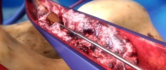
Modern method of treating pelvic vein dilatation
Today, treatment of pelvic varicose veins using modern embolotherapy techniques is the most effective and patient-friendly way to correct the venous system in this area.
After conducting a single invasive intravascular examination of varicose veins, specialists at the endovascular surgery center use an innovative method - endovascular embolization of gonadal vessels.
This treatment method involves the introduction of embolization (clogging) coils into a vein through a diagnostic catheter, which allows you to completely block the pathological blood flow of varicose veins. The operation for embolization of varicose veins takes a minimum of time. In this case, the patient can return to her normal life in the near future after embolotherapy on the vessels in the pelvis. The rehabilitation period after the procedure is simple and takes place in a short time.
Indications and contraindications for CT of the pelvic vessels
MSCT is performed using ionizing radiation, so scanning is done only if indicated. CT angiography is recommended for patients if they have the following symptoms:
- pain in the legs of unknown etiology, numbness, swelling, cramps in the area of large muscles;
- signs of pelvic organ dysfunction;
- abdominal pain in the absence of objective reasons;
- signs of internal bleeding in the pelvic area;
- violation of reproductive function against the background of changes in the nature of the blood supply to the organs of the genitourinary system;
- post-traumatic conditions when injuries are localized in the pelvic area.
A referral for tomographic angiography of the pelvic vessels is given by the attending physician: surgeon, phlebologist, gynecologist, urologist and other specialized specialists.
The use of modern equipment when performing MSCT angiography allows us to reduce the radiation dose to the patient. But the combination of side effects of X-ray scanning and the administration of a contrast solution determines a number of contraindications to this examination method:
- pregnancy in the first trimester;
- conditions and diseases that exclude the possibility of scanning organs and systems using ionizing radiation;
- early childhood (due to difficulties with catheter installation and the need to remain still during the study);
- allergy to iodine preparations;
- severe renal failure;
- diabetes mellitus during therapy with Metformin and analogues;
- hyperfunction of the thyroid gland.
A relative contraindication for CT angiography of the pelvic vessels is a patient weight of more than 150 kg or a chest/abdominal circumference of more than 150 cm, which is associated with the size of the annular part of the tomograph and the load capacity of the scanner table. If there are one or more restrictions, the doctor will select another method of hardware examination of the bloodstream.
MSCT angiography of the pelvic vessels: acute arterial thrombosis with 3D reconstruction (A) and on frontal images (B)
Benefits of using endovascular embolization
Thanks to the endovascular embolization method, varicose veins of the small pelvis can be successfully treated. The main advantages of this method in the fight against dilated veins are:
- High efficiency. Endovascular embolization helps patients whose veins are dilated and pelvic blood flow is impaired in 94.8% of cases.
- High speed of the procedure. Embolization of varicose veins of the pelvic venous system takes only 20-30 minutes.
- No general anesthesia. For pelvic vein embolization, only local anesthesia is used. Any vein is treated using proven local anesthetics, which avoids the long recovery associated with general anesthesia.
- Conducting minimally invasive intervention on an outpatient basis. Women with pelvic varicose veins spend a minimum of time in the hospital after surgery. In some cases, patients can go home immediately after surgery, but usually operated patients remain in the hospital under the supervision of a doctor for several hours.
- Minimum rehabilitation period. Within a few weeks after the intervention, reproductive function will be restored and pain in the lower abdomen will disappear. And after 1-2 months, the patient will be able to completely forget about what varicose veins of the small pelvis are.
Treatment with embolization, carried out by experienced specialists, is truly the most effective and safe. No matter how much the veins are dilated, the operation will not cause discomfort. During the intervention and insertion of emboli into the vein, patients do not experience pain: all unpleasant sensations are relieved by modern medications. In the postoperative period, to fully restore health, the patient will need to meet several easy conditions:
- strengthen the drinking regime (increase the frequency and volume of liquids drunk daily);
- avoid intense physical activity (sports, heavy lifting);
- exclude visiting a pool or bathhouse and taking baths in the first postoperative week, while using a shower is allowed.
Prevention of varicose veins of the small pelvis
Despite the fact that treatment of an altered vein using embolotherapy is accessible and effective, it is better to prevent any disease. In order for each vein to remain in its natural state, it is necessary to ensure normal blood circulation in the body. To do this, you should follow the simplest rules:
- To live an active lifestyle. Even if you have to spend the whole day in the office at the computer, you can always take a leisurely walk in the evening. Exercises for the abdominal area, dance movements of “waggling” and “swaying” with the pelvis, which accelerate blood flow in this part of the body, would be appropriate. Of course, it should be remembered that excessive exercise can provoke the disease, rather than prevent it.
- Be able to relax emotionally. Constant stress often becomes the main cause of problems with blood circulation and stagnant processes. It is very useful to take baths with various aromatic oils, experience positive emotions, sing, and dance.
- It is advisable to have a regular intimate life. Of course, if you have already discovered signs of the disease, you should consult a specialist.
By properly combining work and rest, it is easy to prevent the occurrence of many unpleasant diseases, as well as significantly improve your health.
Request a call back Get a free consultation
Ultrasound of the pelvis in men: what is included, how to prepare for the study at MEDSI
Table of contents
- How is the procedure performed?
- What is included in the examination?
- Indications for examination
- Types of research
- What diseases can be detected
- Preparation for the procedure
- Survey results
- Advantages and disadvantages of ultrasound
- Advantages of carrying out the procedure at MEDSI
Ultrasound diagnostics
is a type of examination of the patient’s body using sound waves of a certain frequency. The basis for the analysis is the difference in the reflection of such waves from tissues of different structures.
This procedure allows you to identify diseases and pathologies and determine compactions in organs. It is accurate and completely safe, so it can be used repeatedly even for a short period of time. Such a study does not require surgical intervention, and the result is immediately displayed on the monitor of the doctor who performs the examination.
How is the procedure performed?
Ultrasound of the pelvic organs in men does not take much time and does not require recovery procedures afterwards. The procedure for conducting the examination depends on its type and on the organs that need to be examined.
When ordering an abdominal test or Doppler ultrasound, the following steps must be taken:
- The patient lies on his back on the couch and removes clothing from the lower abdomen
- The doctor applies a special agent to the area being examined that improves the passage of sound waves through the body tissues
- Then he moves a special sensor over the patient’s abdomen, and the results are displayed on the monitor and recorded in the protocol
With this type of pelvic ultrasound, a man must first undergo the procedure with a full bladder and then with an empty one.
For transrectal examination, the procedure is slightly different:
- The patient should remove clothing from the lower part of the body, sit on the couch in a position lying on his side and bend his knees
- The doctor puts a protective device (condom) on the sensor and lubricates it with gel for better ultrasound transmission
- The sensor is inserted into the patient's rectum, the data is transmitted to the screen and noted in papers
Doppler examination differs in results - in this case, vessels are displayed, marked in different colors, which allows you to determine the quality and speed of blood flow. It can be done using one of the above methods.
The time spent on the procedure is no more than 20 minutes.
What is included in the examination?
Ultrasound of the pelvic organs in men includes a comprehensive analysis of several organs. This:
- The prostate is an endocrine gland that protects the bladder from infection and also produces a number of hormones and secretions that support reproductive health.
- Seminal vesicles are glands with a cell-like structure in which seminal fluid is formed.
- The bladder is an organ that is necessary for storing and removing urine from the body.
- Adjacent structures and lymph nodes
When examining the condition of the seminal vesicles and ducts, abdominal and transrectal examinations are used. They determine the presence in an organ of elements uncharacteristic of its structure. The transrectal method allows you to identify the smallest formations and compactions.
An ultrasound of the bladder is performed abdominally, first with the organ filled with urine, and then a second time with the organ empty. This is necessary to determine how completely this fluid is removed from the body. The study helps determine the presence of stones and other pathologies or neoplasms.
When examining the prostate, its volume is measured, its shape and structure are determined, as well as the presence or absence of any pathologies. In men over forty years old, it often increases in size more than the body needs for proper functioning.
Also, studies make it possible to determine the patency of the vessels of the pelvic organs and the speed of blood flow in them, which is also important for the proper functioning of the structural elements of the genitourinary system.
Ultrasound allows you to examine the tissues and lymph nodes adjacent to the bladder and prostate. This possibility is especially important when inflammation is diagnosed, but its nature is unclear or undefined.
The pelvic organs are located quite close to each other. There is a high probability that an infection that gets into one of them will quickly affect all the others. That is why it is necessary to undergo a comprehensive examination regularly.
Indications for examination
Doctors prescribe pelvic ultrasound for men with the following symptoms:
- Pain when urinating
- Inability or excessively frequent urine output (especially at night)
- Discomfort and pain in the lower abdomen
- The appearance of blood or pus in the urine
- Problems with potency
- Impossibility of conception
- Presence of sexually transmitted diseases
- There are abnormalities in urine tests
- Unexplained pain in the groin
Also, men from 40 years of age need to regularly undergo a comprehensive ultrasound of the pelvic organs, since it is at this age that the likelihood of problems and pathologies in the genitourinary system increases.
Types of research
Three types of ultrasound examinations are used to diagnose the male pelvic organs:
- Abdominal
- Transrectal
- Dopplerographic
The first type of examination is carried out using a sensor that allows you to examine organs through the wall of the abdominal cavity - the doctor moves the device over the patient’s abdomen, and an image is projected onto the monitor. This is a precise and painless procedure. It allows you to identify many pathologies and diseases, including detecting stones (calculi) in the bladder, changes in the size of walls in organs, and the presence of neoplasms.
Transrectal ultrasound is necessary when an abdominal examination cannot be used (due to an abdominal wound or the inability to completely fill or empty the bladder) or does not provide sufficiently detailed results. At the same time, inserting a sensor into the patient’s anus can be quite painful, so they try not to use this method unless absolutely necessary. It can be used to look for very small tumors or cysts.
Dopplerography allows you to detect disorders and pathologies in blood vessels and evaluate their ability to transfer blood to the cells of the body: diameter, patency, blood flow speed, wall thickness. The study visualizes them in different colors on the screen. It represents an additional scan when using either of the two methods described above. This type of ultrasound with Doppler is called duplex.
What diseases can be detected
Ultrasound of the pelvic organs in men can help in diagnosing a number of disorders and diseases. This:
- Inflammation of the prostate gland (prostatitis) in acute or chronic form
- Urolithiasis and the presence of stones (stones and sand)
- Seminal duct disease (vesiculitis)
- Bladder problems (cystitis)
- Oncological formations (malignant tumors)
- BPH
- Cysts, polyps, benign tumors
- Vascular diseases of the testes and testicles
- Problems with blood circulation in organ tissues
To identify such ailments at an early stage, it is recommended to undergo a preventive examination from time to time, and if the slightest symptoms appear, immediately consult a doctor.
Preparation for the procedure
For different types of pelvic ultrasound in men, preparation is somewhat different.
Before performing an abdominal examination, you must:
- Stick to the diet for two or three days. This is necessary to eliminate the possibility of excessive gas formation in the intestines, since it interferes with a high-quality view during the examination:
- On preparation days, food should be low-fat (fish, meat and poultry are acceptable), you can eat cereals, hard cheeses and drink weak tea
- Dairy products, fatty meat and fish dishes, vegetables, fruits, coffee, alcohol should be excluded from the diet.
Transrectal examination requires different preparation:
- The day before it is necessary to use drugs to clean the rectum. When choosing a product, it is recommended to consult with your doctor
- Another preparation option is to use an enema on the day of the examination (2-3 hours before it)
- During the standard procedure, a full bladder is not required, but occasionally (when it is necessary to identify the causes of infertility, erectile disorders, etc.), the doctor may prescribe an examination with a full bladder. In this case, you need to drink 0.5-1 liter of water
Doppler ultrasound requires preparation similar to that for the examination method that will be used.
If an urgent examination is necessary due to injury or other reasons, it is carried out without preparation.
Survey results
During an ultrasound, the data is displayed on the monitor and recorded by the doctor in a special protocol, which contains a comparison of the obtained indicators with the normative ones. The diagnostician evaluates:
- The size and shape of the pelvic organs: they should not be pathologically enlarged or reduced, and their shape should remain clear, uniform and clearly distinguishable. There should be no cysts or tumors
- Their location: organs should not be displaced
- Tissue echogenicity (their ability to reflect ultrasound). There are normal, reduced and increased types of echogenicity, and it is also possible to have it completely absent
In addition to the above data, the compliance of the actual sizes of organs with normal indicators is assessed:
- Seminal vesicles – no more than 1 cm
- Prostate volume – up to 30 cm3
- Prostate sizes:
- Transverse – 27-43 mm
- Anteroposterior – 16-23 mm
- Upper anterior – 24-41 mm
Advantages and disadvantages of ultrasound
The use of pelvic ultrasound as a diagnostic method in men has a number of pros and cons.
Advantages:
- Painlessness (in the case of the transrectal method - low pain)
- No abdominal incisions required
- High accuracy and information content
- Safety (no x-rays used)
- Results are immediately visible on the monitor
- Gives a complete picture of both the structure and structure of organs and their localization
- Displays neoplasms and vascular dysfunctions
- Allows you to diagnose disorders at an early stage
- The procedure is short - takes up to twenty minutes
The disadvantages of ultrasound examination include:
- Discomfort during transrectal ultrasound
- Difficulty in performing an abdominal examination in the presence of wounds or traumatic rashes in the abdominal area
- In case of severe pain, it may be difficult for the patient to take the required position for examination
Advantages of carrying out the procedure at MEDSI
- The latest devices models Pro Focus 2202, Philips iU22 for performing pelvic ultrasound in men allow diagnosing diseases most accurately
- Leading doctors (candidates of medical sciences) will expertly interpret the results and give recommendations for the prevention of diseases of the genitourinary system
- You can consult with a professional at a convenient time and in a convenient place, since there are more than 20 MEDSI clinics in Moscow, and you can make an appointment by phone
- Ultrasound machines are suitable for adults and children
- Possibility of urgent research
Features of therapy at the center of Professor Kapranov
Professor S. A. Kapranov offers modern and effective treatment for vascular pathology. His students and colleagues have accumulated (over more than 10 years) experience in carrying out such minimally invasive interventions, unique for Russian medicine, having operated on several hundred patients. Possessing unique knowledge and skills, they will quickly and effectively help in the treatment of varicose veins.
When treating gynecological diseases (including varicose veins of the small pelvis), the endovascular surgery center uses the latest equipment and innovative instruments. This allows you to treat pelvic varicose veins as carefully and accurately as possible.
An important factor is the optimal cost of treatment for a disease such as pelvic varicose veins.
Sergei Anatolyevich was awarded a high award (Russian Federation Government Prize) for restoring the reproductive health of the fair half of humanity.
The Center for Endovascular Surgery offers an innovative approach to treatment - embolization of pathological veins of the small circle with detachable coils. You can choose the clinic for the intervention yourself. Experienced specialists will do everything so that you can avoid discomfort and numerous risks. You will get rid of varicose veins in the shortest possible time and can return to your normal lifestyle.
Our doctors
Malakhov Yuri Stanislavovich
Doctor - cardiovascular surgeon, phlebologist, Honored Doctor of the Russian Federation, Doctor of Medical Sciences, doctor of the highest category
Experience 36 years
Make an appointment
Drozdov Sergey Alexandrovich
Cardiovascular surgeon, phlebologist, Doctor of Medical Sciences
47 years of experience
Make an appointment