5 / 5 ( 3 voices)
Anasarca is the most severe degree of edematous syndrome and is accompanied by fluid retention in the tissues and internal organs of the human body:
- lower limbs (from foot to thigh);
- external genitalia;
- torso;
- upper limbs (from shoulder to fingertips);
- neck and face;
- pleural cavity and lungs;
- abdomen;
- heart sac (pericardium).
Edema of the lower extremities
The concept of “anasarca” was specifically introduced into medical science in order to focus the attention of doctors on the critical condition of the sick person’s body, which requires emergency and resuscitation actions.
Normal tissue swelling does not pose a threat to the patient’s life, while anasarca is the accumulation of excess fluid in all body cavities and tissues, which leads to compression of internal organs and disruption of their basic function. A particular threat to the life of a sick person is edema of the pleural cavity and lungs, as it leads to insufficient gas exchange, increasing hypoxia and inhibition of the functioning of the brain and nervous system.
Abdominal edema in severe heart failure
Important! Anasarca is a consequence of decompensated pathologies and conditions of the body. The accumulation of fluid in organs and tissues is a protective reaction of the body, which is triggered to relieve the heart muscle and indicates a danger to human health and life.
What is anasarca?
Anasarca is total swelling.
Edema is defined as a palpable swelling on the body caused by an increase in the volume of interstitial fluid. This accumulation of fluid in the interstitial space occurs when capillary filtration exceeds the amount of fluid removed by lymphatic drainage. When the swelling is massive and generalized, it is called anasarca. Anasarca is caused by a variety of clinical conditions such as heart failure, kidney failure, liver failure or problems with the lymphatic system. Clinically, edema usually appears when the volume of the interstitial space exceeds 2.5–3 liters.
Classification
Anasarca is classified based on the etiological factor:
- traumatic - develops against the background of mechanical damage to the skin, in most cases does not require specific treatment, goes away on its own as the integrity of the blood vessels is restored;
- inflammatory – develops against the background of the development of certain inflammatory processes in the body, which can cause the development of serious complications;
- as a result of impaired blood flow or lymphatic system;
- as a consequence of metabolic disorders in the body.
The form of this pathological process can only be determined through diagnostic measures. Self-medication is strongly discouraged.
Sign and symptoms
In most cases, swelling affects 1 or 2 areas of the body (for example, one or both lower extremities).
Anasarca affects the entire body and is more severe than regular swelling. With anasarca, a person's entire body - from head to toe - will look very swollen.
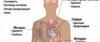
Symptoms of anasarca include:
- skin that will dimple after pressing with your finger for a few seconds;
- high or low blood pressure;
- slow or fast pulse;
- organ failure, especially liver and kidney failure.
Anasarca can be debilitating. The disorder can render the patient immobile, as the swelling makes it nearly impossible to walk or move the limbs. Swelling in the face can also impair vision by making it difficult to open your eyes.
In some cases, anasarca may require emergency treatment. If you experience the above symptoms, as well as shortness of breath, difficulty breathing, or chest pain, seek emergency care immediately. These may be signs of pulmonary edema, i.e. accumulation of fluid in the lungs. The condition can quickly become life-threatening.
Treatment of swelling using folk remedies
To treat edema caused by heart disease, take birch leaves and buds in equal quantities, crush them and add to them the same amount of bedstraw grass and parsley seeds. Stir and brew one tablespoon of this mixture in a glass of boiling water. The infusion is taken warm 3-4 times a day, a quarter glass.
An infusion of May dandelion leaves helps relieve swelling.
Causes and risk factors
The most common causes of anasarca seen by your doctor are heart failure, cirrhosis of the liver, kidney failure, and pregnancy. Other causes of anasarca are venous obstruction, burns, trauma, malignant neoplasms, etc.
Main symptoms of pathology
The development of peripheral edema is entirely associated with the course of heart failure. The first signs of anasarca may appear in the early stages of cardiac pathology. Stagnation in the systemic circulation is not yet pronounced, so tissue swelling will not be significant and can be easily relieved.
Initially, this pathology appears to the doctor in the form of persistent edema, first local and then widespread. Heart failure is characterized by the presence of edema in the lower extremities in the evening and at night. As the disease progresses, edema may spread to new areas and may not go away without therapeutic intervention.
In addition to appearance, a patient with anasarca is characterized by shortness of breath at rest. This is due to the sweating of fluid from the vessels into the lungs and pleura. In severe decompensation, the presence of free fluid is often detected not only in the abdominal and pleural cavity, but also in the cardiac sac.
The presence of fluid in the area of the heart can cause various complications, ranging from the development of pericarditis to acute cardiac arrest.
Diagnosis is made based on studying the medical history, taking an anamnesis, performing ultrasound and x-rays of the heart and lungs. The results of laboratory tests are also important.
We recommend reading the article about fluid in the lungs in heart failure. From it you will learn about the causes of edema, signs, first aid to the patient and treatment.
Pathophysiology
Edema develops as a reaction to an increase in hydrostatic pressure in the capillaries, an increase in capillary permeability, a decrease in plasma oncotic pressure, or a combination of these changes. Edema may also be secondary to lymphatic obstruction, resulting in fluid retention in the interstitial space. Below are the clinical conditions for the various mechanisms described.
- Increased capillary hydrostatic pressure: Heart failure, kidney disease, early cirrhosis of the liver, pregnancy, medications.
- Venous obstruction or insufficiency conditions such as deep vein thrombosis, hepatic venous congestion.
- Burns, trauma, sepsis, allergic reactions, malignant ascites.
- Malignant neoplasms, lymph node dissection.
- Nephrotic syndrome, liver disease, poor diet.
In the first stage, when fluid moves from the vascular space into the interstitium, it reduces plasma volume. This reduces tissue perfusion. Poor tissue perfusion causes sodium and water retention by the kidneys. Some of the resulting excess fluid will be retained in the intravascular compartment. However, changes in capillary hemodynamics cause most of the retained fluid to enter the interstitium and ultimately manifest as edema.
Symptoms of anasarca in heart failure
The symptoms of swelling and the degree of its progression are influenced by the underlying disease. Anasarca in heart failure develops slowly, affecting the distal parts of the lower extremities. This is especially noticeable in the evenings. Why does swelling occur in this area? Only here the hydrostatic pressure is high due to the distance from the heart.
If severe heart failure occurs, significant amounts of edema also begin to affect the lower back, the front of the abdomen, the genitals, and the chest. Swelling in bedridden patients occurs most strongly in the area of the back and sacrum, which is explained by the influence of gravity. If treatment is ineffective, anasarca provokes ascites (fluid accumulation in the abdominal cavity) and pulmonary edema (hydrothorax).
Diagnostics
The history should include timing of swelling and position changes, unilateral or bilateral, as well as medication history and evaluation of systemic diseases.
A physical examination can help make the diagnosis. The examination should be aimed at determining the nature of the edema—peripheral edema versus pulmonary edema, pitting edema versus non-jugular venous edema, and the presence of jugular venous distention. Below are the results of a survey of typical clinical conditions that can cause anasarca.
Patients with pulmonary edema primarily complain of dyspnea on exertion and orthopnea. Physical examination usually reveals moist rales, possible diastolic gallop and heart murmurs. Heart and kidney diseases are common causes of pulmonary edema. Peripheral edema is usually detected by the presence of pitting after pressure is applied to the swollen area for at least 5 seconds. Pitting reflects the movement of excess interstitial fluid in response to pressure. It is usually seen in dependent areas such as the lower extremities in ambulatory patients and over the sacrum in patients who are bedridden. Scrotal swelling is also common in men. Non-pitting edema suggests lymphatic obstruction or hypothyroidism. The acute onset of unexplained unilateral leg swelling should increase the likelihood of deep vein thrombosis (DVT). Patients with cirrhosis may develop ascites and then swelling of the lower extremities due to increased venous pressure below the diseased liver. The presence of other signs of portal hypertension, such as distended abdominal veins and an enlarged spleen, also indicates primary liver disease.
The cause of swelling can be determined by noting changes in skin temperature, color, and texture. Acute DVT and cellulitis can cause increased warmth in the affected area. Hemosiderin deposition or chronic venous insufficiency often causes the skin to take on a muscular, reddish hue and usually affects the medial malleolus. As venous insufficiency progresses, it can lead to lipodermatosclerosis, which is associated with markedly sclerotic and hyperpigmented tissue and is characterized by fibrosis and hemosiderin deposition, which can lead to venous ulcers over the medial malleolus. Ulcers can progress to deep, weeping erosions. Myxedema from hypothyroidism presents with generalized, dry, thick skin with nonpitting periorbital edema and yellow to orange discoloration of the skin on the knees, elbows, palms, and soles. Pretibial myxedema occurs in patients with thyroid disease and is characterized by bilateral, asymmetrical, non-pitted, scaly thickening and induration of the skin. These sores may be purple or lightly pigmented (yellow-brown) and often have an orange peel appearance. The most common location of pretibial myxedema is over the lower legs, especially in the pretibial areas or dorsum of the foot.
— Analyzes and visualization.
Routine laboratory tests, such as a comprehensive metabolic panel, can help evaluate kidney function, albumin, and liver function tests. All children with edema should have a urine test. Urine test strips primarily detect albumin and require an additional sulfosalicylic acid protein precipitation test to detect globulins and Bence Jones proteins. Urine protein/creatinine ratios or 24-hour urine protein tests may eliminate the need for sulfosalicylic acid testing. The finding of an apparently positive protein test in combination with hypoalbuminemia and clinical edema is in fact diagnostic of nephrotic syndrome. Measuring natriuretic peptide in the brain may provide clues to the underlying cause of CHF. Albumin and liver function tests can help diagnose cirrhosis.
Chest x-ray is useful in detecting heart failure, pulmonary edema, and pleural effusion. For reasons of safety, ease of use, and information provided, the most commonly used radiographic method in patients with renal failure is renal ultrasound. Ultrasonography allows the doctor to determine the size of the kidneys and evaluate for cystic kidney disease and hydronephrosis. Cardiac echocardiography can evaluate ventricular function, detect pericardial effusion, and help diagnose heart disease.
Echocardiography to assess pulmonary artery pressure is recommended in patients with obstructive sleep apnea and edema. Vein ultrasound is the preferred imaging modality for suspected DVT. Duplex ultrasound can also be used to confirm chronic venous insufficiency. Magnetic resonance angiography with venography of the lower extremity and pelvis can be used to evaluate internal or external DVT of the pelvis or thigh. Compression of the left iliac vein by the right iliac artery (May-Turner syndrome) should be suspected in women aged 18 and 30 years with swelling of the left lower extremity. Lymphoscintigraphy is the test of choice for the evaluation of lymphedema when the diagnosis cannot be made clinically. MRI can help diagnose musculoskeletal etiologies such as a gastrocnemius tear or popliteal cyst.
Edema of the subcutaneous tissue
Swelling of the subcutaneous tissue may be one of the signs of any pathological processes occurring in the human body. Without a special medical examination, it is not possible to establish exactly what caused the swelling in any part under the skin. The history and etiology of the development of this pathology are very diverse.
Swelling of the subcutaneous tissue is visible even to the naked eye, and this phenomenon negatively affects the aesthetic appearance of a person. It is worth knowing that ordinary swelling of the skin, which is not a serious pathology, can be combined with swelling of the deeper tissues of the epidermis, as a result of which a person should treat such a problem not as a fleeting cosmetic defect, but as a possible presence of serious diseases in his body .
Swelling or edema of the subcutaneous tissue represents excessive accumulation of fluid in the deep soft layers of the epidermis.
The development of such a pathological process can be observed not only in adults, but also in children. In addition, this pathology can occur in a fetus developing in the womb of a pregnant woman. This process is known in medical practice as subcutaneous fetal edema.
Causes of subcutaneous fluid accumulation
Depending on the causes of the disease, the following types of subcutaneous edema are distinguished:
- traumatic. They arise as a result of mechanical damage to the tissues of the epidermis, which leads to disruption of the integrity of its blood vessels, resulting in the formation of subcutaneous hemorrhages. Due to the fact that the damaged vessels can no longer pump blood, swelling of the soft layers of the epidermis occurs, and in advanced cases, the pathological accumulation of fluid spreads to the entire anatomical area. After some time, the vessels are gradually restored, the swelling goes away over time;
- inflammatory. They are formed as a result of the development of various kinds of inflammatory processes in the soft tissues of the skin, which leads to compression of the vessels, resulting in a pathological accumulation of blood in their cavities. Then, due to the resulting leakage of fluid through the walls of blood vessels, a subcutaneous accumulation of fluid is formed, that is, edema. As the inflammatory processes decrease, the swelling of the skin gradually decreases and disappears over time;
- edema that forms as a result of any disturbances in blood flow or activity of the human lymphatic system. This kind of pathological accumulation of fluid in the subcutaneous tissue develops against the background of the presence of such mechanical obstacles in the human circulatory system as calcification and tumor. In the absence of the necessary drug treatment, the pathological process will only worsen;
- accumulation of fluid in the deep layers of the epidermis due to the occurrence of any disturbances in metabolic processes in the human body. Such swelling occurs as a result of a violation of protein metabolism in the human body, which can occur as a result of various pathologies affecting the liver, during prolonged fasting, as well as in the case of an acute lack of various kinds of vitamins and minerals in the body.
Symptoms accompanying swelling of the face and head
Swelling of the face is a serious problem; the cheeks and other areas of the head can get into the affected area, resulting in a pronounced cosmetic defect that can quickly spread throughout the entire face, but does not resolve for a long time. Swelling of the cheeks and other parts of the head can occur as a result of mechanical injuries to the skull, in the presence of kidney diseases, inflammation of the paranasal sinuses and the occurrence of inflammatory processes in the oral cavity to which its mucous membrane is exposed. The main symptoms accompanying the accumulation of fluid in the subcutaneous tissue of the head are:
- swelling of the cheeks and entire face;
- feeling of general malaise;
- nasal congestion, causing the nasal mucosa to swell;
- increase in body temperature.
If swelling of the cheeks and swelling of the face occur in the morning after sleep, most likely the person has some kind of disturbance in the functioning of the body's renal system.
Swelling of the lower and upper extremities
Anasarca
The accumulation of excess subcutaneous fluid in the extremities can occur as a result of any local disorders or the occurrence of serious systemic processes. Swelling of the limbs can occur due to the following:
- the presence of tumors that interfere with normal blood flow;
- various types of heart diseases;
- vascular pathologies, for example, such as varicose veins and obliterating endarteritis.
In the case of a mechanical obstruction, only unilateral loose swelling occurs, while practically no changes in the epidermis are observed. Damage to the vascular system mainly occurs in the lower extremities, especially the feet and legs. Moreover, the skin in these places becomes swollen most often at the end of the day, after a person has performed any physical activity or simply stood on his feet for a long time. In the case of varicose veins, characteristic changes appear on the surface of the epidermis that resemble a mesh or a large accumulation of nodules.
Treatment
When treating edema, one should be guided by its etiology. Causes typically include chronic venous insufficiency, lymphedema, DVT, and drug-induced edema. Pulmonary edema is the only form of generalized edema that is life-threatening and requires immediate treatment. In all other edematous conditions, removal of excess fluid may occur more slowly as it does not pose a serious threat to the patient's life. In patients with generalized edema caused by heart failure, nephrotic syndrome, or primary sodium retention, edematous fluid may mobilize rapidly.
In patients with anasarca, removal of 2 to 3 liters or more of edematous fluid over a 24-hour period can usually be accomplished without a clinically significant decrease in plasma volume. In patients with localized edema due to venous or lymphatic obstruction or malignant ascites, diuretic therapy may result in volume depletion. Diuretic therapy for generalized edematous conditions usually begins with loop diuretics such as furosemide.
In patients with cirrhosis, a combination of spironolactone and a loop diuretic is the preferred initial diuretic regimen to prevent hypokalemia. Because hypokalemia predisposes to increased ammonia production. Patients with nephrotic syndrome may require higher doses of diuretics. Some cases of idiopathic edema are caused by diuretics, and the initial approach for patients with idiopathic edema who are already taking diuretics is to stop the diuretics for at least 2 to 3 weeks and encourage sodium restriction in the diet.
The mainstay of treatment for lower extremity edema due to venous insufficiency is mechanical treatment methods, including compression stockings with leg elevation and pressure of 20 to 30 mmHg. Art. with mild swelling and from 30 to 40 mm Hg. Art. with severe edema complicated by ulcers. Compression therapy is contraindicated in patients with peripheral artery disease. Topical skin and venous ulcer wound care is important to prevent secondary cellulite and dermatitis. Eczematous (congestive) dermatitis, characterized by dry, inflamed, scaly skin over superficial varicose veins, often occurs in patients with chronic venous insufficiency. Treatment includes daily moisturizing with emollients and short courses of topical corticosteroids for severely inflamed skin.
Primary treatment for lymphedema involves comprehensive decongestant physical therapy, including manual lymphatic massage and multi-layer dressings. The first goal is to improve fluid resorption and continue until maximum therapeutic response is achieved. The maintenance phase of treatment includes compression stockings at 30-40 mmHg. Art. Pneumatic compression devices can be used to supplement standard therapy. Surgical tumor removal or bypass procedures are limited to severe refractory cases. Diuretics have not been shown to be effective in treating lymphedema.
Links[edit]
- Kumar Vinay. Pathological basis of Robbins and Cotran's disease
. 8th ed. p.112; Philadelphia: Saunders Elsevier, 2010. ISBN 978-0-8089-2402-9 - Kattula, Sri Rama Surya Tez; Avula, Akshay; Baradi, Krishna M. (2020), "Anasarca", StatPearls
, Treasure Island (FL): StatPearls Publishing, PMID 30085555, retrieved 02 July 2021 - "Anasarca: causes, swelling and treatment". Health line
. 2017-07-11. Retrieved July 2, 2021. - "Anasarca: causes, treatment and definition". medicalnewstoday.com
. 2018-02-13. Retrieved July 2, 2021. - "Nephrotic syndrome - Symptoms and causes". Mayo Clinic
. Retrieved July 2, 2021. - Yasuda, Jessica Lacy; Rufo, Paul A. (02/26/2018). "Protein-losing enteropathy in the setting of severe anemia due to iron deficiency". Journal of Investigations in Medicine.
Significant Case Reports .
6
. DOI: 10.1177/2324709618760078. ISSN 2324-7096. PMC 5833205. PMID 29511696. - Mehta, Ankita; Shah, Mansi (2016). "Case Report: Uncontrolled Anasarca: Capillary Leak Syndrome". Medical forum
.
Medical Forum: Vol. 17, Article 8. 17
. DOI: 10.29046/TMF.017.1.009. Retrieved July 7, 2020. - Madias, J. E.; Bazaz, R; Agarwal, H; Win, M; Medepally, L. (September 2001). "Anasarca-mediated attenuation of electrocardiogram complex amplitude: a description of a hitherto unrecognized phenomenon." Journal of the American College of Cardiology
.
38
(3):756–64. DOI: 10.1016/s0735-1097(01)01429-2. PMID 11527629.