Hypovolemia: a dangerous condition that requires immediate attention
What do we know about hypovolemia?
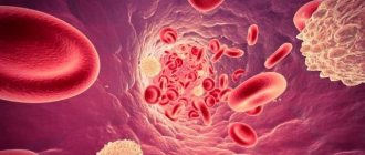
Hypovolemia is a condition when, as a result of fluid loss, the volume of blood that circulates in the human body decreases.
This violation poses a serious threat to the health and life of the patient. A severe degree can lead to hypovolemic shock and the development of multiple organ failure. Therefore, as a rule, the patient requires immediate medical attention: hospitalization and intensive care. Treatment is carried out in a hospital setting.
In rare cases, hypovolemia is caused not by the loss of fluid, but by its redistribution in the tissues of the body. At the same time, the filling of blood vessels with blood is reduced. This phenomenon is called relative hypovolemia. Blood loss and dehydration are absolute.
Reasons and mechanism of formation
The pathological hypovolemic process is based on severe dehydration.
Leads to it:
- Vomiting, characteristic of withdrawal syndrome after binge drinking.
- Diarrhea, with the help of which the body cleanses itself of alcohol toxins.
- Liver diseases characteristic of alcoholism (toxic hepatitis and cirrhosis). In this case, non-functional leakage of plasma into the intercellular space occurs due to a decrease in the oncotic pressure of the blood.
- Insufficient intake of fluid and protein into the body.
Hypovolemia is preceded by increased venous and hydrostatic pressure in small arteries, reflecting the negative influence of factors predisposing to a painful condition. Secondarily, the permeability of the vascular wall increases. Sweating of blood through arteries and veins leads to its general deficiency.
The body tries to compensate by:
- Producing the required amount of plasma.
- Slowing venous return.
- Spasms of small vessels.
The protective reaction allows you to maintain the activity of the brain and heart in a relatively normal state for some time and avoid circulatory complications.
What causes the condition
Most cases are associated with severe dehydration, blood loss, and large burns.
Dehydration can be caused by:
- repeated vomiting and diarrhea, excessive sweating
- insufficient fluid intake,
- taking medications, such as diuretics.
Hypovolemia that is caused by loss of plasma rather than blood is called polycythaemic.
Blood loss occurs as a result of injury, surgery, or internal bleeding. Blood loss associated with internal bleeding is considered especially dangerous, since it is not always possible to immediately determine where it is localized.
Massive blood loss is caused by:
- gastrointestinal bleeding,
- rupture of the fallopian tube during ectopic pregnancy,
- rupture of an aortic aneurysm.
A general decrease in blood volume is called normocythemic hypovolemia.
Forecasts
Foggy. With proper help, or if the pathological process proceeds relatively easily - positive. But decompensation can occur at any time.
The prospects for severe injuries are much worse. Although here everything is ambiguous and depends on the moment of the start of therapy.
Hypovolemia is a syndrome inherent in many dangerous conditions. It is not him who needs to be treated. The main forces should be directed to the fight against the original source. And this must be done as quickly as possible. In this case, there is every chance of recovery without consequences.
Adelina Pavlova
Symptoms
The condition can be recognized by the following signs:
- dry mucous membranes
- pale skin
- decreased skin turgor
- rapid breathing
- lowering blood pressure levels
- tachycardia
- anxiety, confusion
- weak pulse (due to decreased cardiac output)
- thirst
- dizziness
- lack of urination for a long time.
Acute hypovolemia leads to an emergency condition - hypovolemic shock, which can result in coma.
4.Treatment
Depending on the situation, urgent resuscitation measures are taken. The primary objectives are to stop bleeding, restore breathing and replace blood loss through blood transfusion or transfusion of donor blood, and, if necessary, to relieve pain shock. In the future, measures are needed to normalize the acid-base balance, prevent thrombosis (DIC syndrome), prevent renal failure and secondary infections. Further management of the patient is determined by the results of the diagnostic examination and the dynamics of the condition.
Diagnostics
The main means of establishing a diagnosis is examination and interview (if possible) of the patient.
If hypovolemia is caused by visible trauma, large-area burns, its diagnosis is not difficult.
If a patient is suspected of dehydration or internal bleeding, the patient is sent for a clinical and biochemical blood test, a urine test, which will determine whether there is a change in the water-electrolyte balance.
Also, to clarify the diagnosis, the doctor performs CT, X-ray, ultrasound and endoscopy of organs in which bleeding may develop.
The likelihood of developing fibrosis after “crown”
The growth of scar tissue does not occur in all people who have had pneumonia during the coronavirus. The risk group includes patients with:
- complicated course of pneumonia;
- extensive damage to lung tissue (if more than 50% is damaged);
- distress syndrome;
- respiratory failure, due to which it was necessary to use a ventilator;
- the addition of a secondary fungal or bacterial infection.
According to the data that doctors have today (perhaps they will change in a few years when the coronavirus is studied more deeply), fibrosis does not develop in all patients. Persons who have had a mild or moderate form of the virus very often avoid bronchitis and pneumonia altogether.
Hypovolemia in children
Danger of condition
The child's body is not sufficiently adapted to compensate for fluid deficiency. This feature should be taken into account when hypovolemia is suspected, since a state of shock in a child can occur much faster than in an adult. This is even more important the younger the child is. Thus, in children up to one year old, the ductus arteriosus may remain open, which, when fluid or blood is lost, actually deprives the lungs of blood supply. Therefore, the issue of hospitalization of the child is resolved immediately.
Causes
In newborns, hypovolemia can develop due to blood loss through the placenta, internal bleeding as a result of injury, or fluid accumulation in the abdominal or pleural cavity.
In older children, in addition to blood loss, the condition is caused by uncontrollable vomiting and diarrhea due to intestinal infections and intoxication; non-compliance with drinking regime.
Treatment
Treatment is prescribed based on the patient's condition.
Mild degrees require fluid replacement (drip, intravenous).
If the cause is injury, the first thing to do is stop the bleeding. Then restore normal hemodynamic parameters and breathing, while simultaneously treating the injury and pain relief for the patient.
Significant blood loss requires transfusion of plasma, red blood cells, platelets, and donor blood. Further therapy is aimed at preventing thrombosis, associated infections, and preventing renal dysfunction.
Signs of hypovolemic disorder
Symptoms of the disorder depend on its severity.
In the practice of narcology, the following stages of hypovolemia are distinguished:
- Easy. Minor fluid losses (up to 20% of bcc) are accompanied by a decrease in blood pressure, increased heart rate 10-15 beats higher than normal, and slight shortness of breath. The patient is pale, complains of weakness, dry mucous membranes, dizziness, and attacks of nausea.
- Average. The patient loses up to 40% of his total blood volume. In this case, the pressure is sharply reduced (below 90 mm Hg) and the pulse is above 120-130 beats per minute. Breathing is sharply rapid and intermittent. Externally, pallor and cyanosis of the skin in the area of the nasolabial triangle are revealed. There is severe sweating, lethargy, and decreased urine output due to severe thirst.
- Heavy. With a large-scale decrease in blood volume (above 40%), a critical drop in blood pressure is detected. The pulse is barely detectable and reaches 150 or more beats per minute. No urine is released. The patient's appearance indicates that he is in critical condition. There is deathly pale skin, a pointed face, sunken cheeks, and an unconscious state.
Loss of BCC is often accompanied by:
- Hypoglycemia is a serious complication of alcohol withdrawal syndrome. A harbinger of the development of hypoglycemia is any kind of liver disease, a decrease in glycogen reserves, and poor non-caloric nutrition.
- Hyponatremia is a disorder most often found in patients who abuse low-alcohol drinks (cocktails, beer). In this condition, peripheral edema occurs in combination with hypovolemia.
- Hypernatremia is a condition that occurs with encephalopathy, acute liver failure, and a clear deficiency in the amount of circulating blood.
- Hypomagnesemia is a deficiency of magnesium inside cells in people with alcoholism, regardless of its content in the plasma. The most common symptoms: general weakness of the body and drowsiness. The risk of seizures and heart problems (palpitations) may increase.
- Hypokalemia. In people suffering from alcoholism, as a rule, potassium deficiency is detected in the body, even if its content in the blood plasma is at normal levels. In combination with an overexcited nervous system, the lack of this element leads to irreparable disturbances in the functioning of the heart with a threat to life. Anyone suffering from alcohol withdrawal syndrome is prescribed potassium supplements.
This pathology manifests itself:
- Fatigue quickly.
- Depression.
- Attacks of tachycardia.
- Muscle weakness.
Alcohol withdrawal syndrome may be accompanied by metabolic acidosis, which occurs due to the accumulation in the body of breakdown products of alcohol and other substances of pathological metabolism, in the process of tissue hypoxia and respiratory alkalosis.
Drugs for hypovolemia
Treatment is carried out in a hospital, often in the intensive care unit. In most cases, infusion therapy is used.
The drugs of choice are solutions of colloids (solutions of gelatin and dextran, for example, rheopolyglucin) and crystalloids (Ringer's solution), blood substitutes (voluven, refortan).
To avoid infections, the patient is prescribed broad-spectrum antibiotics.
To raise the patient's blood pressure, norepinephrine and dobutamine are prescribed.
How to remove negative consequences after “corona”
The rehabilitation process after Covid-19 includes three main steps:
- Establishing oxygen supply to lung tissue.
- Elimination of symptoms of poor lung function.
- Improvement of general physical and psychological condition.
To solve these problems, patients are prescribed:
- Drug therapy. Involves taking medications that increase blood supply to the lungs, accelerate the resorption of fibrous lesions, and relieve inflammation. Medicines should be selected on a purely individual basis. It is unacceptable to take medications unless prescribed by a doctor, since self-medication very often leads to dire consequences.
- Physiotherapeutic procedures. They allow you to destroy pathogenic microflora, remove inflammation, and increase local immunity. In the post-Covid period, the following have proven themselves to be excellent: pulsed and low-frequency magnetic therapy, SMT, the use of an ultra-high frequency electromagnetic field and polychromatic polarized light, electrophoresis, ultrasound, and inductothermy. It is very important that the patient undergoes a whole course of physiotherapy and does not miss sessions. Only then will physical treatment be highly effective and ensure rapid positive dynamics.
- Special diet. After coronavirus, you need to eat a lot of protein foods. The basis of the diet should be low-fat dairy products, meat, fresh vegetables and fruits.
- Therapeutic exercise. Moderate physical activity promotes the resorption of stagnant lesions. You should start gymnastics with the simplest exercises. As performance improves, it will be possible to complicate the techniques used. If the patient is far from sports and does not want to engage in exercise therapy, cycling or long walks in a forested area are suitable for him.
Don't worry if your doctor has diagnosed negative changes in your lungs after coronavirus. Take care of your health, and soon the situation will improve.
List of used literature
- Savelyev V.S. Comparative effectiveness of plasma substitutes for normovolemic hemodilution and correction of acute blood loss / V.S. Savelyev, N.A. Kuznetsov // Vestn. surgery. –1985.
- Bryusov P.G. Emergency infusion-transfusion therapy for massive blood loss / P.G. Bryusov // Hematology and transfusiology. –1991. – No. 2.
- Pshenisnov K.V., Aleksandrovich Yu.S. Massive blood loss in pediatric practice. Hematology and transfusiology. 2020;65(1):70-86
- Pleskov A.P. Invasive monitoring of central hemodynamics. // Intensive care, ed. V.D.Malysheva, M.: Medicine, 2002. P. 175–190.
Frequently asked questions about hypovolemia
What diseases can cause hypovolemia?
The condition occurs with gastrointestinal diseases, accompanied by uncontrollable vomiting and diarrhea, kidney disease, sharp drops in blood glucose levels, and some allergic reactions. However, hypovolemia is most often caused by blood loss or extensive burns.
How does hypovolemia manifest?
The patient has dry mucous membranes, tachycardia, thirst, and impaired skin turgor. As the condition worsens, a drop in blood pressure, weak pulse, and rapid breathing occur. In the acute stage - confusion.
How is hypovolemia treated?
The main method of treatment is fluid replacement. In most cases, intravenous administration of saline or electrolyte solutions is used.
What does hypovolumia mean?
Hypovolumia is a term used in ultrasound examinations. It means a reduction in organ volume due to congenital anomalies, underdevelopment, or formed after certain diseases. Hypovolumia of the thyroid gland is caused by the following pathologies:
- Hypoplasia of the thyroid gland. The organ is not fully formed, often in the womb. When the amount of hormones produced by the thyroid gland is sufficient, a small size is considered normal. However, most often, due to underdevelopment of the gland, a lack of thyroid hormones occurs, which causes chronic hypothyroidism.
- Atrophy of the thyroid gland due to the death of part of the follicular tissue. In most cases it causes hormone deficiency.
Hypovolumia has 2 degrees:
- At grade 1, the required level of glandular tissue is compensated by the body. As a result, the thyroid gland continues to function normally for some time.
- Stage 2 hypovolemia of the thyroid gland is characterized by a very low level of hormones, as a result of which serious disturbances occur in the body.
The older a person is, the smaller the volume of the thyroid gland. A gradual decrease in its size is a physiological phenomenon. This condition has nothing to do with hypovolumia.
Sometimes a similar diagnosis is made - hypovolemia. This is another problem, namely a decrease in blood supply to the organ.


