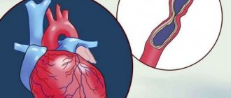
Blood supply to the brain is one of the invisible functions of our body. Strong emotions, physical activity or prolonged mental work cause a feeling of a rush of blood. But when we brush our teeth or make a cup of tea, it seems that nothing happens. However, at the biological level, any, even the most primitive activity is a continuous process of saturation of brain structures with blood. The quality of our life directly depends on the safety of the vascular system. The brain: your own doctor In the blood supply system of the brain, there are two groups of arteries: extracranial (located between the head and heart) and intracranial (vessels of the cranial cavity and bone canals). The brain has protection from mechanical damage to blood vessels - the so-called arterial circle or circle of Willis. This is a closed system of arteries at the base of the brain that provides normal blood supply when one or more arteries are blocked. If nature were not so prudent, boxers would end their careers after the first knockout. In everyday life, it is not only mechanical injuries that pose a danger. Pathologies are associated with blockage or narrowing, changes in the structure of blood vessels and the tone of their walls. In illness, blood flow to the brain deteriorates. Disturbances in natural blood flow cause atherosclerosis, stroke, aneurysm, etc. Chronic pathologies lead to irreversible changes. At an early stage, the disease is often asymptomatic. Cannot be ignored: ringing in the ears or head, heaviness in the head, dizziness, blurred vision, fainting, loss of coordination, spontaneous weakness or numbness of the limbs. If these disorders occur, you should immediately consult a doctor. Smoking harms blood vessels Using ultrasound, the diagnostician determines: - The condition of the walls of the arteries - Altered course of the arteries - The presence of narrowing (stenosis), lack of lumen (occlusion) of the arteries - Disturbances in blood flow through the arteries - The presence of formations inside the arteries (for example, atherosclerotic plaques) The study is performed in lying on your back (sometimes with a cushion under your shoulder blades). The specialist examines the brachiocephalic trunk, subclavian arteries, vertebral arteries, carotid arteries, cerebral arteries (only with a joint examination of the extracranial and intracranial sections; the intracranial section is not examined separately from the extracranial). On the day of your doctor’s appointment, you must stop using substances that affect vascular tone: nicotine, tea, coffee, energy drinks. As with other vascular diseases, the best method of prevention is a healthy lifestyle: giving up bad habits, controlling nutrition and moderate physical activity. You must bring the results of previous studies (if available) with you to your appointment. Ultrasound at OLMED MC is the key to effective treatment without the risk of complications and premature relapses!
Price list
Prices for medical services from April 19, 2021
Ultrasound examinations
| Code | Name of medical service | Cost, rub. |
| 3.1 | Ultrasound scanning of the veins of the lower extremities | 1 400,00 |
| 3.2 | Ultrasound scanning of the arteries of the lower extremities | 1 500,00 |
| 3.3 | Ultrasound scanning of the veins of the upper extremities | 1 300,00 |
| 3.4 | Ultrasound scanning of the arteries of the upper extremities | 1 400,00 |
| 3.5 | Ultrasound scanning of vessels (veins and arteries) of the upper extremities | 2 300,00 |
| 3.6 | Ultrasound scanning of vessels (veins and arteries) of the lower extremities | 2 500,00 |
| 3.7 | Brachial-ankle index (BAI) | 630,00 |
| 3.8 | Ultrasound scanning of the arteries of the lower extremities with PLI measurement | 2 100,00 |
| 3.9 | Measurement of regional systolic pressure (RSBP) of the upper extremities | 1 300,00 |
| 3.10 | Measurement of regional systolic pressure (RSBP) of the lower extremities | 1 300,00 |
| 3.11 | Ultrasound scanning of neck vessels (brachiocephalic arteries) | 1 500,00 |
| 3.12 | Ultrasound scanning of brain vessels and neck vessels | 2 850,00 |
| 3.13 | Ultrasound scanning of the abdominal aorta without visceral branches | 600,00 |
| 3.14 | Ultrasound scanning of abdominal vessels (aorta, renal arteries) | 1 200,00 |
| 3.15 | Ultrasound scanning of abdominal vessels (aorta, celiac trunk and mesenteric arteries) | 1 200,00 |
| 3.16 | Ultrasound scanning of abdominal vessels (aorta, renal arteries, celiac trunk and mesenteric arteries) | 1 600,00 |
| 3.17 | Ultrasound scanning of the inferior vena cava | 800,00 |
| 3.18 | Ultrasound scanning of the left renal and testicular veins | 1 350,00 |
| 3.19 | Ultrasound of the heart | 1 500,00 |
| 3.20 | Ultrasound of the abdominal organs | 900,00 |
| 3.21 | Kidney ultrasound | 700,00 |
| 3.22 | Ultrasound of the abdominal organs + kidneys | 1 400,00 |
| 3.23 | Ultrasound of the bladder | 600,00 |
| 3.24 | Ultrasound of the thyroid gland | 700,00 |
| 3.25 | Ultrasound of the mammary glands | 1 000,00 |
| 3.26 | Ultrasound of one joint | 800,00 |
| 3.27 | Ultrasound of two joints | 1 300,00 |
| 3.28 | Ultrasound of lymph nodes, group 1 | 800,00 |
| 3.29 | Ultrasound of soft tissues (skin formations, subcutaneous tissue, muscle tissue, etc.), 1 anatomical area | 800,00 |
| 3.30 | Ultrasound of the pleural cavity | 1 000,00 |
| 3.31 | Ultrasound of the prostate gland | 1 200,00 |
| 3.32 | Ultrasound of the scrotum | 1 000,00 |
| 3.33 | Ultrasound of the scrotum with Doppler sonography | 1 200,00 |
| 3.34 | Ultrasound of the pelvic organs | 1 200,00 |
| 3.35 | Doppler ultrasound of the pelvic organs | 1 300,00 |
| 3.36 | Obstetric ultrasound - early pregnancy | 1 200,00 |
| 3.37 | Ultrasound Doppler Obstetric | 1 700,00 |
| 3.38 | Folliculometry | 500,00 |
| 3.39 | Doppler | 800,00 |
Ultrasound-guided manipulations
| Code | Name of medical service | Cost, rub. |
| Ultrasonic navigation is not included in the price and is paid additionally according to the price list | ||
| 29.1 | Ultrasound navigation during medical procedures | 900,00 |
| 29.2 | Ultrasound navigation during puncture (diagnostic, therapeutic) of the joint | 900,00 |
| 29.3 | Echosclerotherapy, session | 2 800,00 |
| 29.4 | Endovenous laser coagulation (EVLC) of the perforating vein, session | 6 700,00 |
| 29.5 | Catheter trunk scleroobliteration of the GSV or SSV, session | 14 900,00 |
| 29.6 | LAFOSTherapy (laser associated foam scleroobliteration of saphenous veins), session | 24 300,00 |
| 29.7 | Therapeutic puncture of cysts, hematomas | 3 050,00 |
| 29.8 | Puncture of thyroid formations with puncture biopsy and cytological examination | 1 900,00 |
| 29.9 | Puncture of breast formations with puncture biopsy and cytological examination | 1 900,00 |
1.What is included in the concept of “intracranial pathology”?
Intracranial pathology is a general term that covers the entire spectrum of disorders with intracranial localization.
Intracranial diseases may not manifest significant symptoms. In some cases, on the contrary, they are very painful for the patient. Due to the fact that we are talking about a pathology directly related to the brain, intracranial disorders are often life-threatening and require emergency assistance.
The consequences of intracranial pathology can be: persistent functional failure, mental disorders, organic disorders, including disability. Treatment tactics for intracranial pathology are often developed with the participation of doctors of several specialties. Targeted drug therapy and surgical procedures in close proximity to the brain can have unwanted side effects and risks, so the treatment plan is often approved by a medical council.
A must read! Help with treatment and hospitalization!
Our ultrasound doctors
Garayeva Anna Evgenievna
Head of Ultrasound Diagnostics Department
Lyzhin Dmitry Vyacheslavovich
Kravchenko Nikolay Alekseevich
Ultrasound diagnostics doctor of the first category, author of 15 scientific papers
Naronov Alexey Yurievich
Ultrasound diagnostic doctor
Maklakova Daria Ivanovna
Ultrasound diagnostic doctor
2.Diagnostics and classification
The brain is one of the most private and difficult to access parts of the human body. Diagnosis and treatment for this reason are often difficult. Modern techniques make it possible to conduct research not only through a targeted study of intracranial structures and their functions (using MRI, X-ray diagnostics, angiography, electroencephalography, biopsy), but also indirectly clarify the diagnosis through blood and cerebrospinal fluid studies, bacteriological culture, daily monitoring of brain activity and intracranial pressure.
If intracranial pathology is suspected, further research allows us to clarify its nature, understand the causes of its occurrence and development factors, make a prognosis and provide qualified assistance. All intracranial disorders, depending on the etiology, can be classified into one of the groups:
- inflammatory and infectious diseases (with a primary focus directly in the brain tissue, developed secondarily against the background of general somatic infections or spreading intracranially from neighboring tissues/organs - ear, eye, maxillary sinuses);
- cysts and neoplasms;
- mechanical head injuries (including birth injuries, hematomas, concussion);
- problems of cerebral circulation (intracranial hypertension, aneurysms, strokes, stenosis);
- hypoxic disorders;
- parasitic infection.
Visit our Neurosurgery page
Indications
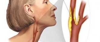
The main symptoms of circulatory disorders, for which the doctor will prescribe a duplex scan of the veins of the neck:
- Frequent headaches.
- Dizziness that occurs when changing body position or turning, throwing back the head.
- Gait disturbances – unsteadiness, involuntary deviation from moving in a straight line.
- Changes in facial expressions and symmetry of facial movements.
- Visual distortions – darkening before the eyes, “spots”, blurred vision.
- Memory impairment, difficulty concentrating.
- Noise or ringing in the ears, deterioration of hearing acuity.
- Numbness in the upper extremities, muscle weakness, deterioration of sensitivity.
- Noticeable strong pulsation on the neck of the blood vessel.
- The difference in blood pressure in the arms is more than 15 mm. rt. Art.
- Sleep disorders.
Duplex scanning of neck veins is recommended even without such symptoms. Direct indications are systemic diseases that impair blood circulation. These are diabetes mellitus, atherosclerosis, high cholesterol, osteochondrosis, heart disease (coronary artery disease, defects), excess weight, hypertension and others. It is also worth examining blood vessels for preventive purposes every year after forty years. This is especially necessary for those who lead a sedentary lifestyle, smoke, and have a history of strokes and cardiovascular diseases in close relatives.
How is ultrasound of neck vessels performed?
Fear in anticipation of the result is a traumatic factor for the psyche; we must remember that modern medicine is able to help many patients, provided that the pathology is detected early.
According to the appointed time (by appointment), the patient arrives at the diagnostic center. The initial position during the examination is lying on your back, with a pillow placed under your head for comfort. The neck and upper shoulder girdle must be freed from clothing. During the procedure, the doctor may ask you to hold your breath, tilt your head back, or change your position. There will be no pain. A special gel will first be applied to the skin to facilitate the sliding of the sensor and enhance visualization due to tighter contact. It is hypoallergenic and does not leave marks on clothes after drying.
Duration of the ultrasound procedure of neck vessels
The duration of the ultrasound procedure depends on a number of aspects, including the human factor: during a collegial examination, more time is required to assess an unclear situation.
Since there is no harmful effect on the body when performing ultrasound with Doppler sonography, the diagnostic procedure continues as long as the doctor needs to detect all pathological changes or make a statement absence of the latter. In municipal institutions, due to the large influx of patients, time is strictly regulated; in a private medical center, the doctor has the opportunity to examine the patient longer. On average, the duration of an ultrasound scan of the neck vessels is 30-40 minutes, and another quarter of an hour will be required to fill out the study protocol.
Preparation for ultrasound of neck vessels
Insomnia, stress, alcohol, smoking and strong coffee contribute to a rise in blood pressure, which can affect the result of an ultrasound scan of the vessels of the neck and head
. No special measures are required before an ultrasound scan of the vessels of the neck, but experts recommend giving up strong tea and coffee, smoking , alcohol, avoid stressful situations. If you plan to take any pills that affect vascular tone, inform your doctor in advance. A skinny stomach is not required to perform Doppler sonography, but you should not eat too much breakfast: blood will rush to the stomach to support digestion, and lying on your back for 30-40 minutes will be uncomfortable. It is necessary to ensure adequate fluid intake, as hypovolemia (decreased circulating blood volume) can lead to unreliable sonograms.
Take with you the results of previous studies (ultrasound, MRI, CT) of this area, a referral from a doctor with a suspected diagnosis, a passport, money or a health insurance policy (if the procedure is paid for by the fund), napkins. Before starting the procedure, remove jewelry: earrings, chain, etc.
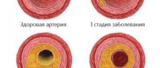
Intracranial atherosclerosis: diagnosis, clinical manifestations, therapy
Atherosclerosis of intracranial arteries is a significant factor in the development of acute cerebrovascular accident. The article discusses pathophysiological aspects, clinical manifestations, methods of diagnosis, treatment and prevention of atherosclerosis of intracranial arteries. It is noted that further progress in the study of this problem is associated with new techniques for visualizing vessels and atherosclerotic plaque and the search for individual approaches to treatment, including determining indications for endovascular interventions.
Rice. 1. Computed tomographic angiography of intracranial arteries: occlusion of the right vertebral artery at the intracranial level (MIP and 3D images)
Rice. 2. Magnetic resonance angiography and magnetic resonance imaging of the brain in T2 mode (magnetic resonance imaging with a magnetic field strength of 3T)
Table. Advantages and disadvantages of imaging methods for intracranial atherosclerosis
Introduction
Atherosclerosis of intracranial arteries is one of the most common causes of stroke worldwide. According to epidemiological studies, the development of acute cerebrovascular accident due to this pathology depends on race. In Caucasians, symptomatic intracranial atherosclerosis (that is, arterial damage causing transient ischemic attack or stroke) occurs in 10% of cases, while in representatives of the Black and Asian races this figure reaches 30 and 50%, respectively [1–6]. This difference is due to genetic predisposition to the development of the disease, as well as differences in lifestyle and typical risk factors [2]. The incidence of asymptomatic atherosclerosis reaches 54% of cases in Asians and 12% among representatives of other races [2, 7–9].
As a rule, severe asymptomatic atherosclerosis of the intracranial arteries is combined with other vascular risk factors, which usually serve as the basis for examining such patients. Damage to the intracranial arteries is associated with a high incidence of recurrent acute cerebrovascular accidents - according to the WASID study, up to 19% over the next two years [10]. The incidence of recurrent stroke is also influenced by morphological characteristics, in particular stenosis > 70%, female gender and time since the previous ischemic episode (less than 17 days).
Pathophysiological aspects of atherosclerosis of intracranial arteries
The immediate causes of stroke in atherosclerosis of intracranial arteries include:
- hypoperfusion of brain tissue with infarction in areas of adjacent blood supply, associated with decompensation of the processes of autoregulation of cerebral blood flow, especially in conditions of impaired systemic hemodynamics;
- arterio-arterial embolism, the consequence of which, as a rule, is a wedge-shaped infarction;
- the presence of an atherosclerotic plaque at the mouth of a small perforating artery, which leads to the formation of small infarctions.
The high frequency of recurrent infarctions, as a rule, is caused precisely by a violation of the autoregulation of cerebral blood flow in conditions of severe stenosis [11]. Knowing the mechanism of development of the first stroke, it is possible to predict the mechanism of recurrent stroke: for example, in patients with small infarctions due to occlusion of a small artery, a recurrent stroke, as a rule, develops of a different type and is localized distal to the affected artery [12]. Most often, atherosclerotic lesions are localized in the middle cerebral artery (up to 27% [13]) and the internal carotid artery (petrosal, cavernous and supraclinoid parts), less often in the basilar artery and the intracranial segment of the vertebral artery [10, 14].
It is believed that even substenotic lesions of the arteries can cause acute cerebrovascular accidents by analogy with atherosclerotic changes in the coronary arteries, which can cause myocardial infarction against the background of relatively mild stenosis. This threat is associated with inflammatory processes that occur during the onset, progression and damage of plaque and lead to rupture of the plaque cap and the formation of thrombosis and infarction.
Diagnostics
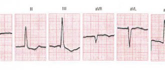
To diagnose atherosclerosis of intracranial arteries, ultrasound methods (transcranial Dopplerography), magnetic resonance (MR), computed tomography (CT) (Fig. 1 and 2) and digital subtraction angiography (table) are used. The latter method is considered the gold standard for diagnosis [15], as it allows one to accurately determine the degree of arterial stenosis. However, digital subtraction angiography is an invasive intervention that involves the introduction of a contrast agent. In addition, the method is associated with the development of transient and persistent neurological deficits, the frequency of which can reach 1.8 and 0.6%, respectively [16].
There is no clear evidence regarding the accuracy of safer and more accessible non-invasive methods. Thus, with MR angiography, visualization of the lumen of a vessel depends on the blood flow in it, while the true severity of vessel damage can be distorted in the case of critical stenosis with very low blood flow (it may look like an occlusion) and in high-degree stenoses with high blood flow velocity.
It is believed that transcranial Doppler ultrasonography and MR angiography can be used as screening to exclude lesions of intracranial arteries, but they are not reliable enough to confirm the presence of stenosis and determine its severity [17]. At the same time, transcranial Doppler sonography provides additional information with which one can assess blood flow through collateral vessels and determine cerebrovascular reactivity.
CT angiography is superior to MR angiography in terms of diagnostic accuracy [18, 19] and has the highest sensitivity and specificity for detecting stenosis after direct angiography, >50% [20]. In addition, CT or MR angiography can be supplemented with methods for assessing cerebral blood flow - perfusion computed tomography or magnetic resonance imaging. As a rule, the latter are included in the standard research protocol and make it possible to identify areas of hypoperfusion that arise in conditions of long-term stenosis and do not manifest themselves clinically.
The classical approach to diagnosis, aimed only at establishing the degree of narrowing of the artery, has a number of disadvantages - in particular, it is impossible to establish the histological structure and, therefore, the degree of “instability” of the atherosclerotic plaque, as well as to differentiate atherosclerotic stenosis from narrowing of the artery lumen caused by other reasons.
Currently, new imaging technologies, such as high-resolution magnetic resonance imaging and intravascular ultrasound, are of particular importance - techniques that make it possible to study the atherosclerotic plaque itself. This is especially important in the early stages of the atherosclerotic process, when the lumen of the vessel has not yet narrowed due to remodeling. Thus, magnetic resonance imaging, performed on a device with a magnetic field strength of 3T, makes it possible to directly visualize a thrombus, hemorrhage into a plaque and determine the composition of the plaque, that is, assess the activity of the plaque [21].
Intravascular ultrasound also makes it possible to detect hemorrhage in the plaque, determine its composition and extent. This may influence the determination of risk and therefore the choice of treatment.
Thus, new research methods are especially important when examining patients with stroke and non-stenotic damage to intracranial arteries, when the cause of the infarction may be plaque damage not detected using classical imaging methods [22].
Clinical manifestations
The most serious clinical manifestations of atherosclerosis of intracranial arteries include transient ischemic attack and ischemic stroke, the symptoms of which correspond to the location of the lesion. Stenosis of the middle cerebral artery is usually characterized by lacunar infarctions and ischemia in the area of adjacent blood supply. Stenosis of the internal carotid artery is accompanied by the development of more extensive lesions and involvement of gray matter. The neurological deficit in this case turns out to be more pronounced than with stenosis of the middle cerebral artery [23].
In addition to motor and sensory disturbances, in cases of damage to the gray matter, thalamus or caudate nucleus, patients may experience cognitive impairment. Cognitive impairment may also develop in the absence of infarcts due to decreased cerebral perfusion and associated changes in the white matter of the brain. Finally, an asymptomatic course of the atherosclerotic process is possible. Clinical symptoms are observed in the presence of a number of factors, among which it is necessary to note the degree of stenosis and the structure of collateral vessels [24]. Symptoms are usually accompanied by damage to the middle cerebral artery, basilar artery and intracranial part of the vertebral artery, while changes in the anterior and posterior cerebral arteries are most often asymptomatic [25].
Atherosclerosis of intracranial arteries can progress, stabilize, or regress [26]. It is generally accepted that asymptomatic patients have a more favorable outcome. Thus, when analyzing data from the TOSS-2 study, progression of the lesion was observed in 13% of cases in patients with symptomatic atherosclerosis and only in 6% of cases with asymptomatic stenosis [27]. In a study of 102 patients with significant middle cerebral artery stenosis or occlusion, the risk of stroke in symptomatic patients was 12.5% per year and only 2.8% per year in asymptomatic patients [28]. Asymptomatic middle cerebral artery plaque can predict a relatively favorable outcome: such plaques are often calcified and, therefore, do not have a high risk of embolism [29].
A number of studies have identified differences between the location of stenosis and the course of the disease [30]. Obviously, the prognosis of the disease with occlusion of the basilar artery is worse than with occlusion of the internal carotid or middle cerebral artery [31].
Prevention and treatment
The main goal of treatment in patients with symptomatic atherosclerosis of intracranial arteries is to prevent recurrent acute cerebrovascular accident. To this end, the following activities are carried out:
- blood pressure control (systolic blood pressure ≥ 140 mm Hg significantly increases the risk of stroke in the corresponding arterial basin);
- elimination of dyslipidemia (total cholesterol ≤ 5.1 mmol/l and low-density lipoprotein ≤ 1.8 mmol/l);
- aggressive correction of other risk factors (normalization of body weight, increased physical activity, smoking cessation, maintenance of normoglycemia) in accordance with recommendations for secondary prevention of stroke [32];
- antithrombotic therapy.
Preference is given to monotherapy with antiplatelet agents. At the same time, there is evidence that dual antiplatelet therapy has a greater effect in preventing recurrent stroke in the early stages after the first vascular episode. In the SAMMPRIS study, patients treated with aspirin and clopidogrel for 90 days followed by aspirin alone plus intensive risk factor management had a 5.8% rate of recurrent stroke in the first 30 days [33].
Based on the results of the largest studies, aspirin monotherapy at a dose of 325 mg/day in combination with intensive correction of risk factors can be recommended for patients with moderate stenosis (
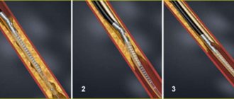
For many years, attempts have been made to surgically treat atherosclerotic stenosis of intracranial arteries and its consequences. The earliest and most studied operation is the application of extra-intracranial anastomosis, but the results of those performed in the 1980s. and in 2011, studies did not confirm its effectiveness and currently this operation is not widespread [35].
Today, such methods of surgical treatment of intracranial atherosclerosis are used as endovascular interventions using balloon angioplasty, balloon angioplasty with stenting and with the installation of self-expanding stents. The latter are characterized by a high rate of technical success and relative ease of installation, since they do not require the use of a balloon and can be delivered to difficult-to-pass areas of the arterial bed. However, in the large randomized SAMMPRIS trial, endovascular intervention was found to be less safe and effective than medical treatment [33]. As a result, no endovascular technique has been approved as the preferred intervention. Possible indications for endovascular surgery in 70–99% of patients with atherosclerosis may be symptoms due to disturbances in systemic hemodynamics, as well as poor development of collaterals and recurrent acute cerebrovascular accidents, despite aggressive drug treatment and correction of risk factors [35].
In patients with asymptomatic atherosclerosis of intracranial arteries, it is advisable to carry out primary prevention of cerebral ischemia, taking into account the identified risk factors. Taking into account the possibility of progression of atherosclerotic lesions, it is advisable to monitor the condition of the arteries at intervals of about two years [36].
The development of stenosis of intracranial arteries is accompanied by a violation of the autoregulation of cerebral blood flow and the formation of zones with reduced perfusion, so it is appropriate for such patients to be prescribed drugs that have a neurotrophic, metabolic and antihypoxic effect. An example is Actovegin, a comprehensively studied drug with a pleiotropic effect and a favorable safety profile.
A number of foreign randomized placebo-controlled studies have shown the effectiveness of Actovegin in elderly patients with signs of mild and moderate dementia of various origins, including vascular [37]. Its use was accompanied by improvements in behavioral characteristics and neuropsychological test results. Extensive experience of Russian researchers confirms the effectiveness of the drug in patients with chronic cerebral ischemia accompanied by cognitive disorders [38]. Actovegin had a positive effect not only on memory and attention indicators, but also improved the psycho-emotional status of patients - reduced the severity of depressive and asthenic symptoms, improved sleep and general well-being [39, 40].
The proven endothelial protective effect of Actovegin and the positive effect on microcirculation likely provide additional therapeutic benefits in patients with cognitive disorders resulting from chronic cerebral ischemia, accompanied by tissue hypoxia, micro- and macrovascular damage [41].
Thus, the inclusion of Actovegin in the treatment regimen for patients with intracranial atherosclerosis, along with measures to prevent vascular events, can help level out the manifestations of cerebral circulatory failure and improve the well-being of such patients.
Conclusion
Atherosclerosis of intracranial arteries is a significant factor in the development of acute cerebrovascular accident, requiring a special approach to diagnosis and treatment. Further progress in studying the problem of diagnosis and treatment of atherosclerosis of intracranial arteries is associated with new techniques for visualizing vessels and atherosclerotic plaque and the search for individual approaches to treatment, including determining indications for endovascular interventions.