Doctors usually distinguish between two forms of oxygen deprivation. First, anoxic injuries which occur when the brain is completely deprived of oxygen due to sudden cardiac arrest, suffocation, suffocation and other sudden injuries. Second, hypoxic damage occurs when this organ receives less oxygen than it needs, but is not completely deprived of it. Because the effects of the two injuries are similar, many brain experts use the terms interchangeably.
A few seconds of oxygen deprivation will not cause long-term harm, so a child suffering from respiratory distress or a diver who needs a few extra seconds to come up for air are unlikely to suffer brain damage. The exact timeline of anoxic injury to this organ depends on a number of personal characteristics, including general brain and cardiovascular health, and the level of blood oxygenation at the time of injury. Generally speaking, injuries begin at the one-minute mark, steadily worsening after that:
- Between 30-180 seconds of oxygen deprivation, you may lose consciousness.
- At the one minute mark, brain cells begin to die.
- After three minutes, neurons suffer more damage and long-term brain damage becomes more likely.
- After five minutes, death becomes inevitable.
- After 10 minutes, even if the brain remains alive, coma and long-term brain damage are almost inevitable.
- After 15 minutes, survival becomes almost impossible.
Of course, there are exceptions to every rule. Some training routines help the body use oxygen more efficiently, allowing the brain to go longer periods without this vital element. Free divers usually train to go without oxygen for as long as possible and the current record holder holds his breath for 22 minutes without suffering any organ damage.
Why does the brain need oxygen?
Gray matter makes up only 2% of body weight, but it uses about 20% of oxygen. Without it, the brain cannot perform even the most basic functions. The brain relies on glucose to stimulate neurons that control everything from conscious functions like planning and thinking to automatic, unconscious processes like heart rate and digestion.
Without oxygen, the cells of this organ cannot metabolize glucose and therefore cannot convert glucose into energy. When your brain is deprived of oxygen, the ultimate cause of brain death is insufficient energy to fuel the cells.
Thiamine deficiency and Korsakoff-Wernicke disease
If a person systematically drinks alcohol, without control over the amount he drinks, and this lasts for quite a long time, then he has every chance of developing brain dysfunction or damage to his cells. Moreover, this can be caused by the consumption of large amounts of alcohol, or there may be serious disturbances in the liver due to chronic alcohol dependence.
For example, most people suffering from chronic alcohol addiction have a lack of thiamine in their bodies, or as it is also called vitamin B1. It may be lacking due to poor nutrition, metabolic disorders in the body and, of course, due to the abuse of alcoholic beverages. It plays an indispensable role in the metabolism of carbohydrates, lipids, and proteins. It supports the normal functioning of the cardiovascular system, digestive tract, and nervous structures.
Most people suffering from chronic alcohol addiction are deficient in vitamin B1. And this can lead to such a serious illness as Korsakoff-Wernicke syndrome, or more precisely, Wernicke syndrome and Korsakoff syndrome.
Wernicke's disease is characterized by paralysis of the eye muscles, disturbance of consciousness and impaired coordination of movements. Sometimes a patient with a similar syndrome cannot independently find the door of the room to leave, he cannot move without someone else's help. If at least one of the signs of the disease appears, you need to urgently seek medical help.
Most people suffering from alcohol dependence, along with Wernicke's syndrome, experience Korsakoff's syndrome, which is also called alcohol paralysis. With this disease, memory impairment is observed, when he forgets everything that happened to him before the disease, but can describe in great detail the events that took place several years before.
In addition, he remembers well everything that happens to him after his illness. The patient begins to invent non-existent events, or talk about those that really happened, but at the same time greatly distorts the facts. All this is accompanied by disorientation. The patient ceases to navigate the world around him and himself. Sometimes, even after seeing his reflection in the mirror, the patient does not realize that it is him.
Call us now:
+7 (812) 454-00-50
Prices for Ultramed clinic services
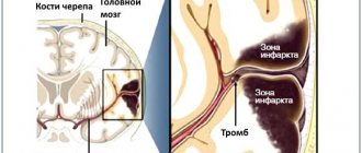
How long does the brain live after cardiac arrest?
Most studies have shown that the process of brain activity after the heartbeat stops is individual for each person. Although the cessation of oxygen flow is almost instantaneous, there is no specific duration of clinical death at which a functioning brain clearly dies. The most vulnerable cells are neurons, which suffer fatal damage in just 10 minutes without oxygen. However, the damaged cells do not actually die for a very long time. If resuscitation is successful, some areas may resume operations. Find out more. what happens to the brain at the moment of cardiac arrest can be found here - https://reactor.space/news/chto-proisxodit-s-mozgom-v-moment-ostanovki-serdca/.
Unravel - and overcome!
The human body is home to a huge number of bacteria - scientists count about 10 thousand of their species, and the mass of these microorganisms can reach 3 kg. When our immune system ceases to function with our last breath, these countless hordes of “little friends” are no longer held back. The microflora begins to devour the deceased from the inside. Bacteria move freely throughout the body, engulf the intestines and then the surrounding tissues, and invade the blood capillaries of the digestive system and lymph nodes. They penetrate first into the liver and spleen, and then into the heart and brain.
Simultaneously with the activity of microbes, cadaveric spots are formed - they appear where stopped blood settles in the tissues. After 12-18 hours, the spots reach their maximum coverage, and after a few days they become dirty green. But it turns out that at the same time some parts of the deceased’s body remain quite viable.
Article on the topic
A light in the end of a tunnel. What do people who have experienced clinical death say? For example, even though the heart has stopped long ago, its valves may still be intact. The fact is that they contain connective tissue cells that live a long time. This means that heart valves can be used for transplantation. And this is after one and a half days after death!
The cornea, the most convex transparent part of the eyeball, lives even longer. It turns out that it can be used for medical purposes for 3 days after a person has died. The reason is that the cornea is in direct contact with the air and receives oxygen from it.
All these facts indicate: the human body does not die at one moment, but gradually. And death as a biological phenomenon - despite the fact that we don’t really like to think and talk about it - still conceals many mysteries. Who knows, maybe by solving them we will overcome death itself?
Consequences after cardiac arrest for 10 minutes
The prognosis depends on how severe the oxygen deprivation is, the degree of neuronal death, and the quality of medical and rehabilitation care. With quality physical therapy, your brain can learn to compensate for damaged areas, which is why even serious injuries require ongoing commitment to physical therapy.
Common long-term effects of oxygen deprivation may include:
- Damage to specific areas of the brain that are deprived of oxygen. Different areas of this organ tend to coordinate different functions, so some of them can be severely crippled while others remain intact. For example, the victim may understand language, but cannot speak.
- Changes in mood or personality.
- Difficulty with memory, including the ability to recall facts, names, objects or people, recognize faces, learn new information, or recall autobiographical facts.
- Changes in motor skills. A number of areas of the brain help coordinate movement, so if these areas are damaged, you may not be able to fight, walk, write, or perform other functions.
- Chronic pain. When the brain is damaged, it can process pain signals incorrectly, causing you to feel pain even when there is no injury.
- Inability to feel pain or respond appropriately to pain signals. For example, pain in your arm may feel like pain in your leg.
- Difficulty with impulse control. Many brain injury survivors develop addictions, violent behavior, or sexually inappropriate compulsions.
- Symptoms of mental illness such as depression or anxiety.
- Symptoms associated with dementia, including confusion, memory difficulties and signs of rapid aging of the organ.
Brain death
1. Introduction
Brain death is the irreversible loss of function of the central nervous system, including the cerebrum, brainstem, pons, midbrain, and cerebellum.
The diagnosis of “brain death” is established with a beating heart and artificial ventilation and is the equivalent of human death.
The most common causes of brain death:
- severe traumatic brain injury;
- massive aneurysmal subarachnoid or intracerebral hemorrhage;
- extensive ischemic cerebrovascular accident with edema and dislocation of the brain;
- massive cerebral edema with fulminant liver necrosis;
- hypoxic and ischemic brain damage during prolonged cardiopulmonary resuscitation or asphyxia.
Brain death after restoration of blood circulation during cardiopulmonary resuscitation develops during the first week from the moment of circulatory arrest, which in some cases may be preceded by a period of initial improvement in neurological symptoms.
2. Conditions necessary to make a diagnosis of brain death
The decisive point in diagnosing brain death is proof of the complete and irreversible cessation of brain function, which requires:
2. 1. Determine what is the cause of brain damage, and whether it can lead to a complete and irreversible loss of its function.
2. 2. Exclude all potentially reversible conditions with similar clinical manifestations.
These conditions are:
- Hypothermia.
- Hypotension.
- Effect of alcohol, sedative drugs, muscle relaxants
- Severe electrolyte and hormonal disorders, acid-base imbalances, hypoglycemia.
The diagnosis of brain death cannot be established if:
- body temperature below 32.2 degrees Celsius,
- mean arterial pressure less than 55 mm. rt. Art. In case of hypotension, blood pressure is maintained by replenishing the volume of circulating blood and adrenergic agonists.
- the presence in the blood of toxic concentrations of sedatives, narcotic drugs, alcohol, muscle relaxants. At subtherapeutic doses, the decision is made in accordance with the clinical situation.
2. 3. Determination of brain death is based on the results of repeated clinical examinations of the patient. Only cerebral angiography demonstrating lack of filling of the internal carotid and vertebral arteries above the level of entry into the cranial cavity can be the basis for a diagnosis of brain death. All other instrumental diagnostic methods are of an auxiliary nature and serve to shorten the observation period.
3. Clinical signs of brain death
3. 1. Lack of consciousness. 3. 2. Lack of motor reactions in response to painful stimuli. 3. 3. Absence of brainstem reflexes. 3. 4. Apnea.
4. Instrumental studies confirming the diagnosis of brain death
4. 1. Electroencephalography (EEG).
Conducted according to generally accepted standards. A minimum of 8 leads are used. The distance between the electrodes is at least 10 cm, the impedance is in the range of 100-10000 Ohms. Recording is carried out with a time constant of at least 0.3 seconds. with a sensitivity of no more than 2 µV/mm (the upper limit of the frequency bandwidth is not lower than 30 Hz). The electrical silence of the cerebral cortex under these conditions should remain for at least 30 minutes of continuous recording. If there is doubt about the electrical silence of the brain, repeated EEG registration is necessary. Assessment of EEG reactivity to light, loud sound and pain: the total stimulation time with light flashes, sound stimuli and painful stimuli is at least 10 minutes. The source of flashes, fired at a frequency of 1 to 30 Hz, should be located at a distance of 20 cm from the eyes. Intensity of sound stimuli (clicks) -100 dB. The speaker is located near the patient's ear. Stimuli of maximum intensity are generated by standard photo and sound stimulators.
4. 2. Cerebral hagiography
Contrast double panangiography of the four main vessels of the head (common carotid and vertebral arteries) is performed with an interval of at least 30 minutes. Mean arterial pressure during angiography should be at least 80 mm Hg. Art. If angiography reveals that none of the intracerebral arteries are filled with a contrast agent, then this indicates cessation of cerebral circulation.
4. 3. Nuclear magnetic resonance angiography
The research in its information content is close to classical angiography. Demonstration of the absence of blood flow in the main cerebral arteries can serve as confirmation of brain death.
4. 4. Transcranial Doppler ultrasonography
A bilateral study is carried out. The sensor is applied in the area of the temporal bone above the zygomatic arch. The diagnosis of brain death is confirmed by the absence of diastolic and reflected blood flow, with the presence of small systolic peaks at the beginning of systole. The complete absence of blood flow is not a reliable sign, as it may be associated with technical problems during Doppler ultrasound.
4. 5. Cerebral scintigraphy.
Confirmation of brain death is the absence of accumulation of the isotope in brain tissue.
5. Algorithm for declaring brain death
To determine brain death, all of the following questions must be answered positively:
5. 1. Is the cause of coma known and can it lead to complete and irreversible loss of brain function?
Coma of unknown etiology (no signs of injury, stroke, hypoxic or hypotensive damage) requires a detailed examination before declaring brain death.
5. 2. Is it possible to exclude the effect of alcohol, sedatives, drugs, muscle relaxants, hypothermia, hypotension, hypoglycemia, electrolyte, hormonal disorders as the cause of the coma and the results of the neurological examination?
5. 3. Can all existing movements be attributed to spinal reflexes?
The diagnosis of brain death cannot be established by adopting any specific posture (decerebrate or decortication), the presence of trembling, or defensive movements of any limb or head in response to pain. The motor response to pain upon irritation of the supraorbital region and compression of the nail phalanges by a hard object is studied.
5. 4. Is there no reaction of the pupils to light?
The reaction of the pupils to bright light should be absent on both sides. Round, oval, or irregularly shaped pupils are compatible with a diagnosis of brain death. In most cases, during brain death, the pupils are in the middle position, their size is 4-6 mm, but the diameter of the pupils can range from 4 to 9 mm. Dilated pupils are compatible with the diagnosis of brain death because the innervation of the radial muscle fibers of the iris by the cervical sympathetic nerves is preserved.
5. 5. Is there no corneal reflex?
5. 6. Are oculocephalic and oculovestibular reflexes absent?
The oculocephalic reflex is not examined in the presence or suspicion of traumatic injury to the cervical spine. To assess it, the patient’s head is turned 90 degrees to one side and held in this position for 3-4 seconds. , then in the opposite direction for the same time. If, when turning the head, no eye movements occur, and they firmly maintain a median position, this indicates the absence of oculocephalic reflexes.
To study the oculovestibular reflex, a bilateral caloric test is performed. Before performing this procedure, it is necessary to ensure that there is no perforation of the eardrums. The patient's head is raised 30 degrees above the horizontal level. A small catheter is inserted into the external auditory canal, and the external auditory canal is slowly irrigated with cold water (50 ml at 0 degrees Celsius). When the function of the brain stem is preserved, a deviation of the eyes in the direction of irritation appears. The absence of nystagmus or deviation of the eyeballs during a caloric test for 1 minute indicates the absence of an oculovestibular reflex. The test on the opposite side is carried out no earlier than 5 minutes after the first.
5. 7. Are cough and pharyngeal reflexes absent?
The presence of cough and pharyngeal reflexes is determined by irritation of the posterior wall of the pharynx and aspiration of tracheal contents.
5. 8. Is there any spontaneous breathing?
The test for absence of spontaneous breathing requires special care to prevent hypoxia and possible complications. It is carried out only after all other data confirming brain death have been received. The necessary conditions:
- Body temperature is at least 36.5 degrees Celsius.
- Systolic blood pressure is at least 90 mm. rt. Art.
- Normovolemia (positive fluid balance during the 6 hours preceding the test)
- Eucapnia is the partial pressure of carbon dioxide in arterial blood (PaCO2) before the test is at least 40 mm. rt. Art.
- The level of partial pressure of oxygen in arterial (PaO2) blood is at least 200 mm. rt. Art.
Test technique:
- Before starting the test, PaCO2 and PaO2 levels are measured.
- The endotracheal (tracheostomy) tube is disconnected from the breathing circuit of the ventilator.
- An oxygen flow of 6 l/min is supplied to the patient's respiratory tract through a catheter. The optimal position of the catheter is at the level of the carina.
- Careful monitoring of the presence of respiratory movements of the abdominal and chest muscles is carried out for 8 minutes.
- After which the levels of PaCO2 and PaO2 are re-monitored.
- If there are no respiratory movements and the PaCO2 level increases to 60 mm. rt. Art. or more, or increases by 20 mm. rt. Art. in relation to the initial level, the test result is considered positive.
- If during observation the blood pressure decreases, arrhythmia develops, hemoglobin saturation drops sharply, arterial blood is drawn and the test is interrupted. It can be considered positive if PaCO2 increases to 60 mm. rt. Art. or more, or increases by 20 mm. rt. Art. relative to the original level.
- If there are no respiratory movements, but the PaCO2 value does not reach 60 mm. rt. Art. — the duration of the test can be increased to 10 minutes.
5. 9. The diagnosis of brain death is established when one of the following conditions is met:
- Positive answers were received to questions 5. 1. -5. 7. with two clinical examinations carried out at least 6 hours apart, and the apnea test (point 5. 8.) confirms the diagnosis of brain death.
- Positive answers were received to questions 5. 1. -5. 7. with two clinical examinations carried out at least 2 hours apart. An EEG study demonstrates the absence of electrical activity in the brain, and the apnea test (point 5.8.) confirms the diagnosis of brain death.
- Positive answers were received to questions 5. 1. -5. 7. with two clinical examinations carried out at least 2 hours apart. An instrumental study (one of those listed in paragraphs 3. 2. - 3. 5.) demonstrates the absence of cerebral blood flow and an apnea test (paragraph 5. 8.) confirms the diagnosis of brain death.
- In the case where the answers to not all questions are 5. 1. -5. 7. may be received (for example, due to extensive trauma). The diagnosis of brain death can be established on the basis of two clinical examinations, carried out at least 2 hours apart, giving positive answers to all tests (5.1. -5.8.), which are possible. The results of one of the instrumental studies (item 3. 2.. -. 3. 5.) confirming the absence of cerebral blood flow and a positive test for apnea.
Responsible for diagnosing a person’s death are the doctors who determined brain death at the medical and preventive institution where the patient died.
Detoxification of the body from alcohol in
With prolonged consumption of alcohol, toxic substances formed during the breakdown of ethanol accumulate in the body. To get rid of poisons and toxins, you should detoxify your body. Specialists of the drug treatment center carry out the cleansing procedure in the hospital and at home.
Professionals use IVs, intravenous and intramuscular injections for this purpose. All medicines are certified and highly effective. Only an individual approach from specialists, drug therapy and rehabilitation will help you get rid of alcohol addiction forever.
If the body is accidentally discovered
If an ambulance team finds a victim without signs of life, doctors have no information how long he remains in this condition. In the absence of cadaveric spots, doctors cannot confirm in the field that biological death has occurred. In this case, resuscitation is mandatory.
Resuscitation measures include artificial ventilation of the lungs and closed heart massage. If there is bleeding, it is important to stop it so the victim does not bleed out. Damage to a large artery or head is extremely dangerous. If resuscitation measures are carried out correctly, a person can be brought back to life.
The effect of alcohol on the nervous system
Alcoholic drinks affect the functioning of the central nervous system in two ways:
- Penetrates nerve cells and affects receptors and ion channels.
- Activates the functioning of certain brain receptors.
When drinking alcohol, the nervous system is in an excited state. Ethanol stimulates the release of dopamine, which increases activity and gives a person a feeling of euphoria. After resources are depleted, the opposite effect occurs - weakness, depression, and poor health appear.
Is it possible to restore a brain damaged by COVID-19?
The human brain and central nervous system are able to compensate for impaired functions by delegating responsibilities to healthy departments. Such plasticity allows the brain to partially or fully regain lost abilities. You can help this process with regular exercise. Attention training and cognitive gaming exercises help restore memory function, increase motivation, and restore concentration.
In case of serious brain disorders or severe neurological disorders, complex psychotherapy and pharmacological medications are prescribed.
How does wine affect blood and blood vessels?
Wine, consumed in small doses, can benefit the body. First of all, the drink is beneficial for blood vessels. Wine contains substances that increase the strength and elasticity of the walls of blood vessels.
- Red wine . Dry wines are the most beneficial. They contain magnesium, potassium, antioxidants and fructose. Dilates blood vessels provided you drink no more than 100 ml per day.
- White wine . Contains fewer antioxidants than red wine, but is also rich in nutrients. Reasonable consumption helps strengthen the immune system.
Still have questions? Call us!
8
Free consultation and appointment
Stages of brain damage by alcohol
Alcohol destroys neural connections, due to which there are more and more receiving receptors, and it is more difficult to activate them each time. Brain damage develops in stages and has three stages:
- First . There is a constant desire to drink, the craving for alcohol becomes systematic.
- Average . After drinking alcohol, a severe hangover begins and a strong psychological need for alcohol-containing drinks develops. Diseases of the internal organs begin to develop.
- Heavy . The person degrades, alcoholism becomes chronic.
Symptoms
The following symptoms indicate brain death:
- there is no consciousness, and this process is persistent;
- there are no reactions to tingling, stroking, handling, no tactile sensitivity;
- no movement of the eyeballs;
- the heart has stopped, as evidenced by a straight line on the ECG;
- there is urinary and fecal incontinence;
- breathing is impaired, the chest does not rise.
Determining whether a patient has died is an extremely important task. Doctors do not immediately diagnose death. Even if all of the listed signs are detected, the person is monitored in the hospital for up to 12 hours. Sometimes, although rarely, signs of brain activity may appear. If during this time the patient does not react in any way, there are no trunk reflexes, then biological death can be stated.
It is important how the brain dies, what triggered the death of neurons. If poisoning has led to such serious consequences, the patient should be monitored for at least a day. If the cause is TBI, the observation time is reduced to 6 hours. The specific decision must be made by the neurosurgeon. It is important that the doctor monitors the patient from the onset of this condition, then he will have a complete picture of events that will allow him to make the right decision.
The onset of biological death is determined exclusively by a neurologist. It is based on subjective and objective criteria. If signs of brain death are clearly identified, the person must be disconnected from life support. This difficult decision is especially difficult to make if the disaster happened suddenly. Often relatives are literally horrified by such a message. If a person is sick for a long time, his loved ones eventually become at least a little accustomed to the idea that their loved one will be gone. In any case, this decision is very difficult.
How to understand that there is a problem?
According to studies, people whose brains have been affected by coronavirus infection report the following disorders:
- Insomnia.
- Impaired attention and concentration.
- Anxiety.
- Memory impairment.
- Depressive state.
- Impaired consciousness.
- Panic attacks.
- Post-traumatic stress disorder.
Patients complain of debilitating headaches, dizziness, and convulsions. Elderly people often report a disorder of consciousness with impaired thinking and perception of the world around them. If you experience similar symptoms, we recommend visiting a doctor. Based on the results of the initial examination, the doctor may additionally refer you for an MRI.