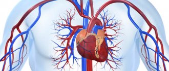
Duplex scanning of head and neck vessels is a highly informative and safe method based on ultrasound technology. Ultrasound waves “reveal” the arteries and vessels of the head and neck and make it possible to assess the state of blood flow according to several basic signs, as well as examine the lumen of the vessel for the presence of a wide range of pathologies. These include aneurysms, blood clots, changes in wall thickness and protrusion, plaques, and tortuosity of blood vessels. In comparative diagnostics, duplex scanning reveals changes in the condition of the walls and blood flow speed, which helps to adjust treatment or decide on its initiation if the patient has previously been under observation.
Doppler ultrasound examination is non-invasive, painless, and does not require prior preparation. The procedure is repeated the required number of times without restrictions to confirm a complex diagnosis.
When is ultrasonography prescribed?
Ultrasound scanning allows you to reliably evaluate such indicators as:
- intensity and speed of vascular blood flow;
- vascular wall thickness;
- the presence of blood clots or plaques in the lumen of blood vessels;
- the structure of the vessel, its width, length, degree of tortuosity.
Duplex scanning is done to identify the following pathologies:
- occlusion of the carotid arteries;
- atherosclerosis of the lower extremities;
- thrombosis and thromboembolism of any location;
- arterial aneurysms;
- aortic diseases;
- varicose veins;
- Raynaud's disease;
- circulatory disorders in the brain.
What is usually revealed during the study
Duplex affects mainly extracranial vessels, and to a lesser extent intracranial ones. The doctor pays special attention to the following vessels of the neck - the common carotid arteries (and bifurcation areas), the extracranial part of the carotid arteries, part of the brachiocephalic trunk, external carotid arteries, veins next to the skull, venous vertebral plexus, temporal arteries, etc.
For transcranial duplex scanning, do an ultrasound taking into account access through the cranial bones - they suppress a large number of ultrasonic waves. Access is through natural openings - the greater occipital, temporal bones, and orbits. An ultrasound scan of a baby's brain can be performed through the fontanelles - they provide a significant overview and help to collect more information. During this type of duplex scanning, the carotid and vertebral arteries inside the skull, the basilar and cerebral arteries are examined.
Duplex scanning is a method that detects a wide range of pathologies of the arteries and veins in the head and neck. If the diagnosis is already known, the doctor is able to monitor the patient’s condition (after surgery, stroke, head and neck injuries).
In patients with pathologies, ultrasound of the head and neck reveals:
- atherosclerosis;
- arteriovenous malformation of the brain;
- compression of blood vessels by neoplasms;
- diseases of the central nervous system;
- blood clots;
- endarteritis;
- stenosis and expansion;
- aneurysms;
- plaques in people with high cholesterol in tests
- bends, loops.
Atherosclerosis can be suspected if you are overweight, have high levels of cholesterol and triglycerides in tests, or have a predisposing heredity. Endarteritis has autoimmune causes, in which the artery wall is damaged, and the damage is visible on the screen. Aneurysms are detected for the first time (with characteristic patient complaints) or confirmed if they were already noticed on a previous ultrasound, CT scan or angiography. On the duplex, they look like thinned and stretched walls of arteries (less often veins), protruding outward.
Some patients require regular vascular monitoring. Once a year, to assess the effectiveness of treatment and create an individual prognosis, duplex is recommended for patients with diabetes mellitus, surges in blood pressure, degenerative changes in the discs in the cervical spine, and strokes in the past.
In addition to patients with diseases, regular diagnosis is indicated for smokers, people over 40 years of age (for men) and over 45 (for women). If you have relatives with vascular pathologies and diseases that provoke them, you should also be examined regularly.
Do you need any preparation for the procedure?
Ultrasound duplex scanning does not require special preparation, the only exception is ultrasound scanning of the abdominal vessels (abdominal aorta).
Ultrasound scanning
of the abdominal aorta
requires additional preparation, including:
- fasting for 8 hours before the procedure;
- a three-day diet excluding raw vegetables, meat, milk and black bread, in addition, the patient is prescribed a drug to eliminate gas formation.
Common advantages and disadvantages of the presented method
Duplex scanning has a number of obvious advantages:
- is highly informative;
- does not require the use of ionizing radiation;
- a cheaper and more accessible research method than alternative ones;
- carried out in real time, does not require waiting for a conclusion;
- helps to reflect the condition of soft tissues that are not sufficiently visualized on x-rays;
- non-invasive, performed without contrast (to which allergies are sometimes found), does not bring any unpleasant sensations;
- Suitable for children and pregnant women.
Any method, along with positive ones, also has negative sides, and in some cases it makes sense to choose other types of research. The decision is made by the doctor on an individual basis, after evaluating the results of other diagnostic procedures and tests, examination, and questioning of the patient.
The disadvantages of the method include:
- difficulty in examining small vessels;
- the possibility of diagnosis only during the research process, and not after it based on the results;
- narrow area to be examined because the bones of the adult skull impede the passage of ultrasound.
An important role in the quality of the diagnostics performed is played by the level of equipment and the professionalism of the doctor. This is one of the diagnostic methods to which increased demands are placed on the professionalism of doctors, so you should choose a clinic for duplex scanning especially carefully, because the result can differ significantly.
Ultrasonic duplex scanning technique
The method is based on the physical properties of ultrasound. When examining blood vessels, ultrasound changes frequency as the speed of blood movement in the vessel changes. This is recorded on the monitor and forms an idea of the state of the vessels and the speed of blood flow.
How duplex scanning is done:
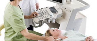
During the procedure, the doctor applies a conductive gel to the area of the body being examined, then examines it by moving a sensor over the skin. The results obtained are compared with normal values.
Duplex scanning of the veins of the lower extremities
During the study, the doctor conducts tests to determine the condition of the valves and the patency of the vessel.
To study these vessels (as with ultrasound of the vessels of the head and neck), three scanning modes are used:
- B-mode (two-dimensional). Helps assess the diameter of the vein, the elasticity of the walls, the nature of its lumen, and the presence of valves.
- The spectral Doppler mode helps to assess the state of blood flow in phases.
- The color mode helps to assess the characteristics of the vessel lumen and allows you to identify pathological turbulence and flow.
Duplex scanning of the brachiocephalic arteries
Duplex BCA examination is indicated for patients with the following symptoms:
- dizziness, headache, noise in the head;
- high blood pressure;
- deterioration of visual acuity;
- memory loss;
- tingling sensation and numbness in the hands.
The patient lies on his back on a couch near the device, and the doctor places a cushion under his neck. The head should be turned away from the device. The doctor lubricates the surface of the skin with a gel that facilitates the passage of the ultrasound signal.
There are no discomforts during the examination. The procedure lasts 20-30 minutes.
Duplex scanning of head and neck vessels
The method allows:
- assess the speed of vascular blood flow in the arteries and veins of the neck and head;
- determine the presence of atherosclerotic formations and the degree of their influence on the blood supply to the brain;
- assess the risk of developing vascular complications (ischemic attacks, strokes);
- identify vascular aneurysms and arterial narrowings;
- assess the hemodynamics of the brain;
- assess the reserve capacity of cerebral circulation;
- identify disturbances in venous outflow.
Duplex scanning procedure
Quite simple and takes no more than 30 minutes.
General description of the study
The essence of the duplex scanning method is to combine the capabilities of ultrasonic waves, which visualize the structure of the vascular walls, and the Doppler effect, which reflects the nature of blood movement using multi-color coding (for clarity).
The software combines the two images and displays the data in a clear form for medical analysis.
Thus, scanning is carried out in 2 modes:
- two-dimensional (B-mode) - vessels and adjacent tissues are viewed, complete data on blood supply is not obtained in this mode;
- duplex (Dopplerography) - a two-dimensional color drawing is constructed, from which an expanded description of the condition of the vessels can be made.
The Doppler effect reflects the change in wavelength for an observer when a signal is reflected from a moving object, which is different types of blood cells. Ultrasound is reflected from them, helping to assess the direction of blood flow and calculate its speed and dynamics. In this case, pathologies will be indicated by turbulence, retrograde movement, and slowdown.
There are 3 main areas of research:
- transcranial ultrasound Doppler ultrasound - examines the blood vessels of the brain;
- Doppler ultrasound of the brachiocephalic vessels - the area of study lies in the neck area;
- Doppler ultrasound MAG is an examination of the main arteries of the head, which include two vertebral and two carotid, forming a circle at the base of the brain (their changes affect the entire body).
Decoding the results
If a comprehensive ultrasound is performed correctly, the doctor will receive complete information about the condition of the patient’s veins and arteries, in particular about:
- cross-country ability;
- anatomical structure;
- blood flow speed;
- the presence of stenosis or any formations that narrow the lumen of the vessel.
Duplex scanning is a collection of data that the doctor compares with normal values.
Duplex scanning rate
Artery diameter:
- general carotid – 4.2-6.9 mm;
- internal carotid – 3-6.3 mm;
- external carotid – 3-6.0 mm;
- vertebral – 2-4 mm.
When performing duplex scanning with color mapping, healthy vessels are colored in one tone, there are no gray areas. Any deviations from normal values indicate a potential violation of vascular functions.
Price policy
The cost of a duplex depends on:
- profile of the clinic;
- location;
- rating;
- personnel qualifications;
- technical equipment of the diagnostic room.
The accuracy of diagnosis and timely detection of even minor injuries depend on the quality and experience of the doctor. Do not save on duplex scanning of the head and neck, even if the cost may vary, we guarantee the result and quality of the examination performed.
If necessary, the cost of the study can include a consultation with a neurologist, phlebologist and other specialized specialists. Our medical center has the best doctors, and we also train and improve the qualifications of staff from other clinics.
In our clinic you will receive one of the best services and high quality research. You can also get treatment here. The cost of duplex scanning of the head and neck can be found in the “Price List” section. The service is provided by appointment and 100% prepayment.
What is ultrasound
Doppler ultrasound is an ultrasound examination based on the Doppler effect, which consists in the fact that the length and frequency of the sound wave changes depending on the speed of the object.
When scanning blood vessels, waves are reflected from red blood cells flowing through blood vessels. This information is read by equipment (a sensor that the doctor passes over the area under study) and displayed on the monitor. The following types of ultrasound examination are distinguished:
- B-mode, or duplex scanning, is the simplest, inexpensive and quite informative for preliminary diagnosis. In this study, veins and arteries are visible only in black and white, which allows the doctor to evaluate the main parameters: the diameter and curvature of the blood vessel, the thickness of its wall, the presence or absence of plaques and blood clots in it.
- Color or triplex scanning allows you to obtain an image on the monitor screen in which different sections of blood vessels, depending on the presence or absence of pathology, are painted in different colors. In color mode, the doctor evaluates, first of all, the uniformity of filling a vein or artery with blood, which makes it possible to identify blood flow anomalies.
- In addition to duplex and triplex scanning, in some cases it is necessary to study the hemodynamics of blood flow. In this case, the spectral mode of ultrasound with Doppler is used, through which a spectrogram is formed on the screen. Such an examination is indispensable during pregnancy, when it is necessary to determine whether the placenta is sufficiently supplied with blood.
Indications for use
There are three groups of indications for diagnosis.
Suspicious symptoms, the origin of which needs to be clarified. Among them:
- Pain of unknown origin in the legs.
- Heaviness in the lower extremities.
- Dilated veins. Tortuosity of blood vessels. The appearance of spider veins. The described signs are early visual symptoms of varicose veins.
- Exercise intolerance.
- Intermittent claudication. A typical symptom of atherosclerosis. When an attack of acute pain begins in response to physical activity.
- Edema. In the morning or evening, after a hard day at work.
- Inadequate temperature perception. Chilliness or excessive sweating of the feet, a feeling of heat regardless of the ambient temperature.
- Cramps.
- Paresthesia. Feeling of goosebumps, tingling, numbness of the skin.
- Pallor of the skin of the legs.
The second group of indications are controversial diagnostic findings obtained as a result of other examinations. Including laboratory tests: blood biochemistry, blood lipid studies.
Phlebologists and vascular surgeons prescribe a systematic examination for patients from high-risk groups:
- Persons over 35 years of age.
- People who smoke and drink.
- Patients with diabetes mellitus, hypertension.
- People with a family history.
- Persons with physical inactivity and lack of physical activity.
- Patients who are overweight.
The frequency of preventive examinations depends on the severity of the clinical case. On average, we are talking about a procedure every 6-12 months.
Frequently asked questions from our patients on the Internet about the vein ultrasound procedure
Where is the best place to undergo an ultrasound examination of veins in Moscow?
In Moscow, the best option would be to perform an ultrasound examination of the veins as part of a consultation with a phlebologist. In our clinic you will not only undergo modern research, but also be advised by an experienced phlebologist. You can make an appointment for a consultation at any time convenient for you by phone: +7 (499) 450-71-16.
Ultrasound examination of the veins of the lower extremities, what is it for?
Ultrasound examination of the veins of the lower extremities is a very good method for identifying venous pathology, the gold standard for diagnosis in modern phlebology. The prevalence of venous pathology in modern society is so great, and the complications are serious, that the study of the venous system before many surgical interventions has become a necessary condition. It is worth noting that ultrasound of the veins of the lower extremities also has its disadvantages. The technique has a pronounced operator dependence. That is, the information content of the method largely correlates with the professionalism of the specialist conducting the research. Therefore, it is better to perform an ultrasound examination from a competent phlebologist.
Good modern ultrasound diagnostics of the venous vessels of the lower extremities, where is it done and how much does it cost?
Ultrasound examination of the veins of the lower extremities is best performed in a good phlebological center. Obvious advantages of this solution:
- High level of diagnostic reliability.
- Ultrasound of veins in a modern phlebology clinic is usually cheaper than in other medical organizations.
The cost of an ultrasound scan of the veins of the lower extremities with a consultation with a phlebologist in our phlebology center is 2900 rubles.
Where can I get a free medical ultrasound examination of veins?
Medical ultrasound examination of veins can be done at a public clinic on a first-come, first-served basis. But, there are two nuances:
- The wait for your turn may last more than one month.
- The information content of such research often leaves much to be desired.
How it goes
No special preparation is required to assess the condition of extracranial vessels. On the day of the examination, it is advisable not to smoke or drink alcoholic or caffeinated drinks. You must warn your doctor if you are taking any medications. Diagnosis takes place in the supine position. There may be a small cushion under the neck. In some cases, the study is carried out with functional tests: turning the head, briefly pressing the arteries, holding the breath.
After the ultrasound, a conclusion is issued, which is deciphered by the attending physician. In some cases, other types of ultrasound or vascular examination using MRI may be prescribed.
Cost of ultrasound (USDG, ultrasonography) of the veins of the lower extremities in Moscow
The cost of ultrasound examination of veins in Moscow, as for many high-tech modern services, does not always correlate with quality. You can undergo an examination at a very high cost, but with a fairly low information content. This is possible in some commercial medical centers where there is no modern phlebology. This approach can be called “ultrasound examination for the sake of ultrasound examination.” Things are far from being the best in the public sector. Patients often wait months in line for an ultrasound examination at a public clinic. And very often the results of such diagnostics leave much to be desired. The thing is that in both of these cases, the ultrasound examination is carried out by an ultrasound doctor who does not himself treat venous pathology. The ultrasound examination, which is carried out by the phlebologist himself, is performed in strict accordance with accepted European standards. The results of such an examination may differ radically from those obtained by an ultrasound specialist. The most paradoxical thing in this situation is that ultrasound examination of veins by a phlebologist in Moscow will almost always cost less in relation to commercial medical organizations. At the Moscow Innovative Phlebological Center, we do not logistically separate specialist consultation from ultrasound scanning. An ultrasound of the veins is performed during a consultation with a phlebologist. This is the only way to quickly and accurately obtain comprehensive information about the patient’s venous system.
| Service | Treatment category | Price |
| Appointment with an expert phlebologist with ultrasound scanning of the veins of the lower extremities | 2900₽ 3500₽ | |
| Appointment with an expert phlebologist without ultrasound scanning of the veins of the lower extremities (based on ultrasound results from another clinic) | 2000₽ 2400₽ | |
| Appointment with an expert phlebologist with ultrasound examination based on the results of treatment for one year | for free |
For the convenience of our patients, we work with banks that offer credit lines. Also in our center there is a flexible system of discounts and installments. Payment by Visa and Mastercard is possible. At the end of the course of treatment, the patient is given a package of documents to submit to the tax office for tax deduction.
The price at the Moscow Innovative Phlebological Center for such a specialist consultation with expert-level ultrasound scanning is 2,900 rubles. It should be noted that the price of this examination, which meets the best European standards, has been maintained in our Phlebology Medical Center since 2015.