The importance of the carotid arteries, located in the neck and chest, in the human body is difficult to overestimate. The carotid arteries participate in the general blood flow and play a critical role in supplying the brain with oxygen. Any problems related to the carotid arteries can have a negative impact on your overall health. Therefore, today there are quite a lot of examination methods, including ultrasound of the carotid arteries.
Ultrasound of the carotid arteries
Ultrasound of the carotid arteries reliably shows the condition of the blood vessels and is one of the safest and most painless technologies. Ultrasound waves do not harm the body, and the non-invasiveness of the method negates the risk of vessel damage. Therefore, this type of diagnosis of diseases and pathologies of the carotid arteries is the most common. There are practically no obstacles in the path of ultrasonic waves when examining the carotid arteries using ultrasound, which makes it possible to obtain reliable readings about the condition of the vessel. A pleasant advantage of this research method is its low cost.
Advantages of diagnostics at the Innovative Vascular Center
Our medical center specializes in vascular and endovascular surgery, so it is important for us to obtain detailed information about the condition of the carotid arteries, because ischemic stroke is one of the most dangerous complications in vascular surgery. We perform ultrasound of the carotid arteries on all patients in our hospital and have developed an accurate ultrasound diagnostic algorithm. Prices for research in our center in Moscow are affordable to everyone. An appointment with a vascular surgeon is always accompanied by ultrasound diagnostics.
The advantage of ultrasound of the carotid arteries in our clinic is expert-level ultrasound scanners, evaluation of the results obtained by an ultrasound physician together with the operating vascular surgeon. The accuracy of the results of this study by our specialists is 96%, versus 80% in many other general practice diagnostic centers.
When is ultrasound scanning of the carotid arteries prescribed?
Duplex ultrasound scanning of the carotid arteries is the general name for ultrasound of the cervical and clavicular regions. Due to vague symptoms, diagnosis of many diseases is difficult, which is why ultrasound of the carotid arteries is prescribed. Indications for ultrasound of the carotid arteries may include headache, dizziness, blurred vision, increased or decreased blood pressure, increased cholesterol levels in the blood, as well as fainting and limb dysfunction.
These alarming symptoms can be signs of many other diseases. Therefore, why to do an ultrasound of the carotid artery is decided by the doctor, based on other signs of a possible disease. Ultrasound of the carotid arteries is also prescribed for vascular damage due to neck injuries, osteochondrosis and spinal injuries.
When is it necessary to do an ultrasound of the vessels of the head and neck?
- headache
- fainting
- dizziness
- “floaters before the eyes”, discomfort in the eyes
- numbness of hands
- memory impairment, difficulty concentrating
- unsteadiness when walking
- blood pressure surges
- impaired motor coordination
- when planning heart surgery
- persons at risk (hypertension, diabetes mellitus, ischemic heart disease, stroke survivors, TIA, smokers, persons with cardiac arrhythmias and metabolic syndromes)
- for swelling of the hands or face in the morning.
Ultrasound of the vessels of the head and neck is a mandatory examination item for persons over 45 years of age who have high blood cholesterol, as it is a sign of possible vascular atherosclerosis. This will allow timely administration of lipid-lowering therapy, which plays an important role in stroke prevention. Contact our specialists, we will determine the causes of your complaints and select the optimal treatment for you.
How is the carotid artery ultrasound procedure performed?
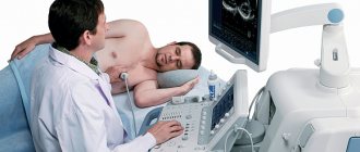
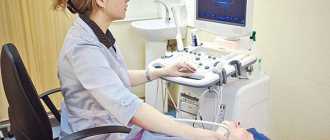
The ultrasound procedure of the carotid arteries does not cause discomfort or pain and is performed with the patient in a supine position. A special pillow is placed under the patient's shoulders so that nothing interferes with the view of the arteries. Throughout the procedure, the sonologist moves the scanner over the skin of the neck, lubricated with gel, and from time to time changes the sensitivity of the device.
There are several methods for ultrasound of the carotid arteries. However, it does not depend on this how ultrasound of the carotid arteries is done - in any case, the sonologist moves the scanner in the neck area, if necessary, telling the patient to turn his head or stretch his neck, examining the subclavian region.
Dopplerography
Using Doppler ultrasound, disturbances in blood flow in the vessels are detected, and ultrasound of the carotid arteries is one of the most effective diagnostic methods. If the blood flow is not visible, the sonologist can apply digital pressure on the artery to obtain more accurate information. In some cases, signals obtained using Doppler ultrasound can be converted into sound, this helps when examining seriously ill patients.
Duplex scanning
Duplex scanning is a combination of Doppler ultrasound, which determines blood flow disturbances, with traditional scanning, which shows the echostructure of the arteries. Duplex scanning of the carotid and vertebral arteries is the most common procedure, since it solves two problems simultaneously.
Triplex scanning
Triplex scanning of the carotid arteries involves research in three modes. The greyscale scanning mode provides information about the structure and shape of the artery, as well as any changes in it. Dopplerography
reveals the quality of blood flow inside the artery. And color Doppler mapping provides a color image that provides comprehensive information about any changes in the structure and blood flow of the artery.
How is ultrasound examination of neck vessels performed?
The procedure for ultrasound examination of the vessels of the neck is not complicated. There are several ways to examine blood vessels, and the choice will be made depending on what disease the doctor suspects and what the preliminary diagnosis was. Each type of examination is non-invasive, which means there is no surgical intervention.
Ultrasound with Dopplerography
Doppler ultrasound is based on the Doppler effect. In this case, the sensors are placed “blindly”, so standard Doppler sonography as a separate study is most often not performed. The ultrasonic waves that the device sends into the tissue change their frequency in proportion to the speed of movement of objects. With the help of such a study, you can learn about the condition and structure of all soft tissues near the blood vessels, and also examine the latter absolutely safely.
Duplex scanning
The most common type of examination, it often complements Doppler ultrasound, which is combined with two-dimensional ultrasound. In this case, the doctor will know where the vessels are located and where to install the sensors for Doppler ultrasound. In this case, assessing the patient’s health status is much easier, and the results of the study will provide much more information.
Triplex scanning
This is the most informative research method, but it is not always needed. If you don’t know what an ultrasound of the neck vessels is called, when the procedure combines conventional Doppler ultrasound, two-dimensional ultrasound and color differentiation, then this is just a triplex scan. This diagnostic method will allow us to identify almost any pathology of blood vessels and soft tissues.
Decoding the results
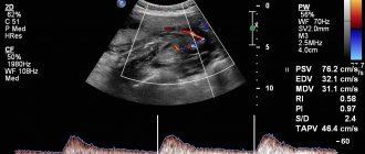
The interpretation of the diagnosis of ultrasound of the carotid arteries is carried out by the attending physician based on the results obtained. However, a sonologist can also clarify the situation by recording the examination results at the end of the procedure. Typically, a sonologist comments on the visible echostructure of the vessel during scanning and blood flow.
The norm for ultrasound of the carotid arteries is that the blood flow in the internal and external arteries is equal in power, as well as the location of the carotid artery to the left of the aorta. The thickness of the vessel walls should not exceed 1.2 mm, and the pulsation in a healthy artery should be continuous.
What diseases can be detected by ultrasound of the carotid arteries?
Ultrasound of the carotid arteries allows you to examine not only the brachiocephalic arteries, but also simultaneously assess the condition of the vertebral vessels of the neck. The norm for ultrasound of the carotid arteries implies an internal and external diameter of the walls sufficient for good blood flow. Ultrasound of the carotid arteries most often reveals atherosclerotic plaques and kinks, which cause strokes. Ultrasound of the carotid arteries allows you to see even the structure of plaques, for example, those that have calcium impurities and are especially difficult to treat.
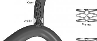
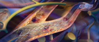
No less dangerous are blood clots and aneurysms, which are clearly visible using ultrasound. Stenosis visible on ultrasound of the carotid arteries may explain ischemic attacks, since blood flow worsened due to narrowing of the artery does not allow the brain to receive enough oxygen. An alarming symptom is a change in the level of the bifurcation of the carotid artery, visible on ultrasound. However, one must understand that ultrasound of the carotid artery can only identify these problems; subsequently, it is recommended to conduct a more in-depth study.
Pathology detected by ultrasound of the vessels of the head and neck:
Atherosclerotic changes in the BCA:
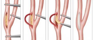
Atherosclerosis can be expressed as thickening of the inner wall of a vessel or as an atherosclerotic plaque. Atherosclerotic plaques are the most unfavorable, as they can cause strokes and heart attacks. To assess the risk of rupture, with duplex vessels, the shape and stability of the plaque, the degree of narrowing of the lumen, impaired blood flow, changes in vessel tone, irregularities, and calcifications are assessed. Small atherosclerotic plaques do not significantly affect cerebral blood flow, but are an indication for taking a lipid profile, regulating blood lipid levels, and correcting them with diet or statins. Plaques that narrow the lumen by more than 70% require surgical treatment. Plaques significant to blood flow with stenoses of more than 50% also require observation and treatment aimed at compensating for impaired blood flow.
Angiopathy on ultrasound of the vessels of the head and neck:
Damage to blood vessels due to disruption of their nervous regulation. Angiopathy is manifested by angiodystonia (reversible spasms and vascular paresis). Angiopathy develops against the background of other diseases (diabetes mellitus, hypertension, coronary artery disease, smoking, etc.) and requires the earliest possible diagnosis and treatment.
Pathological tortuosity of blood vessels:
Vascular tortuosity can be significant (affecting cerebral blood flow) or not significant. There are C-shaped and S-shaped tortuosity, looping, and kinks of blood vessels. In mild cases they have local significance (causing surges in blood pressure), in severe cases they require surgical treatment.
Vascular hypoplasia detected by ultrasound:
Hypoplasia is a congenital pathology in which the diameter of the vessel is smaller than normal. This pathology is sometimes compensated by the body and the blood flow copes, but sometimes it causes hemodynamic disturbances. Hypoplasia of the vertebral artery is the most common: clinically they are manifested by numbness of the hands in the morning, rare dizziness; with the development of any problems in the cervical spine (osteochondrosis, myositis, disc herniation, etc.), and increasing impact on an already narrowed artery, more pronounced disorders arise that require urgent correction.
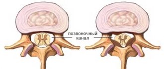
Dystopia of blood vessels of the head and neck:
The most common dystopia is dystopia of the vertebral artery in the form of a high entry into the spinal canal. At the same time, in the “bare” area, the vertebral artery is subjected to muscular action, which leads to a change in blood flow. Dystopia of the vertebral artery is a congenital pathology, but it can be influenced by choosing the right treatment and set of exercises.
Changes in cerebral blood flow:
Changes in cerebral blood flow can be caused by atherosclerotic changes, angiodystonia, stenosis, aneurysms, extravasal compression of the arteries, changes in the pressure gradient (differences between arterial and venous blood flow). To assess tone, functional tests are performed with breath holding and hyperventilation. This reveals a vasoactive reserve.
A test is also carried out to study blood flow when turning the head, which makes it possible to identify compression of the vertebral arteries and refer the patient for the necessary further examination, identify the causes of these changes and select effective individual treatment.
Preparing for the study
No special preparation is required for ultrasound of the neck and head vessels. However, if the patient is taking medications that lower blood pressure or affect blood clotting, the doctor must be informed. He may recommend that you avoid taking medications in some cases.
On the day of the examination, it is recommended to refrain from smoking, drinking alcohol, energy drinks and physical activity. In general, it is not recommended to perform actions that may affect the tone of the vascular wall.
Advantages of performing ultrasound scanning of the carotid and vertebral arteries in CELT
The multidisciplinary clinic CELT invites you to undergo a diagnostic test in accordance with international standards. Our name is well known in the capital, since we have been working in the domestic market of paid services since 1993. During this time, our specialists correctly diagnosed and restored health to thousands of patients, earning positive reviews and a good reputation.
Our diagnostic department is staffed by doctors of the highest category and candidates of medical sciences with at least fifteen years of experience. They know how to scan in such a way as to detect pathological changes even in the initial stages. This is important because timely treatment increases the chances of success, and especially when it comes to vascular diseases and cerebrovascular disorders.
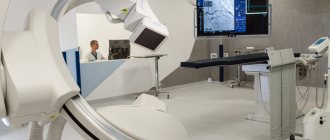
Our specialists have two ultrasound devices in their arsenal: “Electric Medical System” (USA) and “Philips” (Holland), which allow duplex and triplex diagnostics in real time and provide high-quality images.
You can find out prices for services in this section of the website by going to the “Services and Prices” tab. To avoid misunderstandings, we ask you to contact our operators to clarify the cost: +7 (495) 788-33-88.