Open oval window - what is it?
Due to increased blood pressure in the left atrium (the pressure is lower in the right atrium), the oval window valve closes tightly.
Over time, it becomes overgrown with connective tissue. However, there are situations when the hole between the right and left atria closes partially or does not close at all. Then, when sneezing, coughing, screaming, or significant tension in the anterior abdominal wall, blood is thrown through it from the right chamber to the left. If such a pathology is detected, the patient is diagnosed with an open oval window.
Possible complications
Most parents and pediatricians rightly believe that the presence of an open oval window does not threaten the child’s health. This opinion is supported by the statement of a popular pediatrician, Evgeniy Olegovich Komarovsky: this condition is not a malformation of the heart. Indeed, with a small hole, a person can play sports, work and study, and young men are even subject to conscription for military service.
But under certain conditions, a patent foramen ovale can cause the following unfavorable conditions in adults:
- Paradoxical venous embolism is the entry of a microthromb, a bubble of fat or air from the venous system into the left atrium, and then its penetration into the systemic circulation. This condition can provoke a transient ischemic attack or stroke.
- Migraine with a patent foramen ovale often occurs with an aura. It usually occurs in older women when blood is shunted from right to left.
- Platypnea-orthodeoxia is a syndrome characterized by shortness of breath that occurs when standing and subsides when lying down. It is usually observed when a patent foramen ovale is combined with deformation of the IV.
- Obstructive sleep apnea syndrome.
Some people with a patent foramen ovale develop transient global amnesia after a migraine or ischemic attack - a syndrome of memory impairment and inability to perceive new information.
Causes of an open oval window in the heart in newborns and adults
The reasons why the oval window does not close completely are still not clear. It is believed that the occurrence of the disease is contributed to by:
- prematurity;
- hereditary predisposition;
- connective tissue dysplasia;
- Congenital heart defect;
- drinking alcohol and smoking during pregnancy;
- the impact of unfavorable environmental factors on the expectant mother’s body.
Also, due to genetic factors, the size of the valve may be smaller than the diameter of the oval opening. Then complete closure of the oval window in the child’s heart becomes impossible.
It has been noted that the pathology accompanies:
- defects of the tricuspid and mitral valves;
- open ductus arteriosus.
An open foramen ovale in the heart can form in athletes due to severe physical exertion. Thus, persons involved in athletic gymnastics, wrestling, and weightlifting are at risk. The disease is especially common among divers and divers who regularly dive to great depths.
In patients with thrombophlebitis of the pelvis or lower extremities with a history of episodes of pulmonary embolism, pressure in the right side of the heart increases due to contraction of the vascular bed of the lungs. As a result, a functioning open oval window is formed.
In other words, the disease is not necessarily congenital. It can develop due to exposure to adverse factors on the body.
Diagnostics
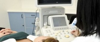
Ultrasound of the heart is of primary clinical significance. The doctor clearly sees a small hole in the projection of the left atrium, as well as the direction of blood flow.
When listening to a heart murmur, the pediatrician will definitely refer your baby for this type of examination.
According to new standards, at 1 month all newborns must undergo ultrasound screening, including the heart.
As a rule, there are no pathological changes on the ECG with LLC.
Symptoms of the defect
A patent foramen ovale is a heart defect that has no specific external manifestations. Usually the disease occurs in a latent form. Sometimes accompanied by scant symptoms.
Indirect signs of the disease include:
- cyanosis and severe pallor of the skin in the area of the nasolabial triangle, lips (the symptom becomes noticeable when crying, screaming, straining, coughing, while bathing);
- child's lagging behind in physical development (low weight gain, poor appetite);
- tendency to bronchopulmonary and colds;
- increased fatigue when performing physical work;
- respiratory failure that occurs when running, walking, or physical labor;
- signs of impaired cerebral circulation;
- sudden fainting;
- migraine, frequent headaches;
- postural hypoxemia syndrome.
If you notice similar symptoms, consult a doctor immediately. It is easier to prevent a disease than to deal with the consequences.
Symptoms
There are 3 options for the development of the oval window:
- It is completely overgrown.
- The size of the hole remains the same throughout life.
- The hole enlarges with age.
The first 2 scenarios are the most common. Clinically, they usually do not manifest themselves in any way. Therefore, it is impossible to even suspect the presence of an open oval window. Sometimes children hear noises. However, they can also appear in the absence of a patent opening.
The last option is the rarest. The mechanisms of window enlargement are not fully understood. There are suggestions about the influence of individual characteristics of the structure of the interatrial septum. In addition, with age, the load on the atria increases, which changes the pressure in them, often upward. Because of this, they stretch, and accordingly the opening of the open oval window increases.
At an older age, when there is a possibility of developing venous thrombosis, a patent foramen ovale can contribute to the occurrence of embolisms (clogging of arteries):
- cerebral arteries, leading to ischemic stroke with the development of paralysis, decreased sensitivity, speech impairment, swallowing, hearing and other symptoms;
- coronary arteries - myocardial infarction;
- mesenteric - intestinal ischemia with severe abdominal pain that does not go away even after narcotic analgesics;
- splenic artery - splenic infarction, manifested by pain of varying intensity depending on the degree of damage, and others.
A patent foramen ovale may be suspected in the following cases:
- the most common symptom is slight cyanosis of the lips or nasolabial triangle when straining (with a strong cough, severe static physical activity), when a child cries;
- frequent colds and inflammatory diseases of the bronchopulmonary system;
- delayed physical development of the child, poor tolerance to physical activity;
- development of unexplained loss of consciousness, symptoms of transient cerebrovascular accident (temporary disturbances of vision, hearing, skin sensitivity, limb movements, facial asymmetry) especially in young people under 50 years of age;
- increased blood supply to the lungs, a tendency to increase the size of the right chambers of the heart;
- signs of overload of the right chambers of the heart according to the electrocardiogram.
Photo: cyanosis of the nasolabial triangle in a child
In people suffering from migraine with aura, a patent foramen ovale is found in 40-60% of cases. However, there is currently no consensus on the effectiveness of foramen ovale closure in relieving migraine attacks.
Diagnosis of the disease in children and adults
Diagnosis of the disease includes the following procedures:
- Analysis of the patient's medical history. During a conversation with the patient, the doctor finds out whether he suffers from dizziness, fainting, low endurance for physical activity, cyanosis of the extremities, and colds. It is also clarified whether there is a hereditary predisposition to diseases of the cardiovascular system.
- Skin examination, blood pressure measurement, weighing, auscultation.
- General blood and urine analysis, coagulogram (blood test for clotting), blood biochemistry.
- Ultrasound of the heart.
- Electrocardiography.
- Echocardiography, Dopplerography (aimed at determining the presence of a hole in the interatrial septum, studying the direction of blood movement through the heart and determining the volume of blood passing through the defect).
- Contrast echocardiography (makes it possible to detect an open oval window even of a small size). During the examination, the patient is given shaken saline solution intravenously. If he has an open foramen ovale, then small air bubbles almost immediately pass through it from the right atrium to the left.
- Transesophageal echocardiography (to identify the hole in the septum of the heart and the patent foramen ovale valve, a probe equipped with an ultrasound sensor at the end is inserted into the esophagus).
- Chest X-ray.
Additional examination methods
To determine patient management tactics, it is important to know how a hole in the interatrial septum affects heart function. Doctors do electrocardiography at rest and after exercise. This will help eliminate arrhythmia and conduction disturbances.
For adults, x-rays of the chest cavity are recommended, which will make it possible to judge the degree of hypertrophy of the right atrium and reveal congestion in the lungs.
Children with minor anomalies of heart development need consultation with a pulmonologist and immunologist. The presence of chronic foci of infections can aggravate the child’s condition, so you need to go to the otolaryngologist’s office or visit a dentist.
Treatment of open oval window
Children and adults diagnosed with a patent foramen ovale should follow non-drug measures:
- Limit physical stress.
- Sleep at least 8 hours a day (children should sleep 10-12 hours).
- Maintaining a daily routine.
- Balanced diet.
- Taking vitamins.
If there are no symptoms of a patent oval window, there is no need for specific treatment. If signs of illness appear, a high risk of blood clots, or significant discharge of blood from one atrium to another (this does not normally occur), the cardiologist may recommend the patient:
- Take anticoagulants and antiplatelet agents - medications that prevent the formation of blood clots (thrombi).
- Perform endovascular treatment of an existing open foramen ovale. A tube (catheter) is inserted into the artery, at the end of which there is an occluder. Getting into the oval window, it completely clogs it. Surgical intervention is performed under X-ray and echocardioscopic control.
Complications and prognosis
The most serious and only complication is paradoxical embolism. In this case, a blood clot (usually with thrombosis of the deep, superficial veins of the lower extremities, pelvis) or air bubbles (during diving, less often during fractures) from the venous system enter the arteries of the systemic circulation through the open oval window. Thus, ischemic stroke, myocardial infarction, intestinal ischemia, infarction of the spleen and other organs develop.
There are no clear statistics in Russia. According to studies in France and the USA, ischemic strokes account for up to 30% of cryptogenic (unexplained) strokes. Of these, about a little less than half of the cases are associated with an open foramen ovale detected in patients.
Forecast
The prognosis depends on the size of the open oval window. It is favorable for sizes up to 4 mm, low blood discharge and no increase in it over time.
In the case of the development of large blood shunts, which increase the blood supply to the right chambers of the heart and increase the degree of pulmonary hypertension, the prognosis sharply worsens.
Usually, before the development of severe pulmonary hypertension, the patient has time to undergo surgery - to close the functioning oval window with an occluder. The operation dramatically improves the prognosis for the patient.
Why is an open oval window dangerous?
For newborns, an open oval window is considered normal. If it does not heal and persists into adulthood, serious health problems may arise:
- Kidney infarction (the organ dies due to impaired blood supply).
- Myocardial infarction (heart muscle tissue dies due to the fact that the required amount of blood does not flow into it).
- Stroke (cerebral circulation disorder causes damage to brain tissue).
- Transient disturbance of cerebral blood supply (due to a disturbance in the blood supply to certain structures of the brain, there is a temporary disruption of its activity).
Dimensions and standards
Closure of the oval window normally occurs within a period of 3 months to 2 years. But even at 5 years old, such a finding is considered normal.
According to statistics, 50% of healthy children aged 5 and 10–25% of adults have this feature. Separately, it is worth noting that it is not a vice. Doctors call it MARS - minor anomaly of the heart. It distinguishes the structure of the heart from the anatomical norm, but does not pose an immediate threat to health.
In 1930, T. Thompson and W. Evans examined 1,100 hearts, the results were as follows: 35% of those examined had an open foramen ovale, 6% of them had a 7 mm diameter (half of them were children under 6 months). In adults, large-diameter PFOs occurred in 3% of cases.
Window sizes can be different: from 3 mm to 19 mm (usually up to 4.5 mm). First of all, they depend on the patient’s age and the size of his heart. The indication for surgical treatment does not depend on the size of the window, but on how much it is covered by the valve and the degree of compensation.
Prevention
There is no special prevention for an open oval window. To prevent pathology from occurring in newborns, during pregnancy a woman should lead a healthy lifestyle, give up alcohol and smoking. She also needs to avoid contact with:
- chemicals (paints, varnish vapors, some medications);
- ionizing radiation (thermonuclear reactions, X-ray machine);
- patients with infectious diseases (if infected with rubella in the first trimester, the risk of heart disease in a child increases significantly).
This article is posted for educational purposes only and does not constitute scientific material or professional medical advice.
Signs
As already mentioned, there is no clinical picture for this pathology, and the anomaly itself is detected randomly. There are usually no complications or consequences.
Combination of an open oval window with other diseases. Symptoms appear when hemodynamics (proper blood flow through the chambers of the heart) are impaired. This happens when there are combined heart defects, for example:
- patent ductus arteriosus;
- defects of the mitral or tricuspid valves.
The chambers of the heart are overloaded, the interatrial septum is stretched, and the valve cannot perform its functions. Right-left shunting appears.
False chords
In our heart there are so-called “true chords”, consisting of dense muscles. They are located in both ventricles of the heart and are attached by their endings to two valves - the triscupidal and mitral. Their function is to keep the valve leaflets in their natural position and prevent them from turning into the cavity of the heart when the ventricles contract.
The false chords of the left ventricle include anatomical formations that are not attached to the valve leaflets and are located in the cavity of the heart chamber. Pathological chords can be longitudinal and transverse, and when the heart contracts, they produce heart murmurs, sometimes reminiscent of the sound of a violin.
There are additional chords that do not cause serious pathological changes. False chords can be identified using echocardiography and ultrasound. Sometimes the child has no pronounced symptoms, except for the characteristic noise when listening to the heart, but if there are too many false chords and they are located transversely, this can lead to arrhythmias and serious circulatory disorders.
MARS are not classified as heart defects, but sometimes they can pose a real danger to the health and life of a child. That is why timely detection of such pathologies is very important for the work of a cardiologist, as well as any specialist involved in ultrasound and functional diagnostics.

