DlyaSerdca → Heart diseases → Heart attack → Heart attack as a necrotic lesion of the spleen: causes, symptoms and treatment
A heart attack is a fairly common necrotic lesion of the spleen, a problem that can arise as a result of insufficient blood supply to this organ.
Lack of arterial blood supply to the spleen can occur with embolism, stenosis or thrombosis of the vascular bed leading to the organ in question.
Often, the problem occurs in people suffering from bacterial, septic endocarditis, vascular atherosclerosis, and mitral stenosis.
The problem can also develop as post-traumatic syndrome after certain infectious diseases. Moderate necrotic lesions of the spleen lead to the fact that the parts of the organ affected by necrosis gradually become scarred.
Although, in rare cases, necrotic lesions begin to soften, forming false cysts. Further infection of the affected areas of the spleen can lead to the formation of an abscess, which is extremely dangerous for the patient.
To understand what the treatment of the disease should be, it is primarily important to understand what a pathology such as splenic infarction is, to consider the main symptoms of the problem and the causes of their occurrence. This is what we will talk about in today’s publication.
What is this pathology?
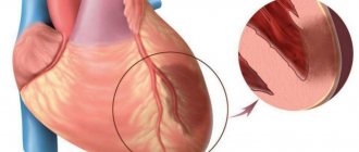
Splenic infarction is considered an ischemic or hemorrhagic lesion of the splenic parenchyma, in which necrotic foci of destruction of spleen tissue occur.
Most often, the problem is associated with occlusion of the vascular bed, designed to nourish (saturate with oxygen) this organ.
The problem can be segmental (focal) or extensive, affecting all tissues of the spleen.
The development of such a pathological condition as splenic infarction is most often associated with primary hematological diseases, with the infiltration of organ cells by pathologically altered cells.
Note that there are many causes of the disease, but all of them, as a rule, are associated with lesions of the body’s vascular system.
Main causes of the disease
Among the main reasons for the development of this pathology, doctors name:
- primary development of malignant blood pathologies - these can be leukemia, lymphoma, leukemia, lymphogranulomatosis or even myelofibrosis;
- blood pathologies of a non-oncological nature - for example, anemia of various kinds, Marchiafava-Micheli disease;
pathologies of the heart and vascular system - such, for example, as atherosclerosis, endocarditis and others;- autoimmune problems that also affect vascular tissues;
- blunt trauma to the spleen, its torsion;
- state of sepsis;
- the problem may arise as a complication of certain intravascular diagnostic or therapeutic procedures - we are talking about the procedure of cardiac catheterization, sclerotherapy, etc.
Kinds
We have already said that splenic infarction can be small-focal or widespread in its distribution, in some sources - single or multiple. In addition, according to the mechanism of development, doctors can classify splenic infarction as:
- Hemorrhagic pathology is a problem that occurs when the vessels supplying a given organ rupture. This is a kind of hemorrhage in the spleen tissue.
- Ischemic pathology that develops when there is insufficient blood supply to a given organ. Pathology occurs when the vascular bed, designed to supply the spleen with oxygen-rich blood, is partially or completely blocked.
It is quite clear that different types of infarction of the organ in question may have slightly different symptoms, suggesting the development of one or another type of pathology.
Symptoms of the problem
It must be said that the pathology under consideration can manifest itself as classical symptoms, or proceed non-standardly, in the complete absence of specific symptoms or with their significant distortion.
That is why we would like to describe the most common symptoms of splenic infarction, which are as follows:
this may be a sudden onset of acute pain in the left upper quadrant of the abdomen;- development of fever, chills, general weakness and other symptoms of intoxication of the body;
- often, the problem is complemented by nausea, vomiting, chest pain radiating to the left side, gastrointestinal disorders;
- in severe cases, the problem develops into the development of collapse.
With moderate necrotic lesions of the spleen, the acute condition can be asymptomatic, and the affected tissues scar quickly enough. In this case, sometimes cysts can form in areas of primary necrosis, which, in turn, can degenerate into an abscess.
Diagnosis and treatment
Diagnosis of spleen diseases begins with a thorough examination by a doctor. After palpation and history taking, additional studies will be required - ultrasound, radiography, MRI or puncture. Laboratory diagnostics are also required.
The primary task is to correctly diagnose in order to prescribe effective treatment. It should only occur under the supervision of an experienced specialist.
Preventive measures to maintain the organ in a healthy state are very simple: proper nutrition and a healthy lifestyle. The spleen begins to work better with regular physical activity, as well as with special breathing exercises.
Pain in the spleen occurs when the organ has significantly increased in size. This may indicate various diseases, which can only be diagnosed by a qualified doctor. It is highly recommended not to make a diagnosis on your own, much less select a treatment.
After all, the spleen is one of the important organs responsible for supporting human immunity.
Timely contact with specialists will help you solve the problem at an early stage with minimal damage.
For early diagnosis of health problems, you can undergo a comprehensive diagnostics of the body at our center.
The best diagnosis of pain in the spleen is MRI of the spleen
You can sign up for a consultation right now: online or by phone
Diagnostics
As we have already said, with relatively small lesions of the organ, diagnosis can be difficult, since not all patients pay attention to barely noticeable symptoms - no one seeks advice and diagnosis.
Extensive organ damage, as well as all sorts of consequences of primary tissue necrosis, can be successfully visualized using computed tomography performed with special contrasting of the vascular bed.
Magnetic resonance imaging is also successfully used to diagnose this pathology, which perfectly identifies foci of necrosis in the splenic parenchyma.
Somewhat less commonly, the following can be used to diagnose the described condition:
- standard radioisotope scan of the organ;
- ultrasound examinations (ultrasound).
It cannot be said that in this condition, differential diagnosis is no less important, which makes it possible to distinguish necrotic lesions of the organ from splenic abscess, acute pancreatitis, pyelonephritis on the left, and kidney abscess on the left.
After all, it is precisely these diseases that can often mask an atypical ischemic infarction of the spleen.
Since the pathological condition in question can be provoked by serious (oncological, parasitic, infectious and other) blood diseases during its surgical treatment, for diagnostic purposes, a microslide can be prepared from tissues affected by necrosis. Microslides are then used for diagnostic, informational, educational and scientific purposes.
Useful information! A micropreparation is usually called a glass slide on which special objects are placed for further research using a microscope. They usually try to cover the micropreparation with a thin top cover glass layer for ease of use.
The micropreparation allows you to determine the presence or absence of pathological microorganisms in tissues. In addition, the creation of microspecimens for splenic infarction is very important from a scientific point of view. After all, it is on demonstrative microslides that it is most convenient to explain to medical students the possible variants of pathology.
Description of drugs in Lesson No. 01.02
Description of drugs in Pathological Anatomy in Lesson No. 1,2
(This is an indicative description, not a cathedral one, some drugs may be missing, as is the description of previous years)
LESSON No. 1 HISTORY OF THE DEPARTMENT OF PAT. ANATOMY OF MMA NAMED AFTER I.M.SECHENOV
See lectures
LESSON No. 2 NECROSIS. APOPTOSIS.
Electronogram No. 20 ISCHEMIC INFARCTION OF THE SPLEN
There is swelling of mitochondria with destruction of the crypts, with the appearance of calcium deposits on them. Destruction of lysosomes.
Microslide No. 7 NECROSIS OF THE EPITHELIUM OF THE CONVOLVED PROXIMAL AND DISTAL TUBULES OF THE KIDNEY. HEMATOXYLIN-EOSIN STAINING
Distal prox tubules are unchanged. The epithelium and glomeruli contain nuclei. The cytoplasm is coagulated and homogeneous in some places. Destruction of the basement membrane (tubulorrhexis) is observed. Karyopyknosis, karyolysis, and plasmorrhexis are noted. The capillaries of the glomerular loop are anemic, and the vessels of the medulla of the kidney are full-blooded
Microslide No. 6 ISCHEMIC RENAL INFARCTION. HEM COLORS. – EOD
.
The necrosis zone is represented by structureless masses, surrounded by a zone of demarcation inflammation, represented by full-blooded vessels with dilated lumens and polymorphonuclear leukocytes. In the focus of necrosis, the tissue structure is disturbed, the nucleus is not stained, with signs of karyopyknosis, karyorrhexis, and karyolysis.
Microslide No. 8 NECROSIS OF LYMPH FOLLICULS. NODES (SURROUNDING GEM.-EOZ.)
A homogeneous structureless mass is determined in the center of the follicle. Along the periphery there are single lymphocytes of small sizes (as part of karyopyknosis). There are many randomly located clumps of formatin (karyorrhexis)
Microslide PANCREONECROSIS (OCR. HEM.-EOS.)
The glandular tissue is represented by a structureless mass, containing single nuclei in the composition of karyopyknosis. Contains chromatin clumps
Microslide No. 215 ISCHEMIA ZONE IN THE MYOCARDIUM. CHIC REACTION.
The presence of glycogen - cardiomyocytes - crimson areas. Lack of glycogen - light areas.
Macro specimen ISCHEMIC INFARCTION OF THE SPLEN
The shape and dimensions have not been changed. The color is heterogeneous - in general it is brown-red, but from the gate to the periphery of the organ there is a 1-2 cm strip of paler color. The focus of necrosis is triangular in shape, dense in consistency, the base faces the capsule. On the capsule in the area of the infarction there are rough deposits of fibrin. D-z: Acute ischemic infarction of the spleen
Macroscopic specimen of BRAIN INFARCTION. FOCUS OF GRAY SOFTENING.
The lesion is located in the occipital region of the left hemisphere, grayish in color, irregular in shape, flabby consistency. Occurred due to a blood clot or embolism of cerebral vessels.
Macro specimen of GANGRENE TOE
Dry gangrene. The fabrics are black (due to iron sulfide deposits). Reduced in volume, with a well-defined zone of demarcation inflammation.
Macro-drug of GUNS GANGRENE
Gangrene is wet. The intestinal wall is thickened, edematous, flabby consistency, black-red in color. The serous membrane is dull with fibrin deposits. Thrombosis of the superior mesenteric artery.
Macrodrug TUBERCULOSIS LYMPH. KNOTS
In the lymph area there is a zone of caseous (cheesy) necrosis. Yellowish-gray color, dense consistency, crumbling.
Macropreparation PETRIFICATIONS IN THE LUNG (for tuberculosis)
Round shape, whitish-gray color, rocky density (due to calcium deposition)
Getting rid of illness
Speaking about the treatment of splenic infarction, first of all, it is important to say that the therapeutic techniques used always depend on the type of pathology - ischemic or hemorrhagic pathology, extensive or small-focal infarction.
A specific choice of treatment tactics can be made only after a full diagnosis, identifying all the characteristics of the pathology. Treatment for splenic infarction can be either conservative or surgical.
Until the characteristics of the pathology are clarified, as treatment, patients need rest and a cold compress on the left hypochondrium. In the future, patients may be prescribed the following treatment:
powerful antibacterial therapy;- sometimes anti-inflammatory treatment;
- antiparasitic drugs, etc. may be prescribed.
Any therapeutic methods for splenic infarction are always performed with dynamic monitoring of the pathology.
If suppuration develops at sites of primary necrosis, patients may be recommended splenectomy or opening of the abscess with drainage of the existing abscess.
Unfortunately, extensive splenic infarction may require urgent surgical treatment, complete or partial removal of the affected organ, and intensive care.
Inflammatory diseases
Acute splenitis in ultrasound diagnostics is indicated by enlargement of the organ with rounding. The echostructure is homogeneous, fine-grained. Sometimes there are foci of acute necrosis. In chronic splenitis, the enlarged size of the spleen remains due to the proliferation of fibrosis. The echogenicity of the tissue is higher than normal. Foci of necrosis may be calcified. Calcifications are visible on ultrasound as small hyperechoic formations.
Often, ultrasound reveals splenomegaly - a pathological increase in the size of the spleen. It can be a sign of various diseases: Gaucher disease, amyloidosis, tuberculosis and others. In 75% of cases, splenomegaly is a symptom of diseases of other organs: liver cirrhosis, active hepatitis. In the initial stage, ultrasound diagnostics shows an increase in the diameter of the splenic vein. Fibrosis gradually sets in with an increase in echostructure.
Splenomegaly with normal echostructure is observed with:
- infections,
- liver pathologies,
- sickle anemia,
- congenital spherocytosis,
- leukemia,
- hemolysis,
- chronic anemia,
- Still's disease
- Felty's syndrome,
- Wilson's disease
- reticulocellular sarcoma.
Hypoechogenicity with an enlarged spleen:
- hepatocellular disease,
- lymphogranulomatosis,
- myeloma,
- non-caseating granulomatous inflammation,
- leukemia.
Abscesses are rare but possible. They appear as anechoic or hypoechoic lesions with poor borders but visible walls. There are gas bubbles inside, creating zones of high echogenicity. To diagnose abscesses, the clinical picture is also used; hematoma and splenic infarction must be excluded.
Forecasts
It must be understood that survival prognosis for splenic infarction depends on many factors:
- degree of prevalence of necrosis;
- speed of contacting doctors and detecting a problem;
- patient's age;
- general health status – presence/absence of concomitant pathologies.
In the absence of oncological pathology, survival prognosis after splenic infarction is in most cases positive.
But if the emergency is caused by malignant blood diseases, the survival prognosis, on the contrary, turns out to be the most negative.
Spleen pain
General information
The spleen is an unpaired parenchymal organ that performs immune, filtration and hematopoietic functions, taking part in the metabolism, in particular iron and proteins. The spleen is located in the abdominal cavity, in the left hypochondrium.
On average, the length of the spleen in adults is 80-150 mm, width 60-90 mm, thickness 40-60 mm, weight 140-200 g. The place where arteries and nerves enter the organ and veins and lymphatic vessels exit it is called the hilum of the spleen.
The peritoneum, covering the spleen on all sides, with the exception of the gate and the area to which the tail of the pancreas is adjacent, forms ligaments. Due to them and intra-abdominal pressure, the spleen is fixed.
The spleen is also covered by a capsule. The section reveals numerous trabeculae, which diverge radially from the hilum of the spleen. They contain arteries, veins, lymphatic vessels and nerve fibers. This connective tissue skeleton and a few smooth muscle cells make up the musculoskeletal apparatus of the spleen, capable of withstanding a significant increase in its volume.
Immune diseases
One of the most important functions of the spleen is immune function. Phagocytic mononuclear cells of the spleen capture harmful substances and cleanse the blood of bacteria, viruses and other foreign agents. In addition, lymphocytes and plasma cells of the spleen participate in the immune response, promoting the elimination of antigens foreign to the body.
In some autoimmune diseases (eg, thrombocytopenic purpura, autoimmune hemolytic anemia), autoantibodies are produced in this organ. These may be the following diseases:
- thrombocytopenic purpura;
- autoimmune hemolytic anemia.
Spleen cells produce immunoglobulins. The spleen controls circulating blood cells, primarily aging and pathologically altered red blood cells, which are destroyed by phagocytes of the spleen (filtration function).
The spleen not only destroys, but also accumulates formed blood elements :
- red blood cells;
- leukocytes;
- platelets.
In particular, the spleen contains up to 50% of platelets, which, if necessary, are released into the bloodstream. The spleen (as well as the liver) has no pain receptors; it cannot hurt. Pain occurs only from stretching of its capsule, as a result of rapid enlargement.
Pain in the spleen in diseases
Injuries to the spleen can be open or closed . Open injuries occur when penetrating cut, stab or gunshot wounds of the abdomen or left half of the chest with simultaneous injury to the diaphragm. The causes of closed injuries are most often a blow to the left hypochondrium, a fall on the stomach, compression of the abdomen and left half of the chest with a simultaneous fracture of the lower ribs on the left.
Possible cracks and wounds of the spleen of varying depth and extent, separation of part or the entire spleen. Splenic ruptures are often complicated by bleeding into the abdominal cavity and shock. Immediately after the injury, the following symptoms appear:
- sharp pain in the spleen in the left hypochondrium;
- pale skin;
- cold sweat;
- frequent small pulse;
- drop in blood pressure;
- nausea, vomiting;
- strong thirst.
The victim takes a characteristic position on the left side with the hips brought to the stomach or in a semi-sitting position. Abrasions may be found on the skin of the abdomen . Respiratory excursions of the left half of the abdominal wall are limited, it is painful, tense, the Shchetkin-Blumberg sign is positive. Percussion reveals dullness in the left hypochondrium. With significant accumulation of blood in the abdominal cavity, the zone of dullness of percussion sound moves with a change in the position of the patient’s body.
Pain in the spleen can occur with various diseases. The spleen is involved in the pathological process in many infectious diseases, namely:
- typhoid and typhus;
- sepsis;
- anthrax;
- infectious mononucleosis;
- acute viral hepatitis;
- infectious lymphocytosis;
- cytomegalovirus infection;
- malaria;
- visceral leishmaniasis;
- tularemia;
- listeriosis;
- brucellosis;
- syphilis
Infarction and abscess of the spleen
Splenic infarction can develop as a result of thromboembolism of the branches of the splenic artery or its local thrombosis in leukemia, diffuse connective tissue diseases, a number of infections, atherosclerosis, lymphosarcoma.
The clinical picture of splenic infarction depends on its size. It is important to consider that small heart attacks are asymptomatic. With more extensive lesions as a result of the development of perisplenitis (inflammation of the spleen capsule), pain appears in the spleen in the left hypochondrium, often radiating to the back and intensifying with inspiration. A splenic abscess can develop due to bacteremia due to endocarditis or salmonellosis, as well as due to infection of splenic infarctions and subcapsular hematomas; for hemoglobinopathies, sickle cell anemia, rupture of a subdiaphragmatic abscess into the spleen.
Symptoms are often characterized by:
- fever;
- pain in the upper left half of the abdomen and chest;
- Tension of the muscles of the anterior abdominal wall.
Other diseases of the spleen
Splenic tuberculosis may be a manifestation of miliary tuberculosis. Isolated tuberculosis of the spleen is not very common; it occurs with scant clinical symptoms. Echinococcus is one of the parasitic diseases of the spleen. The most common lesion is unilocular echinococcus. Possible rupture of the echinococcus bladder and contamination of the abdominal cavity with daughter scolex. Recognizing the disease is quite difficult. Ultrasound and computed tomography play an important role in diagnosis. Primary tumors of the spleen, both benign and malignant, are quite rare. Of the benign tumors found in the spleen:
- hemangioma;
- lymphangioma;
- fibroma.
Among primary malignant tumors of the spleen, lymphomas are most often observed . At the beginning of the development of spleen tumors, as a rule, there are no clinical manifestations. As tumor nodes and the organ enlarges, patients feel heaviness and dull pain in the spleen in the left hypochondrium.
If you have severe pain in the spleen, contact your physician. Having established a preliminary diagnosis, the therapist will refer you to the appropriate professional: an infectious disease specialist, hematologist, traumatologist, surgeon or oncologist. If treatment does not give the expected results, then the issue is resolved through surgery. Many doctors recommend eating right and leading a healthy lifestyle to prevent such serious consequences.