Diagnosing various pathologies of cerebral vessels is a pressing issue, since cases of strokes, which are accompanied by loss of ability to work, disability and death, have become more frequent. Statistics show that only 40% of people after a stroke can return to a full life.
Doppler ultrasound is the leading examination method that can be used to identify various lesions of the brachiocephalic arteries.
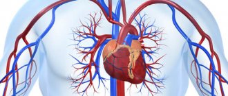
To understand what is meant by the words “brachiocephalic arteries,” you need to trace their path to the brain from the aorta. The blood flow that comes from the aortic arch is distributed into 3 large arteries: the common carotid, the left subclavian artery, and the brasiocephalic trunk. The latter is divided into 3 more arteries, which are located on the right side: subclavian, carotid, vertebral.
All of these vessels, in one way or another, take part in the blood supply to the shoulder girdle and, directly, to the brain. At the same time, they form a complex system in the form of a closed circle, which allows evenly distributing the entire volume of blood in order to ensure adequate blood supply. Impaired blood flow can lead to blood redistribution.
Ultrasound diagnostics
Content:
- Ultrasound diagnostics
- Reasons for the study
- Causes of vascular system disorders
- Indications
- Decoding the results
Examination of arteries using ultrasound is the most informative diagnostic method that allows you to identify physiological changes. In addition, you can determine the qualitative and quantitative parameters of blood flow.
Ultrasound scanning, which is used to diagnose pathologies of the above arteries, is divided into 2 types: Doppler ultrasound and duplex scanning.
The basis of the two methods is the Doppler effect. Its essence is to capture ultrasonic waves from moving objects. Moving objects that move and reflect ultrasound are red blood cells.
The ultrasound monitor displays the change in frequency as a color image. Red is a positive shift, blue is a negative shift.
The positive side of the BCA ultrasound method is the ability to examine the arteries that are located inside the cranium. However, classical ultrasound does not have such capabilities.
The negative side is the inability to accurately determine the position of the vessel. Therefore, diagnosis is made based on the probable location and changes in scanning depth.
Ultrasound of the brachiocephalic arteries is performed by specialists using an ultrasound scanner, which combines classical research in β-mode and Dopplerography.
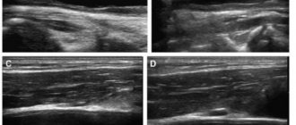
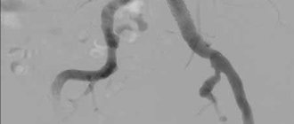
The condition of the arteries and the quality of blood flow can be assessed based on two-dimensional and three-dimensional images. The vessel can be seen both in the transverse plane and in length.
It is also possible to use extracranial ultrasound. This method allows you to examine vessels that are located outside the cranium and obtain the following information about them:
- condition of the walls;
- thickness, structure and number of atherosclerotic plaques;
- the size of the lumen;
- blood flow speed;
- the presence of aneurysms and other pathologies.
Duplex scanning of the brachiocephalic arteries: why is it needed?
Diagnosis and treatment of vascular pathologies, in particular atherosclerosis, are issues that are relevant at all times. One of the widely used methods for studying the vessels of the neck and the blood flow in them is duplex scanning of the brachiocephalic arteries. What kind of research is this and who is it for?
Ultrasound diagnostics doctor at Clinic Expert Stavropol Lusine Nersesovna Nersesyan talks about the features and capabilities of this diagnostic method.
— Identifying changes in blood vessels is not an easy task. But still, a number of modern research methods make it possible to do this. Lusine Nersesovna, what is duplex scanning of the brachiocephalic arteries?
— Let’s start, perhaps, with what the brachiocephalic arteries include. This is the brachiocephalic trunk, which is divided into the right subclavian and right common carotid arteries, as well as the left subclavian and left common carotid arteries. These vessels perform one of the most important tasks in our body - they supply blood to the brain. Duplex scanning of the brachiocephalic arteries is a type of modern ultrasound examination of these vessels, which, due to its informativeness, safety and accessibility, is widely used today in practice.
Along with this name of the method, you can also find another one - ultrasound scanning of the BCA (ultrasound duplex scanning of the brachiocephalic arteries).
— For what symptoms and diseases is it necessary to do a duplex scan of the brachiocephalic arteries?
“The range of symptoms for which this research may be useful is extremely wide. These include headaches of unknown origin, dizziness, tinnitus, blurred vision, hearing impairment, complaints of memory problems and changes in concentration.
With an increase or decrease in blood pressure, as well as people with a family history of hypertension, smokers with more than 15 years of experience, an ultrasound scan of the BCA is required to assess the condition of these vessels and prevent possible diseases and their complications.
Duplex scanning of the brachiocephalic arteries is recommended for diseases such as hypertension, previous myocardial infarction or stroke. Some endocrinological pathologies (in particular, diabetes mellitus, metabolic syndrome), injuries to the head and neck area, and toxic damage to the nervous system also require such a procedure. In these cases, the study can be performed repeatedly to assess the effectiveness of treatment, and in chronic processes, to monitor the degree of pathological changes in the arteries.
Experts recommend undergoing this test for preventive purposes after 40 years of age, even in the absence of pathological signs.
You may be interested in our materials on the topic: What to do when the head is cast iron? Spinning until you drop! Where did the glasses go again? We talk about the causes of memory deterioration. Speculation about hypertension. How to prevent myocardial infarction? How to prevent a brain catastrophe?
— What does duplex scanning of the brachiocephalic arteries show?
“It allows us to evaluate the structure of blood vessels, their patency, as well as functional hemodynamic parameters, in particular, blood flow speed. During the examination, you can see the course of the arteries, their deformations (for example, in the form of a loop, a wave). We also see abnormalities in the development of blood vessels in the form of aplasia (absence), hypoplasia (reduced diameter) or duplication of arteries. In addition, this diagnosis makes it possible to detect disturbances in the location of the brachiocephalic arteries, as well as signs of compression of the vessel from the outside by a tumor or hematoma.
An important nuance is that ultrasound scanning makes it possible to detect narrowing of the lumen or blockage of the brachiocephalic arteries due to atherosclerosis, thrombosis or embolism, or inflammation of the vascular wall.
More materials on the topic: Atherosclerosis: what does modern medicine know about it? Find and neutralize. How to protect against blood clots?
— Which specialists prescribe this study?
- Almost any. First of all, these are, of course, neurologists and cardiologists. In addition, general practitioners, endocrinologists, ophthalmologists, and audiologists can refer ultrasound scans of the brachiocephalic arteries. Since these arteries supply the brain, any changes associated with the possible involvement of the central nervous system in the pathological process may be a reason to conduct this study.
— Lusine Nersesovna, does ultrasound scanning of the brachiocephalic arteries require special preparation?
— No, no preparation is provided for this procedure.
— How is duplex scanning of the brachiocephalic arteries done?
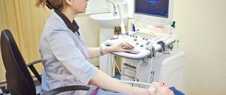
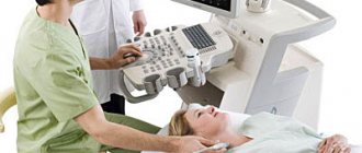
— This procedure is performed in an ultrasound diagnostic room. The patient lies down on the couch in a supine position, turning his head slightly to the side. Using ultrasonic sensors of different frequencies, the specialist passes over the surface of the neck and receives an image of the arteries on the monitor. He can measure their diameter, assess the condition of their walls and all the necessary indicators of blood flow in them.
The study is usually carried out first on the right, then on the left side of the neck. Some functional tests may require a change in head position. The procedure lasts on average from 20 to 40 minutes.
Subsequently, the doctor analyzes all the data received, writes a conclusion and recommendations on the necessary consultations with specialists, as well as on the timing of re-diagnosis.
— Are there any contraindications to this study?
- No. You can undergo this diagnosis at any time for any condition of the patient. The main thing is that it is transportable. There are also no age restrictions. Safety, painlessness, high information content and accessibility - these characteristics contribute to the popularization of duplex scanning of the brachiocephalic arteries among specialists and patients.
Interviewed by Sevilya Ibraimova
You can sign up for duplex scanning of the brachiocephalic arteries (USD BCA) here ATTENTION: the service is not available in all cities
The editors recommend:
Doppler, duplex, triplex... What types of vascular ultrasound are there? Transcranial duplex scanning of cerebral vessels: when is this study needed? Are your legs bothering you? Check your blood vessels! Who can benefit from ultrasound scanning of lower extremity vessels?
For reference:
Nersesyan Lusine Nersesovna
In 1994 she graduated from the Pediatric Faculty of Yerevan State Medical Institute. In 1996, she completed her internship in Pediatrics. In 2005, she underwent professional retraining in ultrasound diagnostics, and in 2008, advanced training in complex ultrasound diagnostics of peripheral and great vessels. Currently works as an ultrasound doctor at the Expert Clinic, Stavropol. Receives at the address: Dovatortsev St., 39A.
Causes of vascular system disorders
The main cause of pathological changes in the brachiocephalic vascular system is atherosclerotic lesions of the arteries. A study of people in relatively good health showed the presence of atherosclerosis in the initial stage in 3 percent of those examined. Their age ranged from 40 to 50 years. This result may indicate a fairly high probability of stroke in people after 55-60 years of age.
Atherosclerotic changes are considered an indirect cause of structural changes in blood vessels. For example, the appearance of tortuosity.
Indications for DS BCA
Ultrasound duplex scanning of the BCA is performed on the direction of a competent doctor - a neurologist, cardiologist, phlebologist. Competently interpreted results help to make a correct diagnosis and select adequate antihypertensive, antithrombotic or hypocholesterol therapy. The main indications for referral to duplex are:
- Regular cephalgia with dizziness, which may be associated with cerebrovascular accident.
- Feeling of pulsation in the head, fainting, noise and ringing in the ears. These symptoms are very common in patients with hypertension.
- Neurological symptoms in the periphery - episodic paresthesia (a feeling of numbness) of the upper extremities or certain areas of the skin of the neck or face. The duplex of the brachiocephalic vessels reveals pathological narrowings, including in the brachial and subclavian arteries.
- Heart rhythm disturbances.
- Sensory impairments - decreased visual acuity, loss or reduction of visual fields, diplopia.
- Pathologies of the cervical spine with suspected involvement of the vertebral arteries - scoliotic changes, osteochondrosis, osteoporosis, spondylolisthesis (displacement of the axis of adjacent vertebrae), instability.
- Suspicion of atherosclerosis, thrombosis, hypertension. Long-term elevated cholesterol in peripheral blood can also be an indirect indication for scanning. The brachiocephalic trunk is a favorite location for cholesterol plaques.
- A history of stroke or heart attack.
- Diabetes.
- Ischemic or organic damage to brain substances that were identified during other instrumental studies.
- The preparation stage before surgery.
How does ultrasound examination of BCA differ from other ultrasound techniques?
In addition to ultrasound scanning of vessels, ultrasound dopplerography (ultrasound dopplerography) and triplex scanning are performed.
A Doppler study, also called Doppler, is carried out blindly, that is, there is no visualization of the vessels. This method makes it possible to determine only the patency of the vessel according to the blood flow schedule. The points where the sensor is installed are determined approximately. The cause of the detected violations cannot be determined using this method.
Duplex scanning provides greater opportunities:
- the vessel is visible on the monitor, which means you can not only assess the speed of blood flow and patency, but also find out the reasons for poor patency (thickening of the walls, tortuosity, cholesterol plaques, blood clots, abnormal development, etc.);
- The method allows you to perform a duplex - two functions: assessing blood speed and examining the anatomy of the arteries.
As for triplex scanning, it is the same as duplex scanning, only a color indicator is added. Diagnostics is considered more accurate and of higher quality, since the method makes it possible to detect even minor changes in blood vessels. Triplex scanning allows you to perform three functions. In addition to studying the anatomy and determining the speed of blood flow, the patency of the arteries is assessed in color mode, that is, the blood flow is highlighted in color on a black and white background. Thanks to this, it is possible to more accurately assess the location of the vessel, its anatomical features, and the presence of deformations.
Carrying out an ultrasound
The procedure is performed with the patient in a horizontal position. A special cushion is placed under the neck, the head is turned to the side. The neck area is freed from clothing and jewelry. A medical gel that conducts ultrasound is applied to the skin and transducer. The doctor moves the sensor along the neck according to the location of the vessels. Such movements are carried out in the longitudinal anterolateral and posterolateral planes and the transverse one.
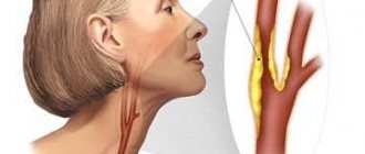
During the study, the patient is prohibited from turning his head or talking. First of all, the common carotid arteries are examined, since it is there that the accumulation of atherosclerotic plaques is most likely to occur. For indications such as genetic or anatomical abnormalities, additional functional tests may be prescribed:
- conducting the examination in a vertical position;
- holding your breath;
- use of antianginal medications (Anaprilin, Nitroglycerin).
The time interval for the procedure is from half an hour to 45 minutes. The examination protocol is given to the patient for further consultation with the doctor who referred for the ultrasound.
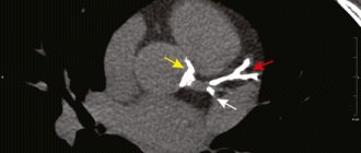
Atherosclerotic plaques are located mainly in the carotid arteries
Possible diagnoses
Using the results of the study, the doctor can make a diagnosis with extreme accuracy. The most common pathologies diagnosed by ultrasound of the BCA are:
- systemic damage to the arteries - atherosclerosis. The primary manifestation is recorded according to IMT indicators;
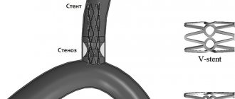
- narrowing of the vessel (stenosis) or blockage (occlusion);
- pathological tortuosity, otherwise deformation of the arterial bed;
- protrusion of the vascular wall (aneurysm);
- fistula (fistula) in the intervascular space;
- dissection or longitudinal tear and dissection of the arterial wall, accompanied by hemorrhage and hematoma;
- an inflammatory autoimmune disease characterized by thickening of the vascular wall, unevenly distributed (nonspecific aortoarteritis);
- steel syndrome (an anomaly in which the blood supply to the arm is carried out through the vertebral arteries);
- functional failure of the vertebral artery canals due to their compression (extravasal compression).
During the examination, the doctor has the opportunity to assess the dynamics of rehabilitation after surgical operations on blood vessels.
If the doctor has prescribed an ultrasound of the BCA, you cannot ignore the recommendations, because this is an examination with which you can predict the patient’s vascular health. The risk of stroke directly depends on the degree of narrowing of the BCA. Today, diagnostics using this method is available to most patients. Where to have an ultrasound done, in a large diagnostic center or a regional hospital, is decided by the patient himself. The cost of diagnostics does not exceed 3,000 rubles.
Preparing for sonography
For standard ultrasound scanning or color duplex scanning of the brachiocephalic arteries (CDS BCA), you need to clear the neck and face area of medicinal and cosmetic products. Before the procedure, you need to sit quietly for about 20 minutes. This normalizes breathing and heart rate.
1 hour to 3 days before the examination (by agreement with the doctor), stop taking medications that affect the arteries. These are antispasmodics, caffeine-containing drugs, eleutherococcus, ginseng, vasodilators, multivitamins, nootropics, medications to normalize blood pressure, and treat the heart.
The day before the examination, exclude from the menu:
- spicy food;
- seasonings, spices;
- energetic drinks;
- coffee Tea;
- alcohol;
- other types of food with tonic properties;
- salty food.
During the day before the procedure, it is not advisable to smoke or sniff tobacco, or be near people who smoke. It is not recommended to stay in stuffy rooms, visit saunas, or take hot baths. These factors affect blood flow and wall tone: duplex scanning will give a false result.
The requirements for preparation for DS are the same for ultrasound Doppler Doppler and examination of the brachiocephalic arteries in the triplex mode, Color Doppler Doppler.
Purpose of the study
Duplex scanning allows you to identify the following vascular pathologies:
- blood clots, atherosclerotic plaques;
- aneurysms;
- elongation, loops, bends, abnormal tortuosity;
- vascular stenosis;
- damage to the walls;
- underdevelopment of blood vessels;
- change in diameter (decrease or increase).
Duplex scanning makes it possible to determine:
- location of cholesterol plaques;
- degree of vasoconstriction;
- thickness, uniformity, surface shape and mobility of the arterial wall;
- blood flow speed and direction.
Thus, ultrasound examination of the BCA makes it possible to diagnose:
- thrombosis;
- vegetative-vascular dystonia;
- angiopathy;
- atherosclerosis.
DS BCA makes it possible to evaluate not only the main arteries, but smaller vessels, as well as the condition of cholesterol plaques and changes in surrounding tissues. Duplex scanning may be prescribed more than once during treatment to assess the effectiveness of the therapy.