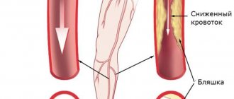
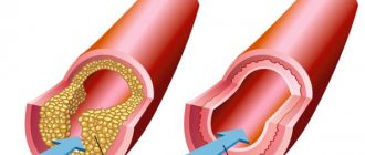
Obliterating atherosclerosis is one of the main diseases that causes impaired blood supply to the lower extremities. The disease is associated with the accumulation of cholesterol in the artery wall, forming plaques that block the lumen of the artery. The arteries become hard and their patency is impaired. This pathology can lead to thrombosis of the affected artery with the development of acute ischemia. The disease usually develops in old age, often against the background of diabetes mellitus, although now relatively young patients also turn to a vascular surgeon.
Lack of blood supply leads to dysfunction of the muscles of the limb, this is manifested by fatigue when walking for some distance, and with severe decompensation of blood circulation, gangrene of the leg can develop.
Treatment of atherosclerosis at the Innovative Vascular Center
Our center was created as a clinic for modern treatment of atherosclerosis and its complications. In our clinic you can consult an experienced vascular surgeon and undergo the necessary instrumental examination. Receive recommendations for conservative treatment of uncomplicated vascular lesions, but the main thing is to fully cure critical ischemia with the help of vascular surgery.
The methods of vascular surgery used in our clinic have no analogues in Russia in terms of their effectiveness in the treatment of critical ischemia against the background of obliterating atherosclerosis. We focus on minimally invasive and microsurgical interventions, which have not yet become widespread in our country. We are able to save a leg in case of critical ischemia due to atherosclerotic blockage in 98% of all patients. We achieve such results thanks to reasonable approaches to treatment, impeccable attitude towards the interests of our patients, collegial decisions taking into account the opinions of related specialists and excellent diagnostic and treatment equipment.
Causes of atherosclerosis of the vessels of the lower extremities
There is a complex of factors that contribute to atherosclerotic vascular damage , including the arteries of the lower extremities. There are non-modifiable (that is, unchangeable) and modifiable (modifiable) risk factors. Non-modifiable risk factors include:
- age (the older you are, the stronger the manifestation of atherosclerosis);
- gender (men suffer from atherosclerosis much more often than women);
- genetic predisposition (if there are strokes and cardiovascular diseases in the family history, atherosclerosis is more likely).
Main modifiable risk factors
:
- disorders of lipid metabolism (increased levels of total cholesterol in the blood. A distinction is made between low-density lipoprotein cholesterol and high-density lipoprotein cholesterol. The threat of atherosclerosis increases with an increased content of low-density lipoprotein cholesterol and, conversely, with a reduced content of high-density lipoprotein cholesterol.);
- smoking;
- diabetes;
- obesity, overweight;
- high blood pressure (accelerates the development of atherosclerosis);
- sedentary lifestyle (leads to metabolic disorders);
- poor nutrition;
- renal failure.
For the development of atherosclerosis of the arteries of the lower extremities, smoking and diabetes mellitus are of greatest importance.
Diagnostics
To confirm the diagnosis and clarify the severity of the disease, additional diagnostic methods are needed:
- Ultrasound Dopplerography of the arteries of the lower limbs with measurement of the pressure ratio on the arm and leg, which normally should be close to unity
- duplex scanning of arteries of the artery is a method that allows not only to assess the blood flow in the vessel, but also to see the vessel itself and changes in its walls
- conducting stress tests (treadmill test, i.e. walking on a treadmill under certain conditions)
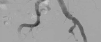
- according to indications, it may be necessary to conduct a contrast study of the arteries of the heart - multislice computed tomography or angiography of the aorta and arteries of the lower extremity
Surgical techniques
Surgery is a last resort and is usually prescribed for severe ischemia and very severe complications. Nowadays, different types of surgical interventions are performed. Some involve a day hospital, some require a long-term hospital stay under observation. Patient rehabilitation plans and post-operative care vary. Our doctors advise patients in detail on all aspects related to surgical intervention and carefully monitor their health in the pre- and postoperative period.
Surgical treatment of atherosclerosis of the lower extremities:
- bypass surgery - an additional “bypass” path for blood flow is created around the area of narrowing of the artery;
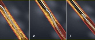
- stenting - a special tubular spacer is placed in the affected vessel, which ensures the required diameter of the artery;
- balloon angioplasty - similar to stenting, only a balloon is inserted into the vessel cavity rather than a spacer, which expands its lumen;
- autodermoplasty - if trophic ulcers are poorly treated conservatively, they are operated on and covered with the patient’s own skin;
- endarterectomy - removal of the affected part of the artery in which the atherosclerotic plaque is located;
- prosthetics – replacement of the affected vessel with the patient’s own vein, taken from another place, or with a synthetic prosthesis;
- amputation - prescribed in severe cases when gangrene occurs, after which prosthetics are performed.
In 75-85% of cases, after surgery, blood circulation is completely restored for an average of 5-8 years.
Indications for surgery:
- an aneurysm that may rupture;
- chronic ischemia of critical stage;
- hemodynamically significant carotid artery stenosis or plaque accompanied by symptoms of cerebral ischemia;
- decompensation of blood circulation in the leg due to embolism, trauma, thrombosis.
Contraindications to surgical treatment:
- wet gangrene with sepsis;
- severe disruption of vital organs - renal and liver failure, circulatory disorders in the brain, myocardial infarction, heart failure, etc.
Treatment
In the early stages of the disease, drug treatment aimed at preventing the development of atherosclerotic plaques and improving the rheological properties of blood is acceptable. For this purpose, statins and antiplatelet agents (aspirin) are used.
If the lumen of the vessel is so closed that the blood supply to the limb is disrupted, surgical treatment is necessary:
- balloon dilatation and stenting of the vessel (expansion of the narrowed part and installation of a stent to prevent re-narrowing of the lumen),
- endarterectomy (removal of plaque with the inner layer of the vessel),
- shunting (creating bypass paths for blood flow around an irreversibly damaged area of the vessel).
Prognosis and prevention
Source: PhotoMIX Company: Pexels
The effectiveness of treatment and the overall prognosis for obliterating atherosclerosis depend on the time of initiation of therapy, the degree of arterial damage and the presence of concomitant pathologies. Therapy is particularly difficult when other vessels, cerebral or coronary, are simultaneously affected.
The most dangerous complication of the disease is gangrene, which has a high probability of death. Its development can only be prevented by emergency amputation of the affected limb.
Prevention measures are aimed at reducing the risk of developing atherosclerosis and include the following recommendations:
- normalize your diet and give up high-fat foods;
- do not smoke or drink alcoholic beverages;
- devote time to moderate physical activity, such as swimming, cycling or even just walking;
- keep your feet clean;
- wear comfortable shoes with low heels;
- Avoid injuries and cuts to the skin on your legs.
Timely treatment can stop the development of the pathological process and significantly improve the patient’s quality of life.
Risk factors for OASNK
Atherosclerosis of the vessels of the lower extremities is characterized by the same risk factors as other arterial diseases, for example: coronary heart disease and cerebrovascular insufficiency.
- High blood pressure (hypertension),
- High blood cholesterol levels,
- Smoking,
- Sedentary lifestyle,
- Obesity,
- Burdened heredity.
A few words about smoking. Complete abstinence from any form of tobacco is necessary. Smoking even 1 cigarette per day of the lightest type is an unfavorable risk factor that causes the progression of obliterating atherosclerosis of the lower extremities and the development of its severe complications. The nicotine contained in tobacco causes the arteries to spasm, thereby preventing blood from moving through the vessels and increasing the risk of blood clots in them.
Vein diseases
Leg pain is accompanied by:
- Varicose veins;
- phlebitis and thrombophlebitis;
- postthrombophlebitic syndrome.
Varicose veins
This disease is widespread and is a very pressing problem in medicine and cosmetology.
Find out more about the promotion >>>
Phlebitis and thrombophlebitis
Phlebitis is inflammation of a vein. Thrombophlebitis is a complication of phlebitis or varicose veins of the lower extremities. With thrombophlebitis, a blood clot forms in the inflamed vein. Pain is a concern and swelling of the legs is possible.
Deep vein thrombosis is fraught with complications, the most dangerous of which are venous gangrene of the limb (with high blockage and blockage of all outflow pathways of venous blood) and pulmonary embolism. The risk of thromboembolism is highest in the presence of an unfixed - floating thrombus. The larger the detached fragment of the blood clot, the greater the likelihood of an unfavorable outcome, including death.
A common complication of deep vein thrombophlebitis is postthrombophlebitis syndrome.
Difference between stenosing and non-stenosing atherosclerosis
Atherosclerosis is a chronic vascular disease that develops with age, and all people are susceptible to it. At the initial stage, the plaques in the vessels are small, but if they do not block the vessel by more than half, this condition is called non-stenotic atherosclerosis. In this case, there are simply no signs of atherosclerosis, and the person feels great. As the cholesterol plaque grows, the vascular lumen becomes blocked—arterial stenosis occurs. In this case, organs deprived of the usual amount of oxygen and nutrients suffer from ischemia. This explains why the diagnosis of “stenotic atherosclerosis” occurs mainly in older and elderly people.
Symptoms
- A sharp weakening or disappearance of the pulse in the groin, popliteal fossa or foot
- Pain in the muscles of the lower leg (more often) and thigh, which regularly occurs when walking and goes away after a short rest (stop) - intermittent claudication
- Constant or periodic pain in the foot, requiring a forced position with the limb lowered down
- Coldness, paleness of the foot; in severe cases – the appearance of pronounced cyanosis (cyanosis), marbled spotted color and/or trophic disorders in the form of necrosis (tissue death)
The most striking symptom is the appearance of pain when walking, which forces the person to stop or slow down their walking speed. Most often, in the initial stages, the patient feels increased fatigue of the lower leg muscles, mainly along the back surface, then a pronounced sensation of painful compression appears. In some cases, similar sensations may occur in the thigh muscles and buttocks. As the disease progresses, walking speed decreases and the distance walked shortens. In addition, young men often experience a decrease in potency. All these symptoms are a reason to contact a vascular surgeon.
Symptoms of atherosclerosis of the vessels of the lower extremities
Atherosclerosis of the vessels of the lower extremities at an early stage can be practically asymptomatic. However, the absence of pronounced symptoms does not guarantee that atherosclerotic lesions are insignificant. Since lipid deposits in the walls of arteries do not immediately lead to disruption of blood flow, the development of atherosclerosis can go unnoticed for years.
If atherosclerosis develops slowly, the body compensates for the difficulty in blood flow through the main arteries with the help of collateral circulation, in which blood flows through the lateral branches of the arterial system. In this case, even a significant degree of atherosclerotic damage may not produce pronounced symptoms. The most striking symptom - intermittent claudication occurs only when the lumen of the artery is blocked by 80%.
The stage of development of atherosclerosis can be determined by how quickly pain occurs:
- Stage I
: pain in the legs occurs with significant physical activity. When walking calmly, pain occurs no earlier than after 1 km. ways. - Stage II
: pain occurs earlier than after 1 km. Moreover, if it arose more than 200 m later, we speak of stage II (A), and if less than 200 m later, we speak of stage II (B). - Stage III
: pain occurs every 25-50 m. At this stage, the legs hurt even at rest, especially at night. Patients usually lower their leg off the bed to improve blood flow and reduce pain. - Discomfort at rest is a critical indicator. The presence of discomfort indicates a high degree of stenosis or occlusion (complete closure of the lumen of the artery).
- Stage IV
is characterized by the appearance of ulcerative-necrotic changes. Feet become cold. Their hair falls out and their nails become deformed. Blackening of the skin occurs on the fingers and heels. Trophic ulcers develop. Possible gangrene.
Intermittent claudication
Intermittent claudication is pain in the calf muscles (less commonly, in the buttocks and thighs), as well as numbness and coldness of the lower extremities, forcing the patient to stop walking. It is enough just to stand in one place, the symptoms go away, and movement can continue.
Obliterating atherosclerosis
Obliterating atherosclerosis
| Obliterative atherosclerosis – occlusive-stenotic damage to the arteries of the lower extremities, leading to circulatory failure of varying severity. Obliterating atherosclerosis is manifested by chilliness, numbness of the feet, intermittent claudication, pain, and trophic disorders. Surgical methods include prosthetics , endarterectomy , thromboembolectomy , balloon angioplasty , and bypass surgery . |
- Obliterating atherosclerosis is a chronic disease of peripheral arteries, characterized by occlusive lesions and causing ischemia of the lower extremities. In cardiology and vascular surgery, obliterating atherosclerosis is considered as the leading clinical form of atherosclerosis (the third most common after ischemic heart disease and chronic cerebral ischemia). Obliterating atherosclerosis of the lower extremities occurs in 3-5% of cases, mainly in men over 40 years of age. Occlusive - stenotic lesions often affect large vessels ( aorta , brachiocephalic trunk , common carotid , iliac arteries ) or medium- sized arteries ( vertebral orifices , tibial , femoral ). In obliterating atherosclerosis of the arteries of the upper extremities the subclavian artery usually affected .
Stenosis of the arteries by more than 70% of the normal diameter leads to a change in the nature and speed of blood flow .
Factors predisposing to the occurrence of obliterating atherosclerosis :
- smoking,
- alcohol consumption,
- elevated blood cholesterol levels,
- hereditary predisposition,
- insufficient physical activity,
- nervous overload, menopause.
Obliterating atherosclerosis often develops against the background of existing concomitant diseases - arterial hypertension , diabetes mellitus ( diabetic macroangiopathy ), obesity , hypothyroidism , tuberculosis , rheumatism .
Local factors contributing to occlusive-stenotic damage to the arteries include previous frostbite and leg injuries. Almost all patients with obliterating atherosclerosis have atherosclerosis of the vessels of the heart and brain.
The appearance of plaques on the walls of the internal carotid arteries is fraught with dire consequences. Plaques are sites where blood clots form. This leads to complete blockage of the lumen of the arteries. A so-called ischemic stroke occurs. In addition, thromboembolism may develop. This condition occurs when a small blood clot breaks off from an atherosclerotic plaque, blocking the arteries of the brain. When blockage of small-caliber arteries occurs, a transient ischemic attack develops.
Classification of obliterating atherosclerosis
During obliterating atherosclerosis of the lower extremities, there are
4 stages:
- 1 – pain-free walking is possible over a distance of more than 1000 m. Pain occurs only during heavy physical activity.
- 2a - pain-free walking at a distance of 250-1000 m.
- 2b - pain-free walking at a distance of 50-250 m.
- 3 – stage of critical ischemia. The pain-free walking distance is less than 50 m. Pain also occurs at rest and at night.
- 4 – stage of trophic disorders. Areas of necrosis appear on the heel areas and on the toes, which can subsequently cause gangrene of the limb.
Taking into account the localization of the occlusal - stenotic process the following are distinguished :
- obliterating atherosclerosis of the aorto-iliac segment,
- femoropopliteal segment,
- popliteal tibial segment,
- multi-storey arterial damage. Based on the nature of the lesion, stenosis and occlusion are distinguished.
According to the prevalence of obliterating atherosclerosis of the femoral and popliteal arteries V types of occlusive - stenotic lesions are distinguished :
- I – limited (segmental) occlusion;
- II – widespread lesion of the superficial femoral artery;
- III – widespread occlusion of the superficial femoral and popliteal arteries; the area of trifurcation of the popliteal artery is passable;
- IV – complete obliteration of the superficial femoral and popliteal artery, obliteration of the fork of the popliteal artery; the patency of the deep femoral artery is not impaired;
- V - occlusive-stenotic lesion of the femoral-popliteal segment and deep femoral artery.
Variants of occlusive - stenotic lesions of the popliteal - tibial segment with obliterating atherosclerosis are represented by type III :
- I — obliteration of the popliteal artery in the distal part and the tibial arteries in the initial parts; patency of 1, 2 or 3 arteries of the leg is intact;
- II - obliteration of the arteries of the leg; the distal part of the popliteal and tibial arteries are patent;
- III - obliteration of the popliteal and tibial arteries; individual segments of the arteries of the leg and foot are patent.
Symptoms of obliterating atherosclerosis
The initial manifestations of obliterating atherosclerosis include chilliness and numbness in the feet, increased sensitivity of the legs to cold, “crawling goosebumps”, burning of the skin. Pain soon appears in the calf muscles when walking long distances, which indicates vasoconstriction and a decrease in blood supply to the tissues. After a short stop or rest, the pain subsides, allowing the patient to resume movement. Intermittent claudication or peripheral ischemia syndrome is the most constant and early sign of obliterating atherosclerosis.
With Leriche syndrome - atherosclerotic changes in the aorto-iliac segment, pain is localized in the muscles of the buttocks, thighs, and lumbar region. In 50% of patients, occlusion of the aortoiliac segment is manifested by impotence .
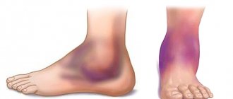
Tissue ischemia in obliterating atherosclerosis is accompanied by a change in the color of the skin of the lower extremities: at the beginning of the disease, the skin becomes pale or ivory; in the late stages of obliterating atherosclerosis, the feet and fingers acquire a purple-bluish color. There is atrophy of the subcutaneous tissue, hair loss on the legs and thighs, hyperkeratosis, hypertrophy and layering of the nail plates. Signs of impending gangrene are the appearance of non-healing trophic ulcers in the lower third of the leg or foot . The slightest damage ( bruises , scratches , abrasions , calluses ) of an ischemic limb can lead to the development of skin necrosis and gangrene .
In acute form of obliterating atherosclerosis (14%), obstruction of the artery site rapidly increases, trophic disorders develop rapidly and rapidly, up to gangrene. Patients require urgent hospitalization and limb amputation . _
In approximately 44% of patients, the clinical picture of obliterating atherosclerosis develops subacutely and occurs with recurrent seasonal exacerbations. In this case, a course of inpatient and outpatient treatment is carried out, which allows to slow down the progression of obliterating atherosclerosis.
The chronic form of obliterating atherosclerosis proceeds relatively favorably : due to the well-preserved patency of the great vessels and the developed collateral network, there are no trophic disorders for a long time. With this clinical variant, outpatient treatment provides a good therapeutic effect.
Diagnosis of obliterating atherosclerosis
The algorithm for diagnostic examination of a patient with suspected obliterating atherosclerosis includes:
- consultation vascular surgeon ,
- determination of pulsation of the arteries of the extremities , measurement of blood pressure with calculation of the ankle - brachial index ,
- Doppler ultrasound ( duplex scanning ) of peripheral arteries ,
- peripheral aorto-arteriography ,
- MSCT - angiography and MR - angiography
Treatment of obliterating atherosclerosis
- medicinal
- physiotherapeutic
- sanatorium
- angiosurgical treatment
To slow down the progression of atherosclerotic changes in the arteries , it is necessary to eliminate risk factors - correction of arterial hypertension , disorders of carbohydrate and lipid metabolism , quitting smoking , preventing foot injuries , hygienic and preventive foot care , wearing comfortable shoes .
Drug treatment of obliterating atherosclerosis is carried out with drugs that reduce erythrocyte aggregation (infusions of rheopolyglucin, rheomacrodex, pentoxifylline), antithrombotic drugs (aspirin), antispasmodics (papaverine, xanthinol nicotinate, no-spa), and vitamins. To relieve pain, analgesics, paranephric and paravertebral blockades are used.
In case of acute occlusion (thrombosis or embolism), the administration of anticoagulants (subcutaneous and intravenous heparin) and thrombolytics (intravenous streptokinase, urokinase) is indicated.
Physiotherapeutic treatment:
- hyperbaric oxygenation,
- physiotherapeutic (electrophoresis, UHF, magnetic therapy, interference therapy)
- balneological procedures (hydrogen sulfide, pine, radon, pearl baths; mud applications),
- ozone therapy,
- ILBI.
When trophic ulcers form , dressings are performed with topical preparations .
Surgical treatment of stage 2-3 obliterating atherosclerosis can be carried out through endovascular or open interventions .
Methods of revascularization the lower extremities include
- dilatation / stenting of affected arteries ,
- endarterectomy,
- thromboembolectomy,
- bypass operations ( aorto - femoral , aorto - iliac - femoral , ilio - femoral , femoral - femoral , axillary - femoral , subclavian - femoral , femoral - tibial , femoral - popliteal , popliteal - foot bypass ),
- prosthetics ( replacement ) of the affected vessel with a synthetic prosthesis or autovenous
Palliative interventions for obliterating atherosclerosis are carried out when radical surgical treatment is impossible and are aimed at strengthening collateral circulation in the affected limb. These include lumbar sympathectomy, revascularizing osteotrepanation, periarterial sympathectomy, etc.
At stage 4 of obliterating atherosclerosis, amputation of a limb to an optimal level that takes into account the boundaries of ischemic disorders most often indicated .
The least invasive method of surgical treatment peripheral artery disease is the technique of angioplasty and stenting . It is used if large arteries have been damaged. Angioplasty involves inserting a flexible catheter into the arterial lumen through the femoral vein. After this, a conductor is inserted, delivering a special balloon to the place where the vessel is narrowed. By inflating the balloon, the normal lumen of the vessel is restored.
In more serious cases, arterial bypass surgery . For this purpose, an additional vessel is created. Blood flow passes through it, bypassing the affected area of the artery. Both artificial prostheses and the patient’s veins are used for the shunt.
The endarterectomy method involves surgical removal of the atherosclerotic plaque. To do this, you need to open the artery. However, it is important to consider that such a procedure may disrupt the overall blood flow through the artery. Consequently, the advisability of using endarterectomy is determined taking into account the location of the lesion and the degree of disruption of blood flow in a particular artery.
The absolute indications for surgical treatment are :
1. Chronic critical limb ischemia with patent arteries of the leg .
2. Aneurysm with threat of rupture .
3. Hemodynamically significant (> 60%) stenosis of the internal carotid artery or ulcerated plaque in the presence of symptoms of cerebral ischemia .
4. Embolism , thrombosis or vascular injury with decompensation of blood circulation in the limb .
Other indications for surgery , such as intermittent claudication corresponding to stage II b , sharply reducing the quality of life and not correctable by other treatment methods , asymptomatic stenosis the carotid arteries more than 60%, small abdominal aortic aneurysms , etc. , are considered relative and determined the general condition patient and the capabilities of the medical institution .
Contraindications to surgical treatment :
- wet gangrene with septic condition ,
- the presence of severe dysfunction of vital organs that makes surgical intervention impossible ( myocardial infarction , cerebrovascular accident , heart failure , low coronary circulatory reserve , respiratory , renal , liver failure )
Endarterectomy , as a rule, is performed on patients with segmental arterial occlusions not exceeding 7–9 cm in length. Open endarterectomy is performed within the arteriotomy wound and involves removing the altered intima along with plaques and mural thrombi.
Endarterectomy can be performed in a semi-closed manner: the obliterating masses are peeled off and removed using long vascular rings or other devices. With a significant spread of the occlusive process and severe calcification, endarterectomy is ineffective. In these cases, shunting of the affected segment , the purpose of which is to restore blood flow bypassing the affected area. Prosthetics are performed in patients who require resection of the altered vascular wall.
For occlusive lesions the branches of the aortic arch open endarterectomy most often performed from the common and internal carotid arteries or from the mouth of the vertebral artery .
Carotid endarterectomy , the operation of choice for atherosclerotic lesions of the carotid arteries, has become the standard of preventive surgery.
The surgical operation consists of removing the atherosclerotic plaque. The operation is performed under anesthesia. Its essence is to dissect the tissue above the carotid artery, isolate the vessel, dissect the wall of the carotid artery, and remove the plaque. After this, the vessel is sutured.
This operation is more traumatic, usually takes longer, is performed under anesthesia and carries more complications. The sutures are removed on the 7th day, patients are discharged with a normal course of the postoperative period on the 9-10th day. According to modern international requirements, the level of complications in a clinic where such operations on the carotid arteries are performed should be no higher than 5%. This means that 5 out of 100 patients experience various complications. The operation is contraindicated in the presence of serious somatic diseases (diabetes mellitus with high sugar levels, arterial hypertension, etc.)
Possible postoperative complications of carotid endarterectomy
As with any other surgical operation, complications are possible after carotid endarterectomy. the most serious of these is stroke. The risk of its development is 1–3%. In addition to stroke, there is a complication such as repeated blockage of the carotid artery, called restenosis . It most often occurs in those patients who have not given up smoking. The risk of developing restenosis is 2-3%. Another complication is nerve damage, which leads to voice disturbance (hoarseness), difficulty swallowing, and numbness in the face or tongue. Usually these complications do not require special treatment and resolve on their own within a month.
In case of occlusion of the subclavian artery, leading to the development of subclavian steal syndrome, the operation of choice is carotid - subclavian bypass or resection of the subclavian artery with implantation into the common carotid .
In case of widespread damage to the main arteries of the aortic arch (brachiocephalic trunk, common carotid), their resection with prosthetics or bypass surgery .
open endarterectomy is also most often performed ; in case of widespread damage bypass surgery ( prosthetics is performed .
In the treatment of occlusive lesions of the abdominal aorta and arteries of the lower extremities, the most popular operations are aorto - femoral ( iliac - femoral ) bypass and femoral - popliteal ( femoral - tibial ) bypass . In the aortofemoral position, the most widely used are synthetic prostheses made of fluorlon-lavsan, dacron and polytetrafluoroethylene (PTFE), the five-year patency of which is 85–90%.
| Endoprosthetics at aneurysm peripheral arteries – a method of minimally invasive X-ray surgical treatment of local asymmetric bulging of the arterial wall by installing an intravascular endoprosthesis. Aneurysms of peripheral arteries are a consequence of inflammatory, atherosclerotic, traumatic changes in the walls of the arteries. The endoprosthesis is installed into the lumen of the peripheral artery using the X-ray endovascular method using a special delivery catheter. After implantation of the endoprosthesis, the aneurysmally altered wall is isolated from the blood flow and is not subject to pressure, which eliminates the rupture of the aneurysm. |
Nowadays, reconstructive surgery of the abdominal aorta, carotid arteries, arteries of the lower extremities with atherosclerosis is considered one of the most developed sections of angiosurgery. Despite this, the results of the operations are still far from perfect. The frequency of early postoperative thrombosis of grafts or reconstructed arteries can be 4–13 % , late reocclusions – 8.5–30 % for the aorto - iliac and 22–60 % for the femoral - popliteal segments . In 10% of patients, attempts at reconstructive surgery end in amputation of limbs in the early postoperative period .
Throughout the world, the last decade has been a time of rapid development of X-ray endovascular surgery - one of the highest priority areas. The endovascular technique has emerged as a beneficial alternative to open surgery.
Intravascular ( endovascular ) stents are intraluminal retention devices that look like a thin mesh of metal threads that are strong enough to withstand the resistance of the arterial wall and maintain good patency of the recanalized area .
Preoperative preparation and postoperative management of patients usually carried out according to the following scheme .
The day before stenting, ticlid is prescribed at a dose of 500 mg/day. During the intervention, rheolytics and anticoagulants are administered, symptomatic therapy is carried out (antispasmodics, atropine); After the intervention, patients receive low molecular weight heparin (fraxiparin 0.3–0.6 2 times a day for 3 days), symptomatic therapy continues for 3–5 days. Patients are discharged 3–7 days after surgery. Patients take antiplatelet agents (ticlid or plavix, and in some patients aspirin) for at least 1 month. after the intervention.
According to indications ( if thrombosis or restenosis is suspected in stenting area or if lesions of previously unoperated arteries are detected ) , CTA and / or control angiography is performed .
If these changes are detected , multiple repeated endovascular interventions can be successfully used , thereby significantly delaying the time when a patient with progressive occlusive lesions will require traditional surgical intervention , or avoiding it altogether .
The range of diseases that are subject to minimally invasive treatment in the first place :
l Lesions of an isolated nature ( segmental stenoses , short occlusions ;
l Lesions that are difficult to access for open surgical interventions ( renal arteries , branches the aortic arch , visceral arteries );
l Restenosis after traditional operations , stenosis of vascular anastomoses ;
l Severe concomitant diseases that increase the risk of traditional operations .
Complicated ( embolic-dangerous ) atherosclerotic plaques , as well as a combination of stenotic lesions of the carotid arteries with kinks , tortuosity and looping , accompanied by significant lengthening of the artery and requiring traditional reconstructive surgery are considered a contraindication to stenting .
Stenting of the vertebrobasilar arteries has proven itself in surgical practice and is already the method of choice . Balloon angioplasty and stenting are indicated for stenosis and occlusion of the proximal part of the subclavian artery, accompanied by significant compromise of the vertebrobasilar blood flow, up to the development of steal syndrome, and damage to the brachiocephalic trunk.
Stenting of the arteries of the lower extremities is indicated in patients starting from stage II of ischemia according to the Fontaine-Pokrovsky classification. The ideal type of lesion for stenting is a short concentric stenosis or an isolated occlusion of less than 5 cm in length for the iliac artery and less than 10 cm in length for the superficial femoral artery. The most commonly performed stenting is the iliac, superficial femoral, and popliteal arteries.
In the late postoperative period, due to the progression of atherosclerotic changes in the proximal or distal vascular bed, repeated stenting is possible. Stenting can be used for anastomotic stenoses after previously performed bypass operations. Renal artery stenting is the most advantageous location
Preparation for carotid artery stenting surgery
Typically, preparation for carotid stenting involves taking aspirin a week before surgery. This is necessary to reduce blood clotting.
Are you a candidate for carotid stenting ?
Currently, carotid stenting surgery is indicated for patients at high risk of complications from endarterectomy. Indications for carotid stenting include significant narrowing (60%) of the lumen of the carotid arteries, symptoms of microstroke and stroke. If you do not have any symptoms, the indications for stenting are significant narrowing (80%) of the lumen of the carotid arteries and a high risk of complications from endarterectomy. In addition, carotid stenting is indicated for patients who have previously undergone endarterectomy in cases of recurrent narrowing of the arterial lumen.
Carotid stenting is not recommended for :
- Presence of abnormal heart rhythm
- Allergy to drugs used during the procedure
- Brain hemorrhages within the previous 2 months
- Complete blockage of the carotid artery
Risk factors for complications of carotid stenting
- High blood pressure
- Allergy to