Probing (or “catheterization”) of the heart is an invasive technique that is practiced in cardiac surgery and is aimed at inserting a catheter into the right or left parts of the organ for diagnostic purposes. In the process, the surgeon creates a puncture in the brachial or femoral artery (to diagnose left cavities) or the ulnar or femoral vein (to diagnose right cavities). A catheter is inserted through it under the control of an X-ray machine and ECG. The procedure allows you to evaluate the condition of the septum and valves of the heart, take blood samples and conduct chamber studies with the introduction of a contrast agent.
The Department of Cardiovascular Surgery CELT invites you to undergo probing of the heart cavities in Moscow. It began its work almost three decades ago and today offers a wide range of services for the diagnosis and treatment of cardiovascular diseases. It welcomes leading domestic specialists, candidates and doctors of sciences with decades of scientific and practical experience. They have modern equipment in their arsenal that allows them to accurately diagnose and carry out treatment in accordance with international standards.
Probing of the heart cavities - 21,000 rubles.
At CELT you can get a consultation with a cardiovascular surgeon.
- Initial consultation – 3,000
- Repeated consultation – 2,000
Make an appointment
Probing of the heart cavities: indications and contraindications
Today, catheterization of the cardiac cavities is one of the most frequently performed procedures in cardiac surgery. In European countries alone, it is performed on almost a million patients every year! The use of modern equipment and the high qualifications of our specialists allow us to minimize the risk of complications after surgery. Depending on the situation, it can be not only diagnostic, but also therapeutic.
Indications
- Assessment of left ventricular function and determination of the degree of ischemic disease;
- Diagnosis of congenital and acquired heart defects;
- Determination of narrowing or insufficiency of the aorta before surgery;
- Confirmation/exclusion of the diagnosis of ischemia in patients who suffer from chest pain of unknown etiology;
- Collection of data during the diagnostic process that confirms and complements the results of other studies;
- Diagnosis of pulmonary hypertension, determination of its type and severity;
- Assessing the possibility of heart or lung transplantation.
Contraindications
There are no absolute contraindications to this procedure. All of them are relative and depend on the individual indications of the patient. Whether to carry it out or not is decided by the doctor himself, assessing and comparing the risks. He may refuse it altogether if:
- The patient's blood electrolyte levels are abnormal;
- There is a diagnosis of acute renal failure;
- The patient suffered an acute stroke;
- Gastrointestinal bleeding was detected;
- The patient has an individual intolerance to contrast agents;
- The level of cardiac glycosides in his blood exceeds the norm.
Our expert Burkert Pieske is director of the Clinic for Internal Medicine and Cardiology at the German Heart Center in Berlin. Private practitioners in Berlin most often recommend this clinic to their patients for inpatient treatment using a catheter for cardiac catheterization (a 2015 survey of doctors conducted by the Tagesspiegel newspaper and the non-profit organization City of Health Berlin). Author: MAGDALENA WEBER
EXPLANATION The cardiac catheter (catheterization) is a very flexible plastic probe up to 1.50 meters long and about three millimeters thick. With this probe, doctors can both recognize and treat heart disease. For example, it is often used for coronary heart disease: If the thin coronary vessels of the heart (coronary arteries) become severely narrowed due to fatty deposits or calcification, they can no longer supply the heart muscle sufficiently with oxygen and nutrients. As a consequence, the function of this vital muscle, the contraction of which makes possible the pumping function of the organ - the heartbeat, so to speak - is increasingly limited. If the vessels are completely clogged, then the muscle tissue for which they are responsible for the blood supply dies and this leads to the development of a heart attack. By using a cardiac catheter, doctors can detect these narrowings and widen them again so that blood can flow freely again. To make sure that the vessels remain patent after the procedure, doctors may additionally implant a stent. In this case, we are talking about a support structure for blood vessels, which, with the help of a catheter, is advanced to the desired area in the heart.
Coronary heart disease is the most common heart disease, and, above all, in industrialized countries the most common disease in general. In Germany, almost five percent of people have such circulatory problems caused by deposits in the coronary arteries. “In the group of people over 60 years of age, this percentage is probably even higher,” says Burkert Pieske, director of the Clinic for Internal Medicine and Cardiology at the German Heart Center in Berlin and at the campus of the Charité University Hospital Virchow-Klinik in the Berlin-Wedding district. However, only a general assessment is possible here, since not every sick person immediately causes symptoms of the disease. However, in general, coronary heart disease is one of the most common causes of death, especially in developed industrial countries.
The catheter (1) is a plastic probe up to 1.50 meters long and with a diameter the thickness of spaghetti. It is injected into the coronary vessels of the heart (2), also called the coronary arteries, through the aorta. Through this access, the doctor can inject a contrast agent, due to which the vessels become visible on an x-ray, including visualization of narrowings (3) in these vessels. Around the catheter is a balloon, above which is a covered stent (white lattice). DRAWING BY FABIEN BARTEL
Around the catheter is a balloon, above which is a covered stent (white lattice). To dilate narrowed blood vessels, the doctor places a catheter with a tiny balloon (4) at the tip to the affected area. There the balloon is inflated and thus the narrowing opens. To prevent the vessel from immediately becoming blocked again, a stent can be used (5). The support structure for the vessel opens together with the balloon, is pressed against the wall of the vessel and secured there. DRAWING BY FABIEN BARTEL
SYMPTOMS A typical symptom of coronary heart disease is the so-called angina pectoris (Angina pectoris). The name Angina pectoris comes from Latin and means “angina pectoris”. Indeed, in addition to pain in the heart area, which often occurs in the form of attacks, for example, during physical activity, in many cases a dull, pressing feeling in the chest also occurs. The pain may radiate to other parts of the body, such as the neck or arms. During such an attack, many patients suffer from lack of air, they feel weak, sweating increases, and pallor appears. Feelings of anxiety may also occur. In addition, angina also manifests itself in the form of intense stomach pain, nausea, poor performance and fatigue, especially in women. Typical chest pain radiating to the left arm is less pronounced in women.
“At first, angina pectoris occurs mainly during physical exertion or in a state of excitement,” says Mr. Pieske. “When the person calms down, the symptoms go away again.” However, if an attack occurs even at rest, this is clearly a warning sign - and an emergency. “In this case, the patient should definitely go to the hospital, as he is at risk of a heart attack.”
CAUSES Coronary heart disease usually develops over a longer period of time. This is why this disease primarily affects older people. Indeed, throughout life, substances such as cholesterol or calcium are deposited on the walls of the arteries - and, consequently, in the coronary vessels. These accumulations of fat and calcium can stick together and form a relatively hard mass that doctors call atherosclerotic plaque. “Atherosclerotic plaques damage the vessel and make it harder, so that it can no longer expand, for example if the heart muscle needs more blood during exercise,” says cardiologist Mr Piske. The more this calcification of the arteries (arteriosclerosis) progresses in the coronary vessels, the narrower the vessels become - and the worse the blood supply to the heart muscle.
For men, age over 45 years is a risk factor that contributes to the disease; for women, it is age over 55 years. This difference is likely hormonally determined. So experts proceed from the fact that the female hormone estrogen performs a kind of protective function in relation to the heart and blood vessels. However, exactly how this process functions remains unclear.
Other risk factors that contribute to the development of coronary heart disease, along with advancing age, are primarily excess weight, elevated blood lipids, high blood pressure and diabetes. “Smoking also has a very negative effect on blood vessels,” says Mr. Pieske. Therefore, smokers often suffer from coronary heart disease at a young age.
DIAGNOSIS To prevent life-threatening consequences such as heart attack, it is very important to recognize coronary heart disease - or an increased risk of developing the disease - as early as possible. Therefore, it is recommended to carry out regular preventive examinations - even in the absence of acute complaints. For example, for patients over 35 years of age, health insurance funds pay for a general preventive medical examination by a family doctor every two years, which also includes important cardiac tests such as measuring blood pressure or determining blood cholesterol levels.
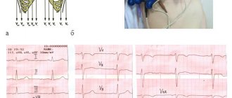
When diagnosing coronary heart disease, we first of all talk about a conversation with the patient (history taking), during which the doctor asks questions about the presence of possible risk factors and complaints. This is usually followed by a physical examination and blood tests, blood pressure measurements, and an electrocardiogram (ECG), which can visualize the heart's electrical currents. To substantiate the initial suspicion, other methods are used, such as an exercise ECG (ergometry) or ultrasound examination of the heart (echocardiography).
To determine whether there is insufficient blood supply to the heart during exercise, doctors may also take magnetic resonance imaging (MRI) images or do what is called a myocardial perfusion (blood supply) scintigraphy. In this process, the blood supply to the heart is visualized using a slightly radioactive substance that is injected into the bloodstream. Under certain circumstances, a computed tomography (CT) scan may be performed.
And finally, another informative diagnostic method is a study using a catheter to probe the heart, the so-called angiography. In this study, doctors use a thin needle to access an artery in the groin or shoulder joint under local anesthesia. “From there, a thin catheter probe is advanced to the heart,” says Mr. Pieske. The entire procedure is carried out under x-ray control - in addition, x-rays also allow you to see possible risk areas. For this purpose, a contrast agent is injected into the blood, making the blood vessels visible on the X-ray screen. If doctors find areas of narrowing in the coronary vessels of the heart, they can immediately treat them on the spot with a catheter.
TREATMENT Incipient coronary heart disease can usually be treated with medications. In addition, patients should minimize risk factors such as smoking and excess weight. However, if the coronary arteries are already narrowed to such an extent that these measures are no longer enough or there is even a threat of a heart attack, then in this case a catheter is used to probe the heart: with its help, doctors can dilate the affected vessel again. “To do this, the empty balloon is advanced with the help of a catheter to the site of narrowing,” says Dr. Pieske. There it is “inflated” under high pressure and thereby pushes the walls of the vessel apart - the artery again becomes passable. To ensure that the vessel continues to remain patent, doctors can use a catheter to install a support structure for the vessel - a stent. Often, dilation of the artery and implantation of the stent are performed in one surgical procedure.
There are several types of vascular support structures: Stainless steel drug-eluting stents are designed to provide long-term prevention of the re-formation of atherosclerotic plaques. Another form is the so-called absorbable stents. They consist of a sugary substance and dissolve on their own over time. This way, the foreign body does not remain in the heart for a long time, however, according to Mr. Piske, this method is not suitable for all patients: “If the coronary arteries are already heavily calcified and hardened, they can crumble the self-resorbable stent.” Therefore, stents made of steel are usually used. To prevent rejection reactions or re-narrowing of the vessel, stents are coated with drugs that are slowly released into the blood. To avoid complications such as thrombosis in the stent and in the long term, after implantation, patients must take medications to inhibit blood platelets (platelets), which are important for the blood clotting process.
Currently, cardiac catheterization and stent implantation have already become standard procedures for cardiologists. “However, there are, of course, certain risks,” says expert Pieske. For example, at the puncture site in the groin or elbow area, minor damage may occur, resulting in bruising. More serious - but also very rare - are possible catheter injuries to large vessels on the way to the heart or to the coronary vessels themselves, which in extreme cases can lead to a stroke or heart attack. In addition, another intervention may be required, either because deposits will reappear in the operated area and re-narrow the vessel (re-stenosis), or because a narrowing will occur in another area in one of the three coronary vessels hearts.
Source - https://www.tagesspiegel.de/themen/arztbriefe/arztbrief-herzkatheter/13465636.html
Types of catheterization of cardiac chambers
There are two of them: catheterization of the right and left parts of the heart.
| Catheterization of the left chambers of the heart | Catheterization of the right chambers of the heart |
Allows you to determine the anatomical features of the structure of the coronary artery and identify coronary artery disease. Thanks to it, you can determine and evaluate the following parameters:
| Allows you to evaluate the following parameters:
The procedure is used for heart transplantation or mechanical support. Thanks to it, you can estimate the pressure in:
|
What diseases can be detected by ultrasound of the heart?
During the examination, the specialist evaluates the following parameters:
- presence or absence of noise;
- quality of the organ's work;
- size and structure of the heart chambers;
- the presence of blood clots, tumors and other formations;
- the presence of inflammation, consequences of infections;
- pericardial condition.
The state of the heart in the contraction and relaxation phases is also analyzed. All received information is processed by a special program.
Ultrasound examination of the myocardium helps diagnose diseases such as:
- hypertension;
- rheumatism;
- myocardial infarction or all existing prerequisites for this dangerous pathology;
- pericarditis;
- hypotension;
- myocarditis;
- various heart defects;
- ischemia;
- vegetative-vascular dystonia;
- diseases of the aorta, arteries;
- rhythm disturbances.
Also, hardware diagnostics will allow you to determine intracardiac formations, heart failure, cardiomyopathy, and dilation of cavities.
Preparation for probing the cavities of the heart
It is necessary to ensure that this procedure does not lead to any complications. Within its framework, CELT specialists prescribe the patient to undergo a number of diagnostic tests:
- General and biochemical blood tests;
- Chest X-ray;
- Electro- and echocardiography;
- Magnetic resonance imaging of the heart.
The above allows you to determine the levels of platelets and hemoglobin in the blood, as well as the correct functioning of the kidneys, liver, heart, lungs, and aorta. The patient must inform the attending physician about his individual intolerance to seafood, contrast agents, iodine-containing and any other pharmacological drugs. In addition, he should be informed if he is taking medications for erectile dysfunction or if he is pregnant.
Additional preparation for cardiac probing is required for those under one year of age or over seventy years of age, as well as for patients who suffer from:
- Type 1 diabetes mellitus;
- Diseases of the blood vessels of the brain;
- Severe pulmonary or renal failure.
Heart ultrasound at home for children
Ultrasound examination is a safe diagnostic method that is allowed for children from infancy. An echocardiogram is prescribed to the baby immediately after birth; the procedure helps to assess the health of the newborn’s cardiovascular system, identify or exclude various dangerous pathologies and defects.
For older children, myocardial ultrasound is prescribed for the following indications:
- the presence of noise, rhythm disturbances detected when listening with a stethoscope;
- weak weight gain;
- shortness of breath;
- the appearance of cyanosis after exertion, strong screaming or intense sucking;
- swelling of the lower extremities;
- coldness of hands and feet;
- fast fatiguability;
- pain in the chest area.
Also, studies are carried out as prescribed by the pediatrician. Preventive examinations are prescribed at 1 month, 1 year, 7 and 14 years.
Ultrasound in pediatrics allows us to identify pathologies such as:
- heart attack;
- congenital and acquired defects;
- inflammatory processes;
- blood clots in the lumens of blood vessels and cavities of the heart;
- tumor formations.
An echocardiogram will help quickly and safely identify dangerous diseases at different stages of development. The procedure does not require special, complex, or preliminary preparation. The main thing is that the child is not nervous and is psychologically prepared for the upcoming medical event.
If the child has left infancy before the study, you should not overfeed him or give him a lot of drink. But it is better to feed the baby right before the examination, this will calm the baby and allow the procedure to be carried out comfortably.
Technique for probing the cavities of the heart
The patient assumes a supine position, the medical staff fixes electrocardiography electrodes on him, installs an intravenous infusion system and starts the administration of a five percent glucose solution or sodium chloride solution. Next steps:
- After anesthetizing the area where the catheter is inserted, the surgeon makes a small incision in the skin and inserts it into the blood vessel, carefully moving it towards the heart and checking his actions through an X-ray machine;
- Once the catheter has reached the desired location, he injects a contrast agent, which allows for good visualization of the cardiac structures and arteries;
- During the procedure, basic physiological indicators (heart rate and contractions, blood pressure and respiratory rate) are monitored every fifteen minutes;
- Upon completion of the diagnosis, the doctor removes the catheter and applies a tight bandage for half an hour.
If the patient begins to feel nauseous during the procedure, they are asked to cough to reduce the effect. Sometimes, after the administration of contrast, arrhythmia may develop, which can be eliminated by deep breathing. It also makes it possible to easily install a catheter into the pulmonary artery and makes visualization of the heart clearer.
How to call an ultrasound diagnostic doctor at home
In order to conduct an ultrasound examination at home, you need to call and leave a request to the operator.
The specialist will ask a number of questions related to the existing symptoms, ask for personal information, ask when it is best to schedule a doctor’s visit, and record the patient’s address. The doctor will arrive at exactly the appointed time and conduct the examination in a comfortable environment for the patient. It is advisable to prepare a place for diagnosis, a medical card, extracts and certificates before the specialist arrives.
Examination at home is practically no different from examination in a hospital. Only in this case the patient is in more comfortable conditions, which can have a positive impact on the accuracy of diagnosis.
Why should you contact the ultrasound department of our clinic?
Expert level doctors
Doctors combine travel with practice in a medical center and hospital, maintaining the skills of complex patient treatment.
The doctor will arrive within 2 hours
You can also schedule a doctor’s visit at any time from 8 am to 11 pm in Moscow and the Moscow region.
Expert class equipment
Ultrasound is performed using expert-class equipment manufactured by General Electric, SONY, Mindray.
Tests and ultrasound on the day of treatment
Tests, x-rays, ultrasound with interpretation on the day of treatment.
Consultations for adults and children
We help adult men and women, as well as babies from the first days of life.
Experienced doctors
Our experienced doctors with over 15 years of experience. Candidates of Medical Sciences.
Reviews of doctors providing the service - cardiac probing
I had surgery on 2 legs with Yuri Stanislavovich Malakhov.
I want to express my deep gratitude to him for a new breath of life, lightness on my feet, one might say, a completely different standard of living. Yuri Stanislavovich, you have golden hands and you are a doctor from God! I recommend it to everyone, women and men - don’t be afraid and... Read full review Irina Nikolaevna G
11.06.2021
I would like to sincerely thank the surgeon-phlebologist Yuri Stanislavovich Malakhov for the high-quality treatment of varicose veins using a modern method: no incision, no hospitalization, high cosmetic effect. The very next day I went to work. Yuri Stanislavovich is a professional... Read full review
Natalia Viktorovna F
18.04.2021
Advantages of performing cardiac ultrasound at home
Paid ultrasound examination of the heart in Moscow has a number of advantages:
- comfortable diagnostic conditions;
- affordable prices;
- no need to contact strangers and wait in line.
The service will become an indispensable option for elderly patients and people with disabilities. It is also the optimal choice for sedentary patients, citizens who have had a heart attack or complex surgery. The hardware procedure at home will be appreciated by parents of small children, business people and constantly working people.
Ultrasound at home eliminates the need for transportation and allows the doctor to conduct a more thorough examination, since at home he will not be distracted by colleagues and other patients.
Calling a sonologist to your home allows you to undergo the procedure in the most comfortable conditions, which will create a positive atmosphere and eliminate additional anxiety and nervous tension.
How to prepare for the procedure?
After prescribing cardiac catheterization, the specialist must tell the patient all the examination techniques and warn about possible complications and adverse reactions.
After this, the patient is given documents confirming consent for catheterization and all the basic recommendations on preparing for the diagnostic procedure.
The importance of preparation
It is especially important to carefully prepare the patient for the examination in the following cases:
- the presence of dangerous pathological diseases (insulin-dependent diabetes, pulmonary and renal failure, serious diseases of the brain and peripheral vessels);
- heart failure;
- severe disturbances in the left ventricle;
- children or old age.
In the presence of the described conditions, it is important to carry out cardiac catheterization with extreme care; in the presence of such lesions, the risk of death greatly increases.
When is it appointed?
Diagnostic cardiac catheterization may be prescribed in the presence of the following diseases:
- congenital heart defects;
- diseases of the cardiac system (valve damage);
- ischemic diseases;
- cardiomyopathy;
- heart failure;
- mild form of hypertension;
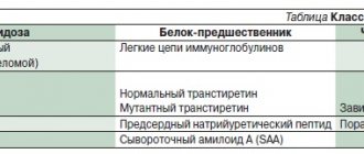
- cardiac amyloidosis.
The diagnostic measure helps to accurately identify the type of damage to the coronary vessels, myocardial tissue and heart valves, which cannot be determined using simpler examinations (or when they show an inaccurate result). In addition, the procedure for probing cardiac vessels helps to fully assess the severity of damage and examine the pathophysiological mechanisms of changes in myocardial function. Most often, this diagnostic method is prescribed to patients undergoing cardiac surgery.
Expert advice
When prescribing venous cardiac catheterization, the patient must inform the doctor about the following factors:
- bearing a child, especially in the early stages;
- use of medications or dietary supplements;
- taking glucose-lowering medications;
- allergies to iodine, radiopaque agents, seafood, rubber or latex;
- the use of Viagra and other medications that are aimed at restoring the condition of the reproductive system.