“Asystole” literally means “absence of systole” or cardiac arrest. Simply put, this is a state of clinical death. There are 2 possible variants of sudden fatal rhythm disturbances - ventricular fibrillation and asystole. It is impossible to distinguish them in the clinic, and there is no time, since there are only a few minutes for survival.
At this time, a person’s life is in the hands of the people around him and depends entirely on the desire and ability to provide first aid. According to research by resuscitators, 2/3 of cases of sudden death that occur in different places on the street occur from ventricular asystole.
Why does the heart stop?
The most common cause of cardiac arrest is chronic or acute diseases with a severe stage of decompensation and organic changes in the myocardium. Depending on the cardiac pathology, all causes are divided into primary and secondary. Primary cardiac diseases include:
- acute widespread myocardial infarction and its complications (cardiogenic shock, pulmonary embolism, aneurysm rupture);
- sudden disturbances in the rhythm of ventricular contraction (paroxysm of atrial fibrillation, ventricular tachycardia, some types of ventricular extrasystoles);
- rupture of aortic aneurysm;
- heart defects in the stage of decompensation;
- acute cardiovascular failure.
Risk factors contributing to asystole include:
- elderly age;
- excess weight;
- concomitant diabetes mellitus;
- drinking alcohol and smoking;
- increased blood cholesterol levels;
- When comparing gender, men are the most disadvantaged.
Secondary causes can occur suddenly in young people, without myocardial damage. They are caused by diseases such as:
- sudden stroke with extensive damage to the cerebral cortex;
- respiratory failure caused by long-term unresolved severe asthmatic condition;
- damage to internal organs in the coma stage with renal and liver failure, diabetes mellitus, leukemia, anemia;
- malignant tumors in the terminal phase.
In addition to diseases, secondary causes can be caused by accidents or injuries:
- suffocation due to foreign body entering the larynx or bronchi;
- poisoning by chemicals and poisons;
- drowning;
- extensive injuries in emergency situations, during man-made and other disasters, in everyday life, accompanied by traumatic shock;
- massive burns;
- large blood loss;
- electrical injury due to high voltage current.
Extracardiac causes
Represented by a large group of phenomena leading to secondary asystole:
- Pneumothorax. Penetration of air into the chest. Normally, it is sealed, which ensures the existence of organs in ideal conditions. Atmospheric gases provoke compression of the heart and lungs. They reduce the efficiency of hemodynamics, and in the long term even lead to the impossibility of normal functioning of cardiac structures. This is not an axiom; there is no 100% probability of such an outcome. Depends on the case and the volume of air penetrated.
- Hypoxia. Disruption of normal gas exchange in tissues. It leads to cardiac asystole relatively rarely, only if the process reaches a certain critical point. This requires a severe disease of the respiratory system or heart. Emergency care requires restoration of normal organ function. But in the case of severe somatic illness, the prospects are pessimistic.
- Electrical injury. Normally, cardiac structures work autonomously: they themselves generate an impulse, conduct it, and contract. As a result of exposure to a significant current, a disruption of the normal rhythm is observed. This ends with the sinus node first generating a signal chaotically, and then stopping producing it altogether. Spontaneous contractions of the ventricles continue for several seconds, then asystole occurs - cardiac arrest.
- Increased blood acid level. Biochemical abnormality. Occurs as a result of metabolic disorders. Relatively rarely leads to such a terrible phenomenon.
- Poisoning with salts of heavy metals, antihypertensive drugs, sleeping pills, tranquilizers, cardiac glycosides, psychotropic medications, insect poisons and others. Intoxication leads to blockage of cellular respiration, acute ischemia of cardiac structures and primary asystole occurs. The organ cannot cope with such loads, activity drops to zero. Recovery is possible, but the likelihood is minimal.
- Decrease in body temperature to critical levels. Thermometer readings below 35 degrees Celsius are deadly. Urgent measures can be effective.
- Excessive amount of potassium in the blood. Not associated with alimentary (food) factor. Normally, excess elements are evacuated from the body naturally. There is also a serious metabolic disorder here. Which one is a question for an endocrinologist.
- Changes in glucose levels. Both a decrease (hypoglycemia) and an increase in the indicator against the background of uncompensated diabetes are dangerous. Cardiac arrest is a relatively common consequence. A drop in sugar carries an even greater danger, since there is much less time for resuscitation.
- Advanced liver failure. As a result of cirrhosis or an oncological process. Ends with multiple organ dysfunction.
- Deviations in kidney function in the terminal phase.
- Cancer in the last stages with many metastases. The reason is intoxication and organ destruction.
What is asystole in medicine? We are talking about cardiac arrest. The prospects for restoration of activity depend on the underlying process. Organic ones have the worst prognosis.
Asystole in the form of attacks
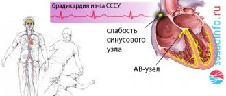
A special pathology of the heart disrupts the conduction of impulses from the sinus node to the ventricles. It is called atrioventricular or atrioventricular block. Similar changes are formed in diphtheria, rheumatic carditis and other inflammatory diseases of the heart muscle, post-infarction scars. In addition, the excessive inhibitory influence of the vagus nerve should be taken into account.
The complete rupture of communication between the atria and ventricles depends on the degree of organic (scar) changes in the tissues. Impulses from the ventricles are not regulated by the sinus node and are characterized by a rare rhythm (bradycardia up to 25–30 beats per minute). The ECG reveals pauses in myocardial contraction.
Arrows indicate missing ventricular contractions
Symptoms of temporary asystole:
- when the heart stops in 3 seconds. the person feels dizzy;
- at 9 sec - fainting appears;
- 15 sec. - epileptiform seizure, clinical death is possible.
Cardiac causes of asystole
This group of factors causes primary asystole, that is, associated with electrical instability of the myocardium itself, often against the background of ischemia (oxygen starvation of tissues).
Heart attack
General acute malnutrition of muscle structures. It occurs as a result of long-term coronary insufficiency, atherosclerosis, thromboembolism and other phenomena of this kind.
The development of cardiac asystole is a consequence of extensive damage, if the area reaches more than 25% or about a similar value. Possible delayed development. Thus, some patients suffer from cardiac arrest within a few days.
The most dangerous period is the first 72 hours. Further, the risks decrease, but remain throughout life, since a heart attack does not go away without a trace.
Cardiosclerosis, persistent heart failure, these are the reasons for the possible development of asystole. Lifelong maintenance treatment and constant monitoring of the condition are indicated.
Decrease in the amount of blood circulating in the body
It has a dual nature. On the one hand, there is a possibility of classic bleeding, when liquid connective tissue leaves the closed system. Then the physical variety takes place.
On the other hand, everything can be more complicated. Against the background of pathologies of the mitral valve and aorta, incomplete ejection and movement in a large circle are possible. Hence the decrease in the amount of circulating blood against the background of a preserved nominal volume.
This is no less dangerous, rather the opposite: transfusion in such a situation is pointless, the problem is of an organic nature.
Cardiac tamponade
Accumulation of blood around. Liquids are also possible. The result of an injury, long-term swelling, and inflammatory process. Recovery is carried out surgically as a matter of urgency.
Pulmonary embolism
Refers to cardiac causes conditionally. The essence of the phenomenon is the blockage of a large vessel with a blood clot.
It can be either a result of the chest injury itself or a consequence of distant damage to anatomical structures (in a large circle the blood clot moves throughout the body and penetrates the pulmonary artery).
It is also possible that spontaneous rupture of small vessels may occur due to the use of anti-inflammatory drugs, corticosteroids, hematological conditions, or excessive thickening of fluid connective tissue.
Such problems are resolved before urgent processes arise. The chances of survival from thromboembolism are almost zero.
Features of asystole in children
All of the above reasons apply to children, but an important difference from adults is a special type of asystole, which is included in the general concept of “sudden infant death syndrome.” In this case, cardiac arrest is recorded in a child more often under the age of 5 months against the background of sleep and the absence of any diseases during this period.
It is believed that ventricular asystole in a baby is caused by underdevelopment of myocardial cells, impulse transmission pathways. Perhaps it is associated with the pathological course of pregnancy and labor in the mother:
- hypoxia (lack of oxygen) in the fetus due to anemia, diseases of the maternal body;
- asphyxia during childbirth;
- vacuum extraction of the fetus;
- multiple pregnancy;
- the birth of a premature baby;
- smoking and alcohol consumption by the expectant mother.
Contributing factors are:
- placing the baby on a soft mattress;
- unventilated hot room;
- many blankets covering the child and causing him to overheat;
- tight swaddling.
Sleeping in the prone position is one of the risk factors for asystole
Many causes of death in a newborn due to asystole are associated with the lack of proper care for the child by adults.
Prognosis and possible complications
The outcome depends on the quality of first aid, the timing of transporting the patient to the hospital and the resuscitation scheme.
Against the background of severe organic pathologies, thromboembolism, cardiogenic shock, intoxication with dangerous poisons, the probability of death is almost 100%. In other cases, the risks are slightly lower. It is possible to return the patient in 30-50% of situations.
Consequences:
- Death is the most common outcome.
- Neurological deficit, its nature depends on the focus. The type of manifestations resembles the period after a severe stroke.
- Brain death. Equal to biological cessation of existence. Cardiac activity is present, natural processes are also carried out, but these are vegetative moments.
Clinical manifestations
Symptoms of cardiac arrest are characterized by a sudden death clinic and are accompanied by respiratory arrest. Let's consider possible options for the conditions for detecting a patient.
- Option 1 - an outwardly ordinary person at work or on the street, in transport suddenly falls, hoarse, rare breathing escapes from the throat, lips and face turn blue, there is no consciousness, attempts to make contact are unsuccessful.
- Option 2 - fatal cardiac arrest occurs during sleep, and the patient lies down and looks like a normal sleeping person. Those around him do not immediately notice his lack of any reactions.
In both cases, the patient does not have time to call for help or complain about deteriorating health.
- Option 3 - the patient is being treated in a specialized department for heart disease in a general ward, he is allowed to walk, or he forces the regime on his own. He can fall in the corridor, in the ward, on the stairs. Symptoms correspond to clinical death.
- Option 4 - the patient is in the acute stage of a heart attack or with severe somatic lesions, trauma, is in the intensive care unit, the condition is monitored by a monitor, the ECG picture is constantly monitored, any deviation in the rhythm activity is indicated by a sound signal.
The last option is the most favorable in terms of providing the necessary assistance.
Doctors in a specialized department do not need to waste time searching for a pulse; all equipment for resuscitation is usually at hand
Not every passerby can find the pulse on the carotid artery (at the angle of the lower jaw) or verify the absence of a heartbeat. Usually the first place is given to:
- loss of consciousness;
- pallor and cyanosis of the face;
- lack of breathing movements.
Characteristic symptoms
Subjectively, the phenomenon makes itself known only in the initial period. Harbingers of organ stoppage:
- Sharp chest pain. It happens that the symptom is absent, depending on the root cause. Breathing disorders. As they say, it takes your breath away. The patient cannot take in air.
- Panic arises, a feeling of fear of death. Motor activity persists for several tens of seconds or a couple of minutes.
- Tachycardia, other forms of rhythm disturbances.
- Weakness, drowsiness.
- Headache, vertigo.
- Nausea and vomiting.
- Loss of consciousness as a result.
In many cases (approximately 50%) there may be no previous symptoms. Then everything happens quickly, the patient does not even have time to react.
Objective signs after fainting can only be tracked by those around you.
- Breathing disappears completely.
- Cardiac activity may be recorded, but it is false activity.
- The blood circulation has already stopped. Possible forms are tachycardia, atrial fibrillation. After a few minutes they disappear too.
- There is also no vascular pulsation.
If this happens in a hospital, the heart rate is assessed. With asystole, the ECG shows minimal physical activity (in the first minutes), then the graph degenerates into a straight line.
The symptoms are specific, but without preparation a person is capable of little. There is too little time to react.
Desirable algorithm for the action of a random passerby
Since a person’s life depends on the speed of first aid, any passerby must be able to navigate the indications and the severity of the victim’s condition.
Recommended:
- Stimulate the victim, shake him, slap him on the cheeks, speak loudly. If a person regains consciousness, this indicates a short-term disturbance of cerebral circulation. Accordingly, you need to try to ensure free breathing, a position with a low head end.
- If there is no reaction to the intervention, then you should try to feel the pulsation of the carotid artery, if it is impossible to listen to the heartbeat, by placing your ear to the chest.
- You need to call for help. Someone must call an ambulance and help carry out emergency measures.
When you see a fallen person, you should not pass by; your family and friends may find themselves in the same situation.
How does cardiac asystole appear on an ECG?
Cardiac asystole on the ECG is detected in the form of an almost straight line reflecting its electrical activity.
Characteristic signs of this condition also include abnormal atrial rhythm and the inability to count the frequency of their contractions when there are no ventricular contractions. In some cases, when the atria continue to contract for some time after the ventricles have stopped, atrial P waves can be detected on the cardiogram.
An auxiliary element for diagnosing asystole can be the arrhythmia preceding this condition. So, if it manifests itself in the form of early polytopic ventricular extrasystoles and ventricular tachycardia, then they will be followed by ventricular fibrillation and flutter, and increasing blockade usually indicates the approach of asystole.
What emergency assistance is needed to restore rhythm?
Emergency care should:
- ensure heart contractions;
- maintain breathing;
- prevent hypoxia of the brain and other organs.
For brain cells, the decisive period is the first 8 minutes, after which the restoration of functions is doubtful.
The person should be laid on a hard surface (on the floor, a board made of boards, the ground) since subsequent compression of the chest on a soft surface is ineffective.
Tilt the victim's head back and check the patency of the airway with your finger; it is necessary to free the oral cavity from food debris, dentures, and other foreign bodies.
Air is blown into the patient's mouth or nose while one of the holes is clamped. With effective passive inspiration, chest rise is visible.
At the same time, for each “inhalation,” 4–5 intense pushes are made with two palms on the chest.
The force must be balanced against the potential for fracture of the ribs and sternum
Indirect massage is carried out until the ambulance arrives or until a pulse appears.
Cardiopulmonary resuscitation in adults
The measures taken in patients with circulatory and respiratory arrest are based on the concept of the “chain of survival”. It consists of actions performed at the scene of an accident, during transport, in the operating room and intensive care unit, as well as during subsequent rehabilitation. The most vulnerable and at the same time very important link is the primary resuscitation complex carried out at the scene of the incident, since 3-5 minutes after cessation of blood circulation and breathing at normal body temperature, irreversible changes in the victim’s brain develop.
Both primary respiratory arrest and primary circulatory arrest are possible. Detection of primary respiratory arrest (foreign bodies of the respiratory tract, electrical trauma, drowning, damage to the central nervous system (CNS), etc.) is unlikely at the prehospital stage, since by the time the ambulance arrives, ventricular fibrillation or asystole has time to develop.
The cause of primary circulatory arrest may be acute myocardial infarction, arrhythmia of various types, electrolyte imbalance, pulmonary embolism, rupture and dissection of aortic aneurysm, etc.
There are three options for cessation of cardiac activity: asystole, fibrillation and electromechanical dissociation. Asystole may be primary or secondary to ventricular fibrillation. In the first case, the chances of successful resuscitation are greater; in the second, when myocardial reserves are depleted, there are fewer. Sometimes an isoline on an electrocardiogram (ECG) is perceived as asystole, but it can also be observed when the electrocardiograph is malfunctioning, accidental disconnection of electrodes, low-amplitude ECG, etc. Electromechanical dissociation is characterized by the presence of electrical output from the heart, but the absence of myocardial contraction.
With fibrillation, scattered, chaotic, ineffective contractions of the myocardium occur. And here the use of precordial shock and early defibrillation are important.
Signs of circulatory arrest are: loss of consciousness; absence of pulse in the carotid arteries; respiratory arrest; convulsions; dilated pupils and lack of reaction to light; change in skin color.
To confirm cardiac arrest, the presence of the first three signs is sufficient.
Cardiopulmonary resuscitation (CPR) is not indicated, and it may not be started in the following cases: if it is determined that more than 25 minutes have passed since cardiac arrest (at normal ambient temperature); patients recorded their refusal of CPR in advance.
In other cases, when providing pre-hospital care, CPR begins immediately.
The reason for stopping CPR is the absence of signs of restoration of blood circulation and breathing when using all available CPR methods for 30 minutes.
Prehospital CPR
It includes basic life support (according to P. Safar) or a primary resuscitation complex (according to A. Zilber):
- restoration of airway patency;
- artificial ventilation (ALV) and oxygenation;
- indirect cardiac massage.
In addition, measures are taken (Fig. 1) of a specialized resuscitation complex (according to A. Zilber), including:
- electrocardiography and defibrillation;
- ensuring venous access and administering medications;
- tracheal intubation.
Restoration of airway patency. When emergency conditions occur, the patency of the airways is often impaired as a result of tongue retraction, aspiration of vomit, and blood. It is necessary to clear the oropharynx and perform the “triple Safar maneuver” - straighten the head in the cervical spine; push the lower jaw forward and upward; open your mouth. If it is impossible to exclude a fracture of the cervical spine and the head cannot be straightened, they are limited to moving the jaw and opening the mouth.
If the denture is intact, it is left in the oral cavity, as this preserves the contour of the mouth and facilitates mechanical ventilation.
If the airways are obstructed by a foreign body, the victim is placed on his side and 3-5 sharp blows are made with the lower part of the palm in the interscapular area, then they try to remove the foreign body from the oropharynx with a finger. If this method is ineffective, then the Heimlich maneuver is performed: the resuscitator’s palm is placed on the stomach between the navel and the xiphoid process, the second hand is placed on the first and a push is made from the bottom up along the midline. After which they also try to remove the foreign body from the oropharynx with their finger.
Due to the danger of infection of the resuscitator upon contact with the mucous membranes of the mouth and nose, as well as to increase the effectiveness of mechanical ventilation, a number of devices are used: a “key of life” device; oral airway; transnasal airway; pharyngotracheal airway; double-lumen esophageal-tracheal airway (combitube); laryngeal mask.
A big step forward was the creation of the laryngeal mask. The laryngeal mask airway is an endotracheal tube that does not pass through the glottis into the trachea, but has a miniature mask at the distal end that is worn over the larynx. A cuff adjacent to the edge of the mask inflates around the larynx, providing a seal around the laryngeal perimeter. The laryngeal mask has many advantages, including the ability to avoid extension of the head in the cervical region if there are contraindications to this.
Every emergency physician should be able to perform tracheal intubation. This method allows you to ensure optimal airway patency, reduce the likelihood of regurgitation during a complex of resuscitation measures, and provide higher intrapulmonary pressure. In addition, some medications can be administered through the endotracheal tube.
Ventilation Artificial respiration is the injection of air or an oxygen-enriched mixture into the patient’s lungs without or with the use of special devices. The air exhaled by a person contains from 16 to 18% oxygen, therefore mechanical ventilation with atmospheric air or an oxygen-air mixture is more effective. Each insufflation should take 1–2 s, and the respiratory rate should be 12–16 per minute. The adequacy of mechanical ventilation is assessed by periodic expansion of the chest and passive exhalation of air.
The emergency team usually uses either an airway, face mask and ambu bag, or tracheal intubation and ambu bag.
Indirect cardiac massage. After stopping blood circulation for 20–30 minutes, the heart retains its automatic and conductive functions, which allows it to be “started.” The main purpose of cardiac massage is to create artificial blood flow. During chest compressions, compression occurs not only of the heart, but also of the lungs, which contain a large amount of blood. This mechanism is commonly called a breast pump.
In patients with ventricular fibrillation and ventricular tachycardia, it is recommended, in the absence of a defibrillator prepared for use, to apply a precordial blow (1-2 sharp blows with a fist to the area of the border of the middle and lower third of the sternum from a distance of at least 30 cm).
When performing chest compressions, the patient must be on a hard surface. One palm of the resuscitator is located on the lower third of the sternum along the midline, the second rests on the dorsum of the first. The time of pressure and release is 1 s, the interval between compressions is 0.5–1 s. An adult’s sternum should be “pressed” by 5–6 cm. When performing any therapeutic measures, a break in traction should not exceed 5–10 seconds. The criteria for the effectiveness of chest compressions are the appearance of pulse impulses in the carotid arteries, blood pressure at the level of 60–70 mm Hg. Art., change in skin color.
If assistance is provided by one resuscitator, then 15 tractions are performed for two air injections; if two resuscitators are working, then 5 tractions are performed for one air injection.
| Figure 2. Emergency care algorithm for ventricular fibrillation |
Electrical cardiac defibrillation (EDC). This is an essential component of SRL. EMF is effective only when the energy resource of the myocardium is preserved, i.e. when large-wave oscillations from 0.5 to 1 mV or more are recorded on the ECG (Fig. 2). If low, arrhythmic, polymorphic oscillations, as well as asystole, are noted, then they begin with mechanical ventilation, indirect massage and drug therapy (Fig. 3), achieve the transition of asystole or small-wave ventricular fibrillation to large-wave fibrillation and apply EMF.
The first discharge for EMF is 200 J, if the second is ineffective - 300 J, if the third is ineffective - 360 J. The break between discharges is minimal - to control the rhythm. Indirect cardiac massage and mechanical ventilation are interrupted only at the moment of discharge. If the first series of three shocks turns out to be ineffective, then against the background of ongoing mechanical ventilation, chest compressions, and drug therapy, a second series of shocks is performed in the same sequence.
Currently, automatic external defibrillators are used in the prehospital stage; in this case, the ECG is recorded from the defibrillator electrodes applied to the chest. The defibrillator records the heart rhythm and performs its automatic analysis; When ventricular tachycardia or ventricular fibrillation is detected, the capacitors are automatically charged and the device delivers a shock. The effectiveness of automatic defibrillators is very high. In addition to automatic ones, semi-automatic external defibrillators are used.
Drug therapy during cardiopulmonary resuscitation. Medications for CPR can be administered: into a peripheral vein; into the central vein; into the trachea.
For obvious reasons, the intramuscular route of administration is not indicated. If possible, a peripheral vein is catheterized. If the resuscitator is experienced and fluent in the technique of central vein puncture, you can use this method. The problem is that in this case it is necessary to interrupt resuscitation efforts, and a break of more than 5–10 seconds is undesirable. The intratracheal route is convenient if the trachea is intubated; in extreme cases, drugs can be administered into the trachea through the cricothyroid membrane. It is permissible to administer adrenaline, atropine, and lidocaine endotracheally. It is better to dilute the drugs in 10–20 ml of 0.9% sodium chloride solution.
Adrenaline remains the treatment of choice for circulatory arrest. During asystole and electromechanical dissociation, it “tones” the myocardium and helps to “start” the heart; it converts small-wave fibrillation into large-wave fibrillation, which facilitates EMF. Doses: 1-2 mg intravenously in a bolus with an interval of 5 minutes, usually up to 10-15 mg in total.
The M-anticholinergic atropine reduces the inhibitory effect of acetylcholine on the sinus node and atrioventricular conduction and possibly promotes the release of catecholamines from the adrenal medulla. It is indicated for bradysystole and asystole. Doses - 1 mg, can be repeated after 5 minutes, but not more than 3 mg during resuscitation.
All antiarrhythmic drugs have a depressive effect on the myocardium and are not harmless to the patient’s body. When ventricular fibrillation has developed, they should be administered only in the case of several unsuccessful EDS attempts, since they, by suppressing ventricular ectopy, make it difficult to restore an independent rhythm. Lidocaine is considered one of the most effective drugs for refractory ventricular fibrillation, sustained ventricular tachycardia and tachycardias of unknown etiology with a wide QRS complex. The dose for saturating intravenous administration is 1.5 mg/kg bolus (usually 75–100 mg). At the same time, administration of a maintenance dose of 2–4 mg per minute begins. To do this, 1 g of lidocaine is diluted in 250 ml of 5% glucose solution.
An indication for the administration of sodium bicarbonate can be considered resuscitation prolonged for more than 15 minutes if cardiac arrest was preceded by severe metabolic acidosis or hyperkalemia. Dose - 1 mmol/kg, intravenously once, with repeated administration it is halved. Some authors believe that with adequate resuscitation measures, sodium bicarbonate should be administered only under the control of the acid-base state, since the body adapts much less well to alkalosis than to acidosis.
It is advisable to use a 0.9% sodium chloride solution as infusion solutions, but the most effective is Ringer's lactate solution according to Hartman, and among colloids - solutions with an average molecular weight containing hydroxyethyl starch - voluven or venofundin.
In all cases, emergency hospitalization for vital indications in the intensive care unit is indicated.
I. G. Trukhanova, Doctor of Medical Sciences, Associate Professor E. V. Dvoinikova , Candidate of Medical Sciences, Associate Professor Samara State Medical University, Samara
Medical assistance
Treatment of asystole with medical methods begins in the ambulance and continues in the intensive care unit. Depending on the equipment, the ambulance doctor continues indirect massage with simultaneous ventilation of the lungs with an Ambu bag, injects adrenaline intracardially and tries to act with a defibrillator discharge.
Intensive activities should be carried out within 30 minutes. In a specialized department, the patient is transferred to mechanical breathing. Other medications are injected into the subclavian vein to support brain functions (alkaline solution, Dopamine, Reopoliglucin, Sodium oxybutyrate).
A cardiac monitor connected to the patient allows you to monitor the results of the staff’s actions. If ineffective, biological death is declared.
Resuscitation measures are not carried out for patients with asystole caused by severe chronic heart disease or malignant tumors.
Prevention measures
It is impossible to prevent an emergency condition with a 100% guarantee. Even healthy people are at risk, although to a lesser extent.
Possible actions:
- Quit smoking, alcohol, and especially drugs. Heroin and cocaine, ethyl alcohol, nicotine, tar, all these substances have a depressing effect on the myocardium. The result may be delayed, against the background of imaginary well-being.
- Adequate physical activity. Hiking, amateur cycling, swimming, exercise therapy. You can't overwork yourself. Lack of training in itself turns out to be an increased risk factor.
- Complete nutrition. Fortification of the diet. From the point of view of diet, the abuse of fatty, fried, in a word, “junk” food is not as dangerous as a lack of vegetables, fruits, and protein in the menu. It makes sense to discuss the diet with your doctor, depending on the underlying disease. Specialist - nutritionist.
- Sleep 8 hours per night. Lack of normal rest leads to overstrain of all the body's forces. Especially in the long term. The body can work like this for a year or two, but at some point it will fail.
It is important to systematically treat the underlying pathology, which can cause ventricular or atrial asystole. Regular (1-2 times a year) visits to a cardiologist are also an important measure.
Diagnostics
Diagnosis is carried out extremely quickly, since within 3-5 minutes it is necessary to restore blood circulation, breathing, and heart activity in order to exclude irreversible changes in the brain. This takes into account the absence of respiratory excursions, pulse and dilated pupil. After which treatment measures begin immediately.
To clarify the nature of the cardiac dysfunction: asystole or fibrillation, an ECG is recorded. Asystole is confirmed by at least two ECG leads.
The appearance of a straight line on the ECG indicates asystole. But when it is recorded through the defibrillator electrodes, straight lines may also appear, which indicates that the electrodes are disconnected or the sensitivity of the monitor is reduced.
If the monitor sensitivity is too high, the interference is mistaken for chaotic electrical activity, which is typical for ventricular fibrillation. Then the ECG assessment is carried out again.
Direct line to ECG
Pansystolic low (rough) murmur over the entire heart is determined by auscultation (by listening to sounds), the “purring cat” syndrome is determined by palpation.
Differential diagnosis is carried out - an ECG is immediately recorded in order to adequately carry out resuscitation. At the same time, it is determined how clinical death developed: the culprit was electromechanical dissociation (asystole) or ventricular fibrillation. If it is impossible to conduct an ECG, then the nature of the onset of clinical death and the victim’s response to resuscitation measures are taken into account.