Bundle branch block is a disorder of intracardiac conduction, characterized by a slowdown or complete cessation of the conduction of excitation impulses along one or more branches of the His bundle. Bundle branch block can be detected only during instrumental examination or symptomatically manifested by rhythm disturbances, dizziness, and attacks of loss of consciousness. Bundle branch block is diagnosed using electrocardiography. Treatment of bundle branch block is reduced to eliminating the causes of conduction disturbances; In some cases, it may be necessary to install an artificial heart pacemaker.
Causes
Blockade of the bundle branches or their individual branches can be caused by the following factors:
- Congenital anomalies - narrowing of the aorta or pulmonary artery, departure of blood vessels from the ventricle.
- Previously suffered cardiovascular diseases - atherosclerosis , myocardial infarction, coronary heart disease, etc.
- Impaired heart function as a result of diseases that reduce immunity.
- Heart surgery.
- Bad habits – alcoholism, drug addiction, smoking.
- Hormonal disorders - diabetes mellitus, diseases of the thyroid and pancreas, adrenal glands.
- Hypoxia is an insufficient supply of oxygen to the body caused by diseases of the respiratory system (for example, bronchial asthma).
| Types of blockade | When does it occur |
| Right bundle branch block | For diseases accompanied by hypertrophy and overload of the right ventricle: high blood pressure, coronary heart disease, narrowing of the mitral valve, acute myocardial infarction. |
| Left bundle branch block | For inflammation and disruption of the heart muscle, aortic valve defects. |
| Blockade of both bundle branches at once | With narrowing of the aortic lumen, incomplete closure of the aortic valve. |
In children, bundle branch block develops in utero for the following reasons:
- mother taking insulin during pregnancy;
- pathological development of cardiac septa;
- diffuse damage to connective tissue in the expectant mother.
There are no reliable data on the prevalence of rhythm disturbances in children. In the structure of cardiovascular diseases in childhood, arrhythmias account for (according to the incidence) from 2.3 to 27%, but they are often detected in healthy children. Heart rhythm and conduction disturbances are found in children of all ages, including newborns; they are detected even in the fetus. The frequency increases during puberty.
Etiology and pathogenesis
Rhythm disturbances can be congenital or acquired and are caused by cardiac, extracardiac and combined causes. Cardiac causes of arrhythmia include congenital and acquired heart defects, rheumatic carditis and non-rheumatic carditis, infective endocarditis, cardiomyopathies and other heart diseases.
A clear relationship between rhythm disturbances and MVP and other minor anomalies of cardiac development has been revealed. Arrhythmias can develop with diseases of the nervous and endocrine systems, many somatic disorders, acute and chronic infectious pathologies, intoxication, overdose or inadequate reaction to medications, deficiency of certain microelements (magnesium, selenium). The formation of rhythm disturbances is influenced by emotional and physical overload, as well as vegetative dystonia syndrome and psychogenic disorders associated with extracardiac pathology.
The main pathogenetic factor of arrhythmias is considered to be disturbances in the formation of impulses and/or the speed of excitation as a result of inhibition of the function of the sinus node, activation of ectopic pacemakers and the functioning of additional pathways. These disorders arise as a result of inflammatory, dystrophic, necrotic and sclerotic processes in the cardiac muscle and cardiac conduction system or as a result of electrolyte imbalance, leading to changes in cellular metabolism and the ionic composition of the internal environment of cardiomyocytes.
In childhood, arrhythmias are more often of extracardiac origin. In this case, perinatal pathology plays an important role (unfavorable course of pregnancy and childbirth, prematurity, intrauterine malnutrition, infection), leading to disruption of morphogenesis and functional immaturity of the conduction system of the heart. Perinatal damage to the central nervous system can lead to disruption of neurovegetative rhythm regulation with a change in the relationship between the sympathetic and parasympathetic parts of the autonomic nervous system, resulting in electrical instability of the myocardium and conduction system of the heart, as well as a decrease in the functional reserves of adaptation of the sympathoadrenal link of heart rate regulation.
Classification
- Automatic disorders (nomotopic - sinus arrhythmia, tachycardia and bradycardia, heterotopic - extrasystole, paroxysmal and non-paroxysmal tachycardia, flutter and fibrillation of the atria and ventricles).
- Conduction disorders (sinoauricular, intraatrial, atrioventricular, intraventricular blockade).
- Combined arrhythmias (sick sinus syndrome, atrioventricular dissociation, premature ventricular excitation syndrome).
Clinical picture
The history of children with rhythm disturbances often reveals an unfavorable course of the perinatal period, a family history of cardiovascular pathology, repeated acute infectious diseases and foci of chronic infection. During examination, hypertensive-hydrocephalic syndrome, residual neurological symptoms, various psychovegetative disorders, manifestations of connective tissue dysplasia, and sometimes delayed motor development and puberty are often detected.
Rhythm disturbances in children are often asymptomatic, which makes it difficult to accurately determine the time of their onset. In approximately 40% of cases, arrhythmias are detected by chance (on an ECG) or during examination in connection with a history of acute respiratory viral infection. Children are much less likely than adults to complain of palpitations, a feeling of interruptions in the activity of the heart, and its freezing, even with severe forms of arrhythmia. Along with this, in prepubertal and pubertal age, rhythm disturbances can have a strong emotional coloring, caused by psycho-vegetative disorders, and be accompanied by other cardiac and extracardiac complaints: pain in the heart, increased excitability, sleep disturbances, and meteosensitivity. With arrhythmias, weakness, dizziness and fainting are possible (with sinus bradycardia, atrioventricular block, sick sinus syndrome, paroxysmal tachycardia).
An objective examination of children with rhythm disturbances can reveal an increase or decrease in the pulse, a change in its character (irregular with periodic drops, alternating periods of increase and decrease, temporary or permanent weakening of the pulse wave, the presence of a compensatory pause). Assessing other basic characteristics of the cardiovascular system (blood pressure, heart size, sonority of sounds, heart murmurs) makes it possible to establish or exclude cardiac pathology as the cause of arrhythmia.
Diagnostics
The main method for identifying and assessing arrhythmias is ECG. With its help, it is possible to detect such asymptomatic rhythm disturbances as single extrasystoles, Wolff-Parkinson-White syndrome, slowing of atrioventricular conduction, and pacemaker migration. Often, when performing an ECG in children, sinus arrhythmia and isolated incomplete blockade of the right bundle branch are detected, which are a variant of the age norm.
Clinical electrocardiographic examination allows us to establish the type of rhythm disturbances (functional or organic), especially extrasystoles. Extrasystoles of functional origin are most often detected in the pre- and pubertal periods; they are not constant, usually disappear or are significantly reduced with changes in body position and physical activity. Extrasystoles most often originate from the right ventricle or are of supraventricular origin.
In order to clarify the origin of rhythm disturbances, cardio-intervalography, daily Holter ECG monitoring, functional tests are performed: stress tests (test with dosed physical activity, bicycle ergometry, treadmill test) and medicinal tests [atropine, with propranolol, isoprenaline (isadrine), gilurhythmal, etc. ]. If organic heart disease is suspected, an x-ray examination and echocardiography are performed. An assessment of the child’s vegetative and psychological status, a neurophysiological examination [EEG, echoencephalography (EchoEG), REG], consultations with a neurologist, otolaryngologist, endocrinologist, and ophthalmologist are also necessary.
Holter monitoring allows much more often than a standard ECG to detect rhythm disturbances even in healthy children (pacemaker migration, extrasystole, etc.) and determine their relationship with periods of the day and night sleep (circadian dependence), which is important for the choice of treatment method.
A study of the state of the autonomic nervous system confirms the significant role of its dysfunction in arrhythmias. Thus, the predominance of parasympathetic with insufficiency of sympathetic influences is found in cases of extrasystoles, slowing of atrioventricular conduction, as well as in children with chronic tachyarrhythmia and even atrial fibrillation.
In difficult cases, in a specialized hospital, electrography of the atrionodal conduction system, surface ECG mapping (for topical diagnosis of rhythm disturbances) and other special research methods are performed.
Flow
A number of arrhythmias (isolated tachy- and bradycardia, rare intermittent monotopic extrasystoles, mild degrees of conduction disturbances, pacemaker migration) are usually not accompanied by organic cardiac pathology and distinct subjective manifestations and proceed quite favorably. Some forms of rhythm disturbances, especially persistent ones, can worsen the patient's condition, adversely affect hemodynamics, cause a decrease in cardiac output and impaired coronary circulation, and as a result lead to an unfavorable outcome. This is possible with ventricular and supraventricular tachycardia, frequent polytopic extrasystoles, significant slowing of heart rate, atrioventricular block, long Q-T interval syndrome (Romano-Ward syndrome).
In newborns and young children, rhythm disturbances can be asymptomatic or severe, with complications. In older children, the prognosis for arrhythmias is usually favorable, but persistent arrhythmias, especially severe forms, can also lead to an unfavorable outcome.
Treatment
Treatment of rhythm disturbances, especially life-threatening ones, is carried out strictly individually depending on their origin, form, duration, impact on the child’s well-being and the state of his hemodynamics. It is necessary to stop the arrhythmia and carry out maintenance therapy to prevent recurrence. For all types of arrhythmias, cardiac and extracardiac causes should be treated simultaneously.
- Basic therapy includes a course (at least 2-3 months) of nootropics [piracetam, pyritinol (for example, encephabol), gamma-aminobutyric acid (for example, aminolone), glutamic acid, phenibut, carbamazepine, hopantenic acid (for example, pantogam), nicotinoyl , gamma-aminobutyric acid (picamilon)] for the correction of neurovegetative disorders and normalization of trophic processes in the nervous system. The effectiveness of the use of cell membrane stabilizers and antioxidants [vitamins E and A, cytochrome C, vetoron, etidronic acid (xidifon)], agents that correct metabolic processes in the myocardium [vitamins C, B1, B2, B15, benfotiamine, potassium orotate, inosine ( for example, riboxin), levocarnitine, orotic acid, magnesium salt (magnerot)], as well as electrolyte imbalance [potassium and magnesium asparaginate (for example, asparkam, panangin), potassium chloride, calcium glycerophosphate]. In complex treatment, vasodilators [vinpocetine (for example, Cavinton), vincamine, pentoxifylline (for example, Trental)], angioprotectors [pyricarbate (parmidine)], bellataminal, and biogenic stimulants (tinctures of aralia, lemongrass, ginseng, zamanikha) are used. Consistent use of these drugs often helps to normalize the well-being of patients, eliminate arrhythmias or improve their tolerability. Fainting and palpitations require a special therapeutic approach, since they can be the result of not only severe rhythm disturbances, but also concomitant hemodynamic and other disorders.
- Antiarrhythmic drugs [procainamide (procainamide), amiodarone, lidocaine, verapamil, etc.] are used in children with certain restrictions due to the fact that they are not always quite effective and can cause adverse reactions (decreased blood pressure) much more often than in adults. pressure and contractility of the myocardium, worsening of arrhythmias, slower conduction). Their use is justified in cases of severe subjective intolerance to arrhythmias, significant hemodynamic changes, as well as in conditions with an unfavorable prognosis (paroxysmal tachycardia, frequent ventricular and supraventricular extrasystoles, atrial fibrillation). Attacks of paroxysmal tachycardia can be stopped by the administration of propranolol (for example, obsidan) or verapamil (for example, isoptin). Determination of indications for the use of antiarrhythmic drugs and their individual selection is carried out in the hospital.
- In case of conduction disturbances (isolated or accompanying other forms of arrhythmias), it is possible to use adrenomimetics [isoprenaline (isadrin), orciprenaline (for example, alupent), norepinephrine), atropine, as well as glucocorticoids (for high-degree atrioventricular blockades). Long QT syndrome requires long-term administration of beta-blockers.
- For some persistent, prognostically unfavorable, life-threatening rhythm disturbances (paroxysmal supraventricular and ventricular tachycardia, atrial fibrillation), cardiac defibrillation is indicated to restore sinus rhythm. In case of high degree atrioventricular blockade, and in some cases also in case of sick sinus syndrome, a pacemaker is implanted. Cardiac surgery may be indicated for patients with treatment-resistant chronic non-paroxysmal and paroxysmal supraventricular tachycardias and atrial fibrillation.
Dispensary observation
Clinical observation should be regular; its frequency is determined depending on the underlying disease (rheumatism, non-rheumatic carditis, congenital heart defects, autonomic dystonia syndrome, etc.), the form of arrhythmia and the characteristics of its course.
Dynamic ECG monitoring is required; 24-hour Holter monitoring and other studies are prescribed according to indications. They also monitor the implementation of medical prescriptions and the effectiveness of therapy. Clinical observation of children with arrhythmias has no time limits, and it is often continued until the patient is transferred under the supervision of an adolescent therapist. Children treated in a hospital are observed monthly in the first quarter, up to 1 year - at least once every 3 months, then - once every six months.
Prevention
Prevention of rhythm and conduction disorders is aimed at eliminating predisposing factors. In order to timely detect arrhythmias, it is advisable to conduct regular ECG monitoring, especially during periods of greatest risk of their development (in newborns, at 4-5, 7-8 and 12-13 years). Secondary prevention involves maintaining normal rhythmic activity of the heart and preventing the progression of arrhythmias and includes a complex of non-drug interventions (psychological correction, restorative measures, exercise therapy) and drug treatment (courses of nootropic, membrane-stabilizing drugs, antioxidants, etc.) depending on the characteristics of a particular case.
Forecast
Most arrhythmias in childhood are benign, reversible and do not affect the prognosis of life. However, in newborns and young children they can cause the development of arrhythmogenic cardiomyopathy or heart failure, which can lead to early disability and even death. Chronic non-paroxysmal and paroxysmal tachycardia, atrial fibrillation, and acquired complete transverse heart block have an unfavorable prognosis. Diseases associated with a high risk of sudden death (usually due to asystole or ventricular fibrillation) include long QT interval syndrome, severe dysfunction of the sinus node, some tachyarrhythmias, especially ventricular ones, accompanied by fainting, myocardial ischemia, acute heart failure, arterial hypertension.
Source: Children's diseases. Baranov A.A. // 2002. Illustrations from the site: © 2011 Thinkstock
Classification
Cardiac impulse conduction disorders are divided into:
- complete – there is no impulse conduction at all;
- incomplete – impulse conduction is difficult.
Bundle branch block often occurs in seemingly healthy people, including athletes.
Also, blockade of elements of the conduction system of the heart is classified depending on the number of areas where patency is impaired:
- single-bundle – only one leg is affected;
- double-bundle – simultaneous damage to two branches of the His bundle occurs;
- three-bundle – impulse conduction is absent in all branches of the His bundle.
How dangerous is the blockade?
Complications can occur at any stage of the pathological process. However, there is no guarantee that death will not occur.
An approximate list of consequences is as follows:
- Heart attack. The death of muscle organ cells and their replacement with scar tissue. The area of the lesion depends on the nature of the conduction disorder.
- Myopathy. Defect in the formation and development of the myocardium. It is determined for congenital reasons or as a consequence of alcoholism and other factors.
- Heart failure. Sudden, without prospects for restoration of cardiac activity.
- Stroke. As a result of a disruption in the supply of oxygen and nutrients to the brain.
- Respiratory failure of varying severity. Characterized by the impossibility of normal gas exchange.
- Pulmonary edema. Emergency condition. Requires urgent recovery in a hospital setting.
- Cardiac asthma. The attack may be isolated. In some cases, a group of episodes is identified over a short period of time.
- Death. As a result of the presented complications.
- Cardiogenic shock. Acute disorder, mortality approaching 100%.
The likelihood of consequences varies. It all depends on the degree of blockade.
Symptoms and signs
The child has
In children, bundle branch block can manifest itself even during intrauterine development, at the stage of formation of the cardiovascular system. The development of pathology is due to both heredity and the woman’s unhealthy lifestyle during pregnancy.
Signs of intracardiac conduction disorders in children:
- decreased physical activity;
- dizziness, weakness;
- chest pressure.
In an adult
The main signs of bundle branch block in adults:
- heart rhythm disturbance;
- dizziness, temporary loss of consciousness;
- shortness of breath, feeling of heaviness in the chest.
In adults, the disease is asymptomatic in most cases. Patients learn about cardiac obstruction when undergoing an ECG during medical examinations.
The appearance of at least one of the above symptoms is a reason to consult a cardiologist. Do not self-medicate by taking medications as recommended by friends.
Diagnostics
At the first visit, the doctor collects a patient’s life history, which includes the following information: complaints, symptoms, presence of heart pathologies in the past and methods of their treatment. Next, the specialist measures the heart rate and determines the boundaries of the heart by tapping and listening to the chest. For final diagnosis, the patient is prescribed cardiography and laboratory tests (blood test: general and for hormones).
To obtain a more complete clinical picture, additional examinations are prescribed:
- Ultrasound is a study that allows you to identify heart diseases that caused the blockage.
- Transesophageal electrophysiological study - simultaneous stimulation of the heart with electronic pulses is performed and the results are displayed on the cardiogram.
- Holter monitoring – the patient wears an ECG device throughout the day and records his actions. The doctor analyzes the situations in which cardiac conduction disturbances occurred.
When collecting a medical history from a child, you will need to provide the doctor with information about the mother’s health during pregnancy and find out whether there is a hereditary predisposition to diseases of the cardiovascular system.
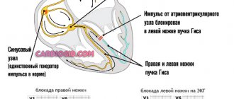
Mechanism of development of deviation
Pathological deviation occurs gradually, suddenly - relatively rarely, we are talking about acute processes. To understand what underlies the development of pathology, you need to turn to anatomical information.
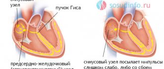
The heart is capable of autonomous operation indefinitely. This is the result of the presence of the so-called sinus node or natural pacemaker. It is responsible for generating an electrical impulse.
Excitation is transmitted through special fibers known as the bundle of His. It is a tree-like, branched structure.
As a result of previously existing inflammatory pathologies, congenital or acquired defects and other conditions, conduction disturbances occur, the electrical impulse slows down the movement (with incomplete blockade) or it becomes completely impossible (complete blockade).
As a result, the contractility of the myocardium on the right side (atrium and ventricle) decreases, and the movement of blood in the pulmonary circle is disrupted. Hence pulmonary problems as an early symptom of the pathological process.
Treatment
Treatment for bundle branch block is prescribed individually for each patient, depending on the final diagnosis, the presence of contraindications and general health.
The child has
If a child is diagnosed with a violation of the conduction of cardiac impulses, the cause should be identified and undergo a course of treatment under the supervision of a cardiologist. If parents notice a sharp weakness in the child, pale skin and complaints of pressure in the chest, they should lay the baby on his back, call an ambulance and perform breathing exercises. Take a deep breath, hold the breath for a few seconds, then exhale slowly. The exercise is repeated several times.
Treatment of diseases of the cardiovascular system is primarily aimed at eliminating symptoms that interfere with a healthy lifestyle. Together with drug therapy, the child is prescribed a course of vitamins and restoratives.
In an adult
Like children, adults, when a bundle branch block is detected, specific drug therapy is prescribed. You should not change or stop taking medications on your own, even if you feel better.
If drug therapy is unsuccessful and the patient’s well-being does not return to normal, surgical intervention to install a pacemaker is prescribed.
During treatment, it is important not only to take the medications prescribed by your doctor, but also to adhere to your diet. Avoid fatty and fried foods and include more fiber-containing foods (strawberries, cereals, bran) in your diet.