Deep vein thrombosis (DVT) occurs when a blood clot (thrombus) forms in one or more deep veins in your body, usually in the legs. Deep vein thrombosis can cause leg pain or swelling, but may be asymptomatic.
- Symptoms of deep vein thrombosis
- Causes of deep vein thrombosis
- Risk factors
- Complications of DVT
- Prevention of thrombosis
DVT may be associated with diseases that affect the blood clotting process. A blood clot in your legs can also form if you have not moved for a long time, such as after surgery or an accident. But walking over extremely long distances can lead to the formation of blood clots.
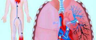
Deep vein thrombosis is a serious condition because blood clots in your veins can travel through your bloodstream and become lodged in your lungs, blocking blood flow (pulmonary embolism). However, pulmonary embolism can occur without evidence of DVT.
When DVT and pulmonary embolism occur at the same time, it is called venous thromboembolism (VTE).
What is thrombophlebitis
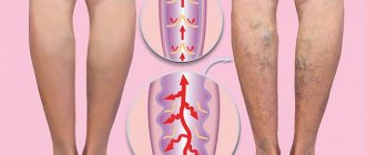
Thrombophlebitis is an inflammatory process of the inner surface of a vein due to the deposits of thrombotic masses on it. They can partially or completely block blood flow. It is worth noting that such a diagnosis will be made if the superficial veins of the lower extremities are affected. If pathology is found in the deep veins, then the diagnosis will be phlebothrombosis.
Thrombophlebitis is a severe pathological condition affecting about 15% of the population, causing damage to the superficial and deep veins. The detachment of part of a blood clot threatens immediate death, submassive pulmonary embolism. In 70% of cases, this disease causes disability. This disease was included in the International Medical Register under code 10, with the designation I80.2, calling it “Phlebitis and thrombophlebitis of the lower extremities.”
Thrombophlebitis: from acute to chronic
Thrombophlebitis, localized in the lower extremities, happens:
- superficial - common in almost 70% of cases, affects the subcutaneous vessels, a feature of this pathology can be called its vivid manifestation in the form of various symptoms, the pathogenic process is accompanied by the involvement of soft tissues and subcutaneous tissue;
- deep - occurs half as often, is characterized by the development of inflammation in the deep veins, unlike the previous type, this type of thrombophlebitis is often asymptomatic and can be treated without immediate surgical intervention.
This disease is also classified according to the severity of its course: it can be acute or chronic. With the development of acute deep vein thrombophlebitis, slight swelling and severe pain occur along the length of the affected vein. Pathology can develop in the lower leg, thigh, foot, or affect the entire vein. Deterioration of the condition is accompanied by an increase in temperature to 39-40C and fever. There are acute thrombophlebitis of purulent, non-purulent type, with abscesses or necrosis of soft tissues.
In the case of acute phlebothrombosis, pain occurs suddenly, intensifying during movement. Further, bloating or heaviness may appear in the left or right leg, and the temperature of the body and the affected area may increase. The pulsation of peripheral arteries is not disturbed, weakened or absent. After 3-4 days, a network of dilated vessels appears on the surface of the calves.
In acute thrombosis of the deep veins of the lower extremities, doctors can observe a blurred clinical picture if the veins in the calves are involved in the pathological process. This disease can be recognized by minor swelling of the ankle or pain in the calves.
The chronic course of the disease lasts from 3-4 months to several years. The severity of inflammation and pain is low in this case. The general clinical picture is expressed by pain in the left and right leg, mainly after active physical activity, after long walking, standing, and lifting large loads. With rest, the pain subsides.
Thrombophlebitis is also isolated during pregnancy - at the beginning of the second trimester. Clinical manifestations of the pathological process are reduced to the formation of spider veins. As the venous channels increase in size, pain develops. Evening time is the period of swelling and periodic cramps.
Post-injection thrombophlebitis is the result of a violation of the procedure used to treat varicose veins.
Clinical forms of deep vein thrombosis
Vein thrombosis of the leg
Complaints of swelling of the foot, pain and tension in the calves, pain when pressing on the calf muscles. If thrombosis does not spread, it is almost asymptomatic. Sometimes there is thromboembolism of small branches of the pulmonary artery with cough and the development of pneumonia (pneumonia). Treatment of thrombosis of the veins of the leg can be carried out on an outpatient basis, under the supervision of a phlebologist with control ultrasound examinations.
Thrombosis of the popliteal vein
Has a clear clinical picture. Severe swelling and tension of the lower leg, swollen saphenous veins, severe pain when walking. Thrombosis of the popliteal vein is very dangerous due to frequent pulmonary embolism, so treatment is best carried out in a vascular hospital. Most often, conservative therapy is carried out with antithrombotic drugs (heparin). If the patient had thromboembolism, then urgent surgical treatment is necessary - ligation of the femoral vein above the thrombus.
Thrombosis of the iliofemoral segment (ileofemoral phlebothrombosis)
It is characterized by a severe general condition, pronounced swelling of the entire lower limb, and severe pain. The saphenous veins are sharply dilated, the leg takes on a bluish color. With ascending deep venous thrombosis, thrombosis of the entire venous bed is possible with a block of venous outflow and the development of venous gangrene (blue phlegmasia), which is accompanied by high mortality. Pulmonary embolism often occurs with a fatal outcome. Treatment of ileofemoral phlebothrombosis is only in a hospital. For occlusive thrombosis, conservative treatment is possible, but it is better to remove the thrombus so that post-thrombotic disease does not develop. In case of floating thrombosis, urgent removal of the thrombus (thrombectomy) is necessary, possibly using innovative methods. A vena cava filter can be installed in cancer patients.
Thrombosis of the inferior vena cava
The most dangerous disease. Clinically, it manifests itself as a severe general condition, swelling of both legs. Kidney failure and blood in the urine often develop. With thrombosis of the hepatic segment, liver failure develops resulting in Bud-Chiari syndrome. Treatment of acute thrombosis of the inferior vena cava should be active. It is necessary to remove thrombotic masses, as surviving patients may develop severe inferior vena cava syndrome. For this, it is good to use our innovative methods and systemic thrombolysis. The effectiveness of this treatment is very high.
Why does the disease occur?
Often the pathological process is triggered by the presence of several factors, which are called “Virchow’s Triad”:
- increased blood clotting (indicator significantly exceeds the norm) - the result of an excess/lack of blood clotting inhibitors, a protein with anticoagulant properties;
- poor condition of the walls of the vascular system (the structure at the cellular level is disrupted);
- phenomena of stagnation, accompanied by a slowdown in blood flow.
The etiology of thrombophlebitis also often involves:
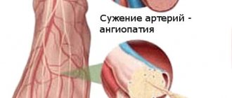
- narrowing/expansion of blood vessels in the area of varicose nodes;
- slowdown of metabolic processes;
- dysfunction of hematopoiesis.
The listed pathological changes in the body, which cause vascular thrombophlebitis, do not arise out of nowhere. The etiology of phlebitis may be associated with:
- excess body weight;
- genetic predisposition to varicose veins, phlebothrombosis;
- sedentary lifestyle;
- dehydration and insufficient fluid intake;
- treatment with medications that negatively affect blood clotting;
- pregnancy, childbirth;
- nicotine addiction;
- injuries of the lower extremities;
- cardiovascular diseases;
- surgical intervention not only on the lower extremities, but also in the peritoneum and pelvic organs;
- age-related changes in the walls of blood vessels (elasticity decreases, blood viscosity increases);
- infectious and purulent-septic infectious processes;
- hypothermia;
- allergies;
- acquired problems with blood clotting;
- malignant tumors.
Also, the risk of developing phlebitis increases if there is information in the medical record about a heart attack, stroke, large blood loss and complications of the postpartum period. The risk group includes people who lead a sedentary lifestyle, suffer from arterial hypertension or have a weakened immune system.
Reasons for development
- Changes in blood composition due to increased viscosity. This happens, for example, with hereditary diseases, oncology, after surgery or serious injuries. Blood viscosity also increases during pregnancy or due to taking medications: hormonal contraceptives, drugs for treating oncology or stopping bleeding.
- Damage to the vessel wall. This happens with injury, burns, serious infection, or after long-term damage to the vascular wall by cholesterol plaques. Radiation or chemotherapy and even the installation of a catheter for administering intravenous drugs also affects.
- Change in blood flow. This can happen with heart rhythm disturbances. Arrhythmias cause turbulence in the blood flow and clots form. Another option is to slow down the blood flow. This happens when there is a lack of movement, for example, after a long flight.
Pathogenesis of the disease
Thrombophlebitis and phlebothrombosis develop according to a simple pattern. First, there is a sharp decrease in blood flow, then the damaged vascular endothelium provokes the release of interleukins (they are responsible for platelet aggregation), while simultaneously increasing their adhesiveness. As a result, blood clots form - pathological blood clots that attach to the walls of veins, arteries and even in the cavity of the heart.
Depending on the type of blood clots, thrombosis is distinguished:
- Parietal - a blood clot is attached to the wall of the vessel, but most of the lumen is preserved. This phenomenon is typical not only for thrombophlebitis, but also for some other diseases (aneurysm of the heart, blood vessels, chronic heart failure, etc.). In the case of mural thrombosis, one part of the thrombus is attached to the wall of the vein, and the other hangs freely into the lumen. With an increase in the speed of blood flow, small oscillatory movements, or awkward getting out of bed, such a clot can break off and provoke a pulmonary embolism.
- Obstructive (occlusive) - complete closure of the lumen by a thrombus is noted, as a result of which blood stops circulating through the affected area of the vein or artery. In this case, the patient must be treated urgently, as there is a risk of gangrene or death.
The thrombus-forming process can progress and lead to the enlargement of veins to impressive sizes. The location of the thrombus occurs along the length of the bloodstream. In the first few days, the clot is weakly fixed on the surface of the vessel. On days 5-6, it becomes more securely attached to the inner wall, which is why it begins to become inflamed.
Within 6 months, 70% of patients experienced an improvement in venous patency. In other cases, damage occurs to the vessels supplying the vein wall. This process is accompanied by gross fibrinous changes in the walls and disruption of the venous valves. As a result of the pathological process, there is a significant increase in pressure in the veins of the lower leg, and the general clinical picture is accompanied by the development of chronic venous insufficiency.
What is the difference
The pathology differs in the size of the blood clot:
- Occlusive . The thrombus completely covers the venous lumen. The outflow of blood from the lower extremities is completely stopped. This condition leads to necrosis and gangrene.
- Non-occlusive . The venous lumen is partially blocked. The outflow of blood is difficult, but it happens.
There is another type of pathology – floating thrombosis. The thrombus is partially attached to the venous wall and dangles in the lumen of the vessels. This form is no less dangerous than the occlusive one, since a blood clot can break off at any time, block the functioning of the heart or lungs, which will lead to death.
Signs of acute thrombophlebitis
Acute thrombophlebitis is manifested by painful sensations in the leg where the veins are affected. Pain is felt on the skin along the inflamed area of the vein. This area can be felt like a painful cord, possibly with nodes. There is swelling of the skin around it with redness. The swelling becomes more noticeable during the day during exercise, and subsides during the night.
Hyperthermia is observed - the inflamed area of the skin becomes noticeably warmer. Body temperature can reach 37.5-38°C, and in case of infection - up to 39°C. Chills are possible. After some time, the body temperature drops to normal or remains only slightly elevated. However, it happens that thrombophlebitis develops without fever.
The general condition is marked by loss of strength, rapid fatigue, even with minor physical activity. When walking, the pain in the leg intensifies.
Signs of thrombosis depending on the location of the thrombus
- The portal vein facilitates the flow of blood from unpaired organs in the abdominal cavity (from the stomach, intestines, spleen, pancreas). From them the blood is sent to the liver (where it is cleansed). If thrombosis occurs in the portal vein, the risk of liver disease increases.
Symptoms of portal vein thrombosis include abdominal pain and bloating, intestinal distress, vomiting, rarely black stools, and an enlarged spleen.
- The pulmonary artery becomes blocked after blood flows from the veins of the legs and pelvis. The number of clots, the reaction of the lungs to them and the action of the homeostasis system are important. The smaller the clot, the less severe the symptoms. Large blood clots interfere with gas exchange in the lungs, and hypoxia develops.
Symptoms: chest pain; the skin turns pale and blue; veins swell in the neck; a cough with bloody discharge and wheezing appears; loss of consciousness.
- Thrombosis of the lower extremities accounts for up to 70% of the total number of thromboses. It is especially dangerous when the deep veins in the thighs and below the knees are thrombosed (it is necessary to monitor for signs of lower leg thrombosis). The first symptoms are invisible.
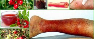
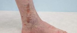
Swelling, pain when walking or bending the lower leg, pain in the inner thigh and foot, reddened skin and cramps are all signs of the development of the disease. If the acute form occurs, the person begins to suffer from shortness of breath, his temperature rises, he becomes dizzy and loses consciousness. You can compare the signs of thrombosis of the lower extremities from the photo, but the diagnosis must be made by the attending physician.
- In the upper extremities, thrombosis is less common (subclavian vein thrombosis), and it is difficult to distinguish its initial symptoms from arm injury. They manifest themselves in swelling, pain and blueness. The patient feels a burning sensation, his hands go numb, and his skin becomes insensitive.
- The vessels of the brain connect with veins and arteries, where blood clots can also appear, and this leads to a stroke. Symptoms of cerebral thrombosis are more pronounced than with thrombosis of other vessels. The disease is accompanied by headaches, dizziness, decreased hearing and visual acuity, loss of consciousness, and periodic convulsions. The person is feeling sick.
- Blood clots also settle in the veins of hemorrhoids, this is considered a complication of hemorrhoids. Signs of thrombosis of hemorrhoids (Haemorrhoids): pain, the affected area itches, fever, swelling.
- The central retinal vein is also affected by thrombosis. Signs of the disease may not be expressed, and the person loses vision as a result.
- Thrombosis of the mesenteric veins of the intestine is manifested by prolonged abdominal pain. Other signs: bloating, high fever, vomiting and nausea. In the early stages, it is difficult to determine the disease by symptoms. Usually they talk about a complication of the pathology.
- If the femoral and iliac veins are affected, then we are talking about ileofemoral thrombosis. It is accompanied by swelling of the legs (the skin changes color from reddish to blue), when pressure is applied, brown marks appear, the legs and groin hurt, and the body temperature rises.
Treatment and diagnosis of venous thrombosis
How is thrombophlebitis diagnosed?
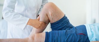
A phlebologist is able to determine and prescribe adequate treatment for venous thrombosis of the lower extremities. In especially severe cases, consultation with a vascular surgeon is required. To diagnose this disease, in addition to visual examination and entering data into the patient’s medical record, they can use:
- Duplex ultrasound angioscanning using color Doppler mapping allows you to determine thrombosis localized below the level of the inguinal ligament. Shows the condition of the vessels, features of the movement of blood flow, thanks to which it is possible to determine the places of blockage and parietal thickenings.
- X-ray contrast venography - a contrast agent is injected into the venous system. This method is often used when the previous research is unreliable. Phlebography is especially good at identifying clots in the groin area.
- Magnetic resonance imaging (MRI) and magnetic resonance angiography (MRA) - allow you to examine peripheral and central veins. MRI is used to visualize blood clots, and MRA is used to visualize blood vessels.
- Radionuclide scintigraphy is the filling of a vein with a radioactive drug, the movement of which through the deep veins is monitored using a gamma camera. The method allows you to determine the speed and nature of blood flow. This technology is used in extremely difficult cases.
- Plethysmography is a study that is most widespread in Western countries. It is used to measure the degree of blood filling of the venous trunk during physical activity and rest. As a result, it is possible to determine the difference in the electrical resistance of the limb.
The use of laboratory tests is additional. They are not able to give a complete picture of the boundaries of the spread of phlebitis and thrombosis.
Great importance is given to differential diagnosis, which makes it possible to exclude other diseases and pathological conditions that have a similar clinical picture to thrombophlebitis:
- cellulite - subcutaneous fat tissue is affected, mainly in the lower extremities, accompanied by frequent cramps, changes in skin color, and dryness;
- lymphedema - depending on whether the disease is primary or secondary, it affects one or two limbs, is characterized by swelling of the lower leg, pain, fatigue;
- compression of the vessel by neoplasms or inflamed lymph nodes;
- stretching, rupture of muscle tissue - intermuscular hematoma puts pressure on the venous trunks, causing them to narrow;
- rupture of Baker's cyst - diagnosis is carried out using ultrasound.
Diagnosis of thrombosis
Primary diagnosis is based on a detailed history and anthropometry (calf or thigh circumference).
The Wells scale is used to diagnose acute thrombosis and diagnose pulmonary embolism [8,9]. Instrumental diagnostics include compression or duplex scanning of veins, Doppler ultrasound with compression of veins, impedance plethysmography, pulmonary angiography, radiocontrast or MRI venography [6,9], CT and MRI angiography [7,9].
To diagnose arterial thrombosis, physical tests are used (6-minute walk test, treadmill test), determination of pulsation of superficial arteries (arteries of the dorsum of the foot), duplex scanning of the arteries of the extremities, angiography (X-ray of a vessel filled with a radiopaque substance) and measurement of transcutaneous oxygen tension [ 7].
Drug treatment
It consists of prescribing anticoagulants, angioprotectors, non-steroidal anti-inflammatory drugs, and certain enzyme preparations. And antibiotics in case of acute purulent thrombophlebitis.
Special ointments are also used: Heparin, Essaven-gel, Ketonal 5% cream, Lyoton-gel and some others. Ointments such as Levomekol, Levosin and others, intended for the treatment of superficial skin lesions and ulcers, only worsen the problem.
Surgery is prescribed when the possible development of pulmonary embolism is suspected after differential diagnosis. Also, the surgical method gets rid of blood clots that have accumulated during acute thrombophlebitis in the bed of the great saphenous vein. The main indication for surgical treatment is signs of movement of part of the clot noticed during ultrasound.
Folk remedies
Along with drug treatment, traditional methods can give good results. However, you should not blindly believe and thoughtlessly try everything on yourself. The doctor should control the treatment regimen, the admissibility and effectiveness of folk remedies.
- White cabbage. Cabbage leaves need to be softened, one side greased with oil and secured with a bandage on the painful area. Leave the compress on all night.
- Kalanchoe pinnate. Grind the leaves and fill the jar halfway with them. Fill the rest of the jar with vodka. Next, leave for 7 days in a dark place, shaking the contents periodically. Strain the finished tincture. Rub the sore leg from the foot to the groin in the direction of the vein. So for a month 2 rubles/day.
- Apple vinegar. Mix 2 teaspoons of vinegar and honey in a glass of water. Drink half a glass before meals 2 times a day.
- Mumiyo. You can make a tincture for internal use and ointment:
- for the tincture you need to mix 10g of mumiyo with 0.5l of water - drink 1 tablespoon/1 ruble per day, 10 days
- For the ointment, you need to mix mumiyo with Vaseline (or peach oil) in a ratio of 1 to 5 - smear 3 times a day, leave for 1 hour, 10 days.
Treatment with leeches
In acute thrombophlebitis of the lower extremities, leech therapy is practiced. They are dispersed in the direction of the affected vein, up to a maximum of 10 pieces. for 1 time. And not on the vein itself, but at a distance of at least 1 cm from it, staggered. in a checkerboard pattern, if leeches are placed in quantities of up to 10 pieces along the painful vein in a checkerboard pattern at a distance of at least 1 cm from the vein. Up to 60 leeches can be delivered per course.
The use of leeches is not suitable for everyone and not always. Minor reactions are also possible - redness, swelling of the skin, itching. This is an allergy to bites, but it is not serious and will go away on its own fairly quickly. Hyperpigmentation rarely occurs. But in case of individual intolerance, Quincke's edema is possible.
Prevention of thrombophlebitis
Prevention of thrombophlebitis of the lower extremities helps prevent relapses. Therefore, patients after acute thrombophlebitis are advised to strictly follow the doctor’s instructions.
- Diet. Normalize the intake of nutrients, fats, carbohydrates, salt. It is worth increasing the proportion of fruits, vegetables, and berries in the diet. Additionally, take ascorbic acid and introduce bioflavonoids. It is worth reducing or even eliminating wheat and pearl barley, potatoes, beets, bananas, grapes, fatty meat and dairy products, confectionery, milk chocolate and candies, fast food, smoked alcohol. Drink up to 2.5 liters/day of clean water.
- Try to distribute the load evenly throughout the day, rest more often. But at the same time, maintain a reasonably active lifestyle. Spend more time outdoors.
- Clothes should be comfortable. Tight jeans, tight belts, corsets, uncomfortable shoes, tight underwear that can put too much pressure in the groin area, etc. will only be harmful. But special stockings or knee socks will do the trick.
- It is better to give up bad habits forever.
- Temperature conditions. Hypothermia, like overheating, is best avoided. It is worth excluding long stays outside in cold and hot weather, swimming in cold ponds, baths, hot baths.
- Take medications prescribed by your doctor.