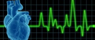
Electrocardiography is one of the most informative ways to obtain information about the condition of the heart. For an uninitiated person, not only the tape itself is incomprehensible, but also the conclusion of a functional diagnostics specialist.
The vertical position of the electrical axis of the heart can often be found in conclusions. Moreover, this situation can be both the norm and a sign of heart disease.
Conducting system of the heart, its role in determining EOS
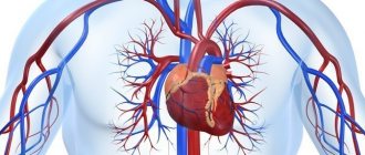
The conduction system refers to the entire set of anatomical elements that ensure contraction of the organ. All these bundles, nodes and fibers consist of special, modified muscle fibers that have automaticity and the ability to conduct excitation to the lower parts of the heart.
The system that ensures the conduction of a depolarization wave through this organ consists of:
- The sinus node, which, in fact, is normal and sets the rhythm of contractions for the entire organ.
- Conducting fibers that transmit electrical impulses from the sinus node to the atrioventricular and atria.
- Atrioventricular node.
- The Hiss bundle, through which excitation should spread through the ventricles.
The sum of the excitation vectors on the first standard lead is the electrical axis.
Determination of the electrical axis of the heart using ECG
Determining the electrical axis of the heart using an ECG can be done in several ways. The simplest and fastest, although the most inaccurate option - it only allows you to navigate the situation in general terms.
In the most simplified, “student” version, it looks like this:
- The R waves are highest in lead 2, which roughly corresponds to the normal axis of the heart.
- If these same teeth turn out to be the largest in the first lead, then this indicates a horizontal variant of the axis location.
- The highest R's in the third lead of the electrocardiogram indicate a vertical electrical axis.
A more accurate determination is possible using another method. To do this, you will need special diagrams or tables, as well as certain calculations. It is necessary to count the algebraic sum of the teeth of the ventricular complex (taking into account the negative teeth) in the first and third standard leads.
Determining the sum itself is not difficult - just measure the size of each tooth in millimeters, and then find their sum, taking into account the negative values of those teeth that are located under the isoelectric line. Next, using the table, find the intersection points of the obtained values - this will be the alpha angle.
EOS deviation to the right
When the EOS deviates to the right with an angle of +1200, the maximum positive sum is R-(Q+S) in lead III, the maximum negative sum is R-(Q+S) in lead aVL. The algebraic sum of the R waves - (Q + S) is equal to or close to zero in lead aVR.
ECG 7. EOS deviation to the right. The angle is +1200
ECG source.
When the EOS deviates to the right with an angle of +1500, the maximum positive sum is R-(Q+S) in lead III, the maximum negative sum is R-(Q+S) in lead aVL. The algebraic sum of the waves R - (Q + S) is equal to or close to zero in lead II.
ECG 8. EOS deviation to the right. The angle is +1500
ECG source.
Vertical position of the electrical axis of the heart - what does this mean?
In most cases, this only refers to the anatomical features of a particular person. However, with a sharp deviation, this situation may indicate a number of diseases.
For example, these could be:
- Pulmonary stenosis, both congenital (hence, such an ECG can be recorded in children, including young children), and acquired. The axis changes due to myocardial hypertrophy.
- Cor pulmonale and primary pulmonary hypertension have a similar mechanism for changing the electrical axis. In such cases, hypertrophy of the right ventricle also occurs, which will lead to characteristic changes in the electrocardiogram.
- A defect of the interatrial septum, with sufficient size of such an opening, can also lead to a change in such an electrocardiographic indicator as the electrical axis. The mechanism of development of changes is approximately the same as in the case of cor pulmonale and pulmonary hypertension.
- It can also be observed in coronary heart disease, in which, due to restriction of the lumen of the coronary arteries, myocardial ischemia occurs, which, with severe stenosis, can develop into a heart attack.
What options for placing an EOS can a healthy person have?
There are three main options for its location:
- Horizontal. This variant is most often found in obese people.
- Normal. Typical for people of average build.
- Vertical. It is often determined in asthenics, in whom the heart literally “hangs” in the chest cavity, which is associated with the characteristics of the physique.
All three options, if this is not a sharp deviation of the axis, in the absence of clinical or detectable abnormalities on the electrocardiogram, are a variant of the norm and do not pose any threat.
This is nothing more than an individual characteristic of a particular organism. However, a sharp deviation to the left or to the right may indicate a number of serious heart diseases, which can only be determined by a combination of clinical manifestations and data from additional research methods.
What positions of the EOS exist normally and what is the difference between them?
The muscle mass of the left ventricle (LV) is commensurately greater than the right. Therefore, the electrical processes that occur in the LV are stronger, and the EOS vector will be directed in this direction. If we project the heart onto a coordinate system, then the left ventricle will be located in the range of values +400+700 (which is considered the normal orientation of the axis).
However, the individual characteristics of the heart structure and the physique of each patient vary the EOS position within the range from 00 to 900.
Variants of the normal position of the EOS
The normal position of the EOS is the angle α from 300 to 690, the height RII≥RI>RIII, and in III and VL the R and S teeth are approximately the same. The cardiac axis is clearly perpendicular to lead III.
Horizontal position of the EOS - the orientation of the axis coincides with the placement of standard lead I (RIII >SIII), angle α from 0 to + 300. Occurs in hypersthenics or short people with a wide chest, as well as at the peak of exhalation, with abdominal obesity, in II and III trimester of pregnancy. The heart “lies” on the dome of the diaphragm.
Semi-horizontal position of the EOS - the cardiac axis is at an angle of 900 to standard lead III (RIII=SIII), angle α=+300.
Vertical electrical position of the heart - the direction of the EMF is perpendicular to lead I (RI=SI), angle α=+900. This type is typical for tall, asthenic people with a narrow chest, at the end of a deep breath. The heart “hangs” between the roots of the lungs on a vascular bundle.
Semi-vertical electrical position of the heart - the direction of the axis is parallel to lead II and vaguely perpendicular to lead I (RII>RIII>RI), angle α from +700 to +900.
The presence of transitional types of EOS position is explained by the fact that asthenics or hypersthenics in their pure form are rare, “intermediate” types of constitution are widespread.
Rotation around its horizontal or vertical axis is also sometimes determined (the rotation of the apex anteriorly or posteriorly relative to its normal position).
The horizontal axis of the heart is a symbolic bisector, laid through its apex and base.
The longitudinal axis is characterized by the location of the gastric QRS complex in the thoracic leads, the axes of which are located frontally. It is necessary to identify the turning zone and evaluate the QRS structure in V6.
Types of heart orientation in the frontal plane:
- Normal position - the turning zone is located in lead V3, the R and S teeth are identical in height. In V6, the QRS complex acquires a qR or qRs configuration.
- Clockwise rotation is a turning zone in the area of leads V4-V5, and in V6 the complex has the form RS. Often combined with the vertical position of the EOS and its deviation to the right.
- Counterclockwise rotation - the turning zone is shifted to V2. In leads V5-V6, a deepening of Q is observed (not to be confused with the coronary one), and the QRS complex takes on the shape of qR. Combined with the horizontal position of the EOS and its deviation to the left.
Rotation of the heart along the vertical axis:
- The apex is anterior - the QRS complex in leads I-III takes the form qRI, qRII, qRIII.
- At the apex posteriorly, the QRS complex takes on the form RSI, RSII, RSIII.
Vertical position of the EOS - is it dangerous?
During pregnancy
The vertical position of the EOS during pregnancy is uncommon. This is due to physiological changes in a woman’s body - the increasing size of the uterus affects the location of other internal organs. In the case of the heart, it usually deviates to the left and acquires a horizontal position.
The vertical location option, especially in the case of late pregnancy, requires additional research, as it may indicate the development of pathology of this organ.
In children
The vertical position of the EOS in children in the vast majority of cases is not a sign of any disorders - it is only an age-related feature, which, as the body develops, will most likely turn into normal (horizontal or remain vertical, it all depends on the individual characteristics of a particular organism).
ECG formulation
The conclusion of the ECG may include the following wording: “Vertical EOS, sinus rhythm, heart rate per minute. – 77” – this is normal. It should be noted that the concept of “rotation of the EOS around an axis”, the mark of which may be in the electrocardiogram, does not indicate any violations. Such a deviation in itself is not regarded as a diagnosis.
There is a group of ailments that differ precisely in the vertical sinus EOS: various types of cardiomyopathy, especially in the dilated form; ischemia; congenital abnormalities; chronic heart failure.
With these pathologies, a violation of the sinus rhythm of the heart occurs.
Will they be accepted into the army if the EOS is in a vertical position?
The army may not cancel the vertical position of the EOS. It all depends on the reason. If such its location is due to the individual characteristics of the body, and is not a manifestation of pathology of the heart or large vessels, then there are no grounds for exemption from military service.
A completely different situation arises when such changes in the electrocardiogram are a sign of a disease (often a slight deviation is a variant of the norm, but a sharp one most likely indicates pathology).
Then this issue is resolved based on the characteristics of the clinical picture and the severity of the degree of cardiac dysfunction.
Concept and specifics
There are several options for the location of the electrical axis of the heart; it changes its position under certain conditions.
This does not always indicate disorders and diseases. In a healthy organism, depending on the anatomy and body composition, EOS deviates from 0 to +90 degrees (+30...+90 is considered the norm, with normal sinus rhythm).
The vertical position of the EOS is observed when it is within the range from +70 to +90 degrees. This is typical for people of thin build and tall stature (asthenics).
Intermediate types of body composition are often observed. Accordingly, the position of the electrical axis of the heart changes, for example, it becomes semi-vertical. Such displacements are not a pathology; they are inherent in people with normal body functions.
An example of wording in the conclusion of an ECG may sound like this: “EOS is vertical, sinus rhythm, heart rate - 77 per minute.” - this is considered normal. It should be noted that the term “rotation of the EOS around its axis,” which may be noted in the electrocardiogram, does not indicate any pathologies. In itself, such a deviation is not regarded as a diagnosis.
The vertical position of the EOS is observed when it is within the range from +70 to +90 degrees.
There is a group of ailments that are characterized by vertical EOS:
- ischemia;
- cardiomyopathy of various nature, especially in the dilated form;
- chronic heart failure;
- congenital anomalies.
Sinus rhythm is disturbed in these pathologies.