One of the most accurate and safe methods for diagnosing veins in the lower extremities is ultrasound scanning. Using the procedure, it is possible to determine the characteristics of blood flow in the legs and identify possible pathological conditions.
Ultrasound scanning of the arteries of the lower extremities RUB 2,900.
Ultrasound scanning of the veins of the lower extremities RUB 2,900.
Ultrasound scanning of vessels (arteries and veins) of the lower extremities RUB 5,400.
All prices Make an appointment
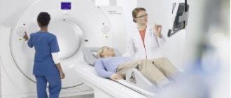
At the International Hemostasis Clinic, duplex scanning is performed using high-precision equipment - a Voluson E8 ultrasound machine (USA). The procedure is carried out by experts in the field of diagnostics, professors and doctors of medical sciences. That is why we can guarantee 100% accuracy and efficiency of the examination.
Types and purposes of ultrasound of the veins of the lower extremities
In recent decades, ultrasound diagnostics has become a simple, inexpensive, and most importantly, accessible research method.
The quality and capabilities of ultrasonic devices are constantly being improved, their classification is constantly expanding. The great advantage of the technique is its non-invasiveness. Vein ultrasound is no exception. Modern ultrasound scanners allow a thorough diagnosis of the venous system.
The main goals of ultrasound of the veins of the lower extremities are to assess the condition of the vessels (their diameter, course, relationship with other structures, wall condition), examine blood flow parameters, and identify blood clots. The article will talk about the types and purposes of ultrasound examination of blood vessels, as well as how ultrasound of the veins of the lower extremities is performed.
Due to the constant improvement of devices, many terms have appeared to denote various components of ultrasound examinations and their combinations.
Let's try to figure out what ultrasound, DS, ultrasound, ultrasound of veins are.
- Doppler ultrasound (Doppler ultrasound) is a method based on the Doppler effect. This effect consists of changing the frequency of an ultrasonic wave when reflected from a moving object. In the lumen of the vessel, such objects are formed elements of blood, for example, red blood cells. Computer processing of reflected signals allows one to obtain a curve of the speed characteristics of blood flow. Currently, ultrasound scanning in its pure form is not used, but is included in the structure of other studies.
- DS (duplex scanning (USD)) - allows you to visually evaluate the vessel - its wall, course, lumen, as well as conduct an ultrasound scan of any part of the vessel - i.e. in addition to examining the vessel, show the characteristics of blood flow at any point. Modern devices also use coloring of blood flows in blue and red colors, based on the Doppler effect (the so-called color Doppler or color Doppler mapping (CDC)). Combining duplex scanning with color flow is also called triplex scanning.
- USAS (ultrasound angioscanning) - a synonym for DS, is the same.
- Ultrasound of veins (ultrasound examination of veins) - this term refers to the entire combination of the above methods. Modern devices provide all the components of the study - ultrasound, DS, and color Doppler mapping (CDC).
Currently, when a doctor prescribes an ultrasound scan of the veins of the lower extremities, an ultrasound scan of the veins of the lower extremities, he means that the patient will undergo an ultrasound scan of the veins according to all possible parameters.
Ultrasound equipment
To conduct research, devices are used that are designed for visual diagnostics of the human body. Ultrasound rooms are equipped with multifunctional equipment designed to meet stringent requirements. Modern models of devices have ultra-precise focusing and compact dimensions. They provide high quality images.
To improve performance, the devices are equipped with various modes. For example, two-dimensional visualization, as well as color and power Doppler. Many models are equipped with glare-free monitors that rotate freely around an axis and tilt in the desired direction. It is possible to connect several sensors simultaneously. The received information is recorded on the hard drive and stored in a database.
When developing ultrasound machines, new technologies are being introduced: grain suppression, image detailing, automatic image optimization, wireless information transfer. The scanners are easy to operate. If necessary, a specialist can adjust the settings. The image is instantly transferred to the monitor. The doctor examines the details and assesses the situation.
There are two types of equipment on sale: stationary ultrasound and portable ultrasound. Stationary devices are installed in rooms designed for ultrasound examinations of leg vessels. They are impressive in size and provide reliable diagnostics. Portable devices are portable and small in size. Such devices are designed to conduct research in any place: ward, ambulance, emergency department. They are indispensable in cases where it is necessary to urgently determine a person’s condition and take the right actions.
Who needs to undergo Doppler ultrasound of the extremities?
An ultrasound examination of the veins is usually prescribed by a doctor (surgeon, phlebologist, therapist). Indications for it can be a wide variety of conditions. Most often, the doctor prescribes a study if there are complaints and suspicions of diseases of the venous system. In other cases, ultrasound of the veins is prescribed to exclude hidden pathology of the venous system (for example, before operations, during pregnancy).
The main indications for prescribing ultrasound examination of veins are:
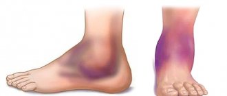
- Swelling of one or both legs
- Leg pain
- Feeling of heaviness and fatigue in the legs
- Night cramps in the legs
- Varicose veins, spider veins
- Before any operations
- Late pregnancy
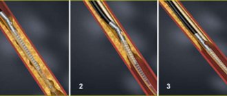
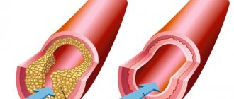
Deep vein thrombosis
Thrombosis in the early stages may be asymptomatic.
The main classic symptoms of deep vein thrombosis are: increased temperature in the affected area. The general body temperature may rise to 39 degrees. Bursting pain and heaviness in the legs appear, the color of the skin is bluish. During the first two days after the formation of thrombosis, the symptoms are mild. As a rule, this is not pronounced pain in the calf muscle, intensifying during movement and when touched, and swelling. Subsequently, the condition worsens and swelling increases. The risk of blood clot rupture increases. Within a few days after the formation of a blood clot, superficial veins appear. Indications for ultrasound scanning for the following symptoms: - Presence of swelling of the legs; -Cold feet; -Pain in the calf and feeling of “heavy legs”; - Convulsions; - Suspicion of changes in the blood vessels of the legs (especially during pregnancy); - Periodic numbness of the legs; -Itching in the legs, without external skin changes; -Diabetes; -Decreased vascular tone; - Suspicion of atherosclerosis; - Suspicion of varicose veins or other changes in venous blood flow; - Trophic changes in the skin; -Heart problems (angina or heart attack); - Migraines and fainting; -Pain when walking or exercising; -Increased cholesterol levels.
How is the examination done?
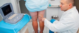
Many patients are concerned about the question of how ultrasound examination is performed, whether there are any contraindications or painful sensations during the examination.
Ultrasound diagnostics is non-invasive, painless, does not cause any discomfort, and has no contraindications. An ultrasound can be performed either by an ultrasound diagnostics doctor or by a surgeon or phlebologist who knows this method and has undergone appropriate training.
The examination of the veins of the lower extremities is carried out from the inguinal fold to the ankles. The patient completely frees his legs (panties can be left on) and lies down on the couch. The doctor applies a special gel to the lower limbs, along which the sensor will be moved during the examination. The gel is needed to ensure good contact between the skin and the sensor so that there is no obstacle to the passage of ultrasonic waves. The examination can be carried out both lying down and standing. At certain moments, the doctor may ask you to hold your breath, strain, bend your leg, etc.
The study lasts from 10 to 40 minutes (depending on the structural features of the veins and the identified pathology). For example, detected varicose veins increase the examination time, because it is necessary to carry out a number of additional tests. At the end of the study, the doctor gives the patient paper napkins to remove any remaining gel. The gel is absolutely harmless and does not leave stains on clothes.
Then the doctor writes a conclusion and hands it to the patient. It should be remembered that the conclusion itself is not a diagnosis, but only describes the identified ultrasound signs. The diagnosis can only be made by a clinician (surgeon, phlebologist), based on a comparison of the ultrasound picture, examination data and other examinations. In many cases, an ultrasound of the veins is performed not by an ultrasound diagnostics doctor, but by a phlebologist who is proficient in vein ultrasound. Then a clinical diagnosis can be made immediately.
Contraindications and restrictions
There are no direct contraindications to Doppler ultrasound of the vessels of the upper or lower extremities. But the information content of the procedure is noticeably reduced if the person being examined makes movements with his arms and legs during an ultrasound. Therefore, for patients who cannot remain stationary for some time due to psychological, neurological or other disorders, it is better to consult a doctor first. He may suggest another diagnostic method or recommend taking sedatives before the procedure.
How to decrypt data
Only a doctor can determine what an ultrasound examination of the veins of the lower extremities shows.
The doctor evaluates:
- Vein diameter
- Condition of the vascular wall (irregularities, thickening)
- The condition of the veins of the right and left legs is compared
- Valve condition
- Presence of pathological blood flows (refluxes)
- Vessel lumen (presence of blood clots, their structure, level of extent, apex)
- Presence of varicose veins
- Presence of incompetent perforators (veins connecting superficial and deep veins)
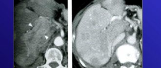
If during the examination the doctor notices changes in other structures that are not related to the veins (for example, pathology of the arteries, Baker's cyst, altered lymph node), he may note this in the protocol or recommend additional research.
Based on the data received, the doctor writes a conclusion; if necessary, photographs are attached to the conclusion.
Example of a conclusion: “Echo signs of varicose veins of the lower extremities. Insufficiency of the great saphenous vein on the left. Incompetent perforator of the left leg." It should be noted that the classification of varicose veins according to this conclusion is impossible - the stage is determined by the attending phlebologist based on a comparison of the ultrasound picture with clinical signs.
results
Based on the results of ultrasound examination of veins and arteries, a protocol is formed in which the doctor indicates the patency of the veins, the degree of blood flow disturbance, wall parameters, the presence of blood clots and cholesterol plaques, their parameters, peak blood flow velocity in different areas and other data.
The results obtained are compared with established standards. When decoding, the patient's age is taken into account. For young and middle-aged people, the norm is:
- absolute patency of veins and arteries;
- wall thickness without seals;
- absence of pathological reflux and valvular insufficiency;
- normal blood flow velocity with a permissible deviation of 10-15 cm/s: in the femoral artery 75, in the distal artery - 56, in the iliac artery 96, popliteal 53, in the tibial artery 46 cm/s.
If any abnormalities are detected, the doctor will inform the patient and give directions for additional tests and studies.
Where to take the study
Currently in Moscow there are many medical centers that offer ultrasound diagnostics. What should you look for in order to get to a good specialist?
The doctor must have extensive experience, work experience, and have positive reviews from other patients. It is very good to have an ultrasound of the veins done by a doctor who not only knows ultrasound diagnostics, but is also a surgeon or phlebologist. Such a doctor has the most complete understanding of the anatomy and functioning of the venous system, knows well how ultrasound of the veins of the lower extremities is performed, and can also make a clinical diagnosis and prescribe treatment immediately after the ultrasound.
Dr. Elshansky Igor Vitalievich fully meets these requirements, is a phlebologist surgeon, and also has advanced training in ultrasound diagnostics in angiology, has been involved in the diagnosis and treatment of vascular diseases of the lower extremities for many years, Ph.D.
Rules for preparing for research
2-3 days before the test, it is important to limit the consumption of alcohol and tonic drinks, including strong coffee, tea, and energy drinks. On the day of the study, you should not smoke, play sports or do heavy physical work, as this causes vascular tone.
It is important to limit medications that affect blood clotting and blood flow. The diagnostician must know about the medications taken by the patient. If they cannot be canceled, the specialist will conduct a study taking into account the effect of the drugs on the blood vessels.
What does ultrasound examination of leg blood vessels evaluate?
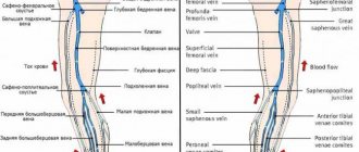
The procedure allows you to determine the functioning of the venous collectors, namely:
- The level of patency of deep and superficial veins and the severity of existing pathology.
- The degree of consistency or insufficiency of valves in the venous vessels.
- Detect a blood clot and its level of mobility in the vein.
- Assess the degree of consistency of the perforating veins.
During the study, the level of blood movement through the arteries, vascular patency, the presence or absence of curvatures and aneurysms are checked.