Infographics for the “bio/mol/text” competition: A clinical blood test is the most common laboratory test prescribed by a doctor when we come to an appointment and complain of feeling unwell. “Blood from a finger, tomorrow from 8:00 to 9:30, on an empty stomach, Nth office,” several generations have invariably heard this phrase. However, blood testing technology has undergone great changes in recent decades and has moved from manual methods to automatic ones. Let's figure out how your grandmother's blood was analyzed and why things are done differently now.
Preparation rules
- To donate blood from a finger prick, you need to come to the laboratory in the morning (usually collection occurs from 7.30 to 10 o’clock).
- The test must be taken on an empty stomach, that is, you cannot eat in the morning, you can only drink plain water. The last meal should take place the night before - no later than 8-12 hours before the procedure.
- You can eat a day earlier, but it is recommended that a day or two before the analysis, in order to avoid getting distorted results, you should avoid fatty foods and alcoholic beverages.
- The day before, you should avoid physical and emotional stress.
- In the morning before the procedure, you should refrain from smoking.
How is blood taken and is preparation needed for the procedure?
Donating blood is a simple and virtually painless procedure that even infants can easily tolerate. Biological material for general examination is taken mainly from a finger, but to check against an extended list of parameters and in some other cases, doctors prescribe venous puncture.
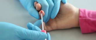
To prevent infection and take clean material, the nurse wipes the pad of one of the fingers of the left hand with a swab soaked in alcohol. Immediately after this, a tiny incision about 3 mm deep is made with a special instrument, a scarifier. The blood protruding from the incision is collected with a collection pipette and poured into a tall tube. A small portion of the liquid is smeared onto a glass slide.
If a doctor prescribes a venous puncture, a rubber tourniquet is placed on the patient's forearm to slow down the blood flow below the dressing. Then the puncture site on the inner surface of the elbow is disinfected with alcohol, after which a hollow needle is inserted into the vein. Through it, blood from the vessel fills a special test tube, and a small amount of biomaterial is applied to a glass slide.
No special preparations are required for the procedure. The only thing doctors can ask is not to have breakfast before collecting biomaterial, since eating food can distort the results obtained.
In some cases, the analysis has to be done several times over a set time period, including to track the effectiveness of the prescribed treatment. To obtain the most accurate data for comparison, repeat blood donation is carried out approximately in the same period of time as the first time.
General analysis
It can be shortened or expanded. The first option includes indicators such as the level of hemoglobin and all blood cells (erythrocytes, platelets, leukocytes), as well as ESR (erythrocyte sedimentation rate).
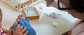
Capillary blood collection from a child
In a detailed analysis, other indicators are added, including:
- hematocrit;
- width of red cell distribution;
- average erythrocyte volume;
- average content of hemoglobin in the red cell;
- leukocyte formula and others.
Times change
Changes are visible already at the stage of blood sampling: if previously the doctor collected blood in several test tubes with reagents, a glass capillary and made a smear on the glass, now very small volumes are used - from 12 to 150 μl [15] of blood is enough to examine it for all parameters.
Let's take a look into a modern hematology laboratory. Wow! Everything is cluttered with equipment, and the laboratory assistant is nowhere to be seen... Maybe he went off to make himself some coffee? He won't make it in time! The blood test will be ready in a minute, and the device will give the result in the form of a paper tape with numbers and abbreviations, behind which all sorts of parameters are hidden.
Modern hemoanalyzers are divided into several classes, depending on what they can do. Each subsequent class is a new stage of evolution - faster, more accurate, more perfect. The use of a combination of technologies works wonders: if the first analyzers could determine eight blood parameters and did not distinguish between types of leukocytes [16], then the latest devices are able to differentiate up to seven populations of leukocytes [17] and in total examine more than 40 blood characteristics.
As Arthur C. Clarke said, “Any sufficiently advanced technology is indistinguishable from magic.” Indeed, the most detailed results in such a short period of time cannot but surprise. But all magic is based on physical laws. And although names like electrical impedance, light scattering and photometry are a little intimidating at first glance, now we will understand what principles underlie each analysis technology.
Population census
In the middle of the last century, Wallace Coulter made a revolution by patenting the technology of automatic cell counting. One of the leaders in the production of hematology analyzers, the Beckman Coulter company, is named after him [18]. The aperture-impedance method (or Coulter method) is based on the registration and analysis of impulses that occur when a cell passes through an aperture from one container to another, each of which contains an electrode. When there is no cell in the hole, current flows freely through the electrolyte between the electrodes under the influence of an electric field. To direct the cells to the aperture, a pump is used that pumps out liquid from one container, and the formed elements rush into it. Passing through the aperture, the cell displaces a volume of electrolyte equal to its volume from one container to another. In this case, a pulsed change in resistance (impedance) occurs - the cell membrane creates an obstacle to the free flow of current. At the same time, the current strength recorded by the meter also changes. The number of generated impulses corresponds to the number of formed elements, and the height of the impulse is proportional to the volume of the cell [19]. Using information about the number and volume of formed elements, the device can calculate hematocrit, the average concentration of hemoglobin in an erythrocyte, the width of the distribution of cells by volume and many other parameters [15].
Divide and rule
Differentiation of leukocytes into populations can be carried out using a Coulter counter, but a problem arises - different types of leukocytes are similar in volume and similar pulse amplitude does not always allow one to accurately determine the cell type. What should I do? To solve this problem, combinations of reagents are selected that change the size of cells to varying degrees so that it becomes possible to separate them [15].
But the most common method of differentiation is flow cytometry [20]. The method works as follows: cells in the flow are alternately irradiated with a laser, and the resulting light scattering and fluorescence signals are recorded by detectors and analyzed. In order to correctly determine membership in a population, several parameters are examined at once. Thus, light scattering at a small angle provides information about the relative size of cells, and light scattering at a right angle allows you to “look” inside the cell and study its internal structure - the presence of granules and the shape of the nucleus. Another parameter - fluorescence - can tell about the number of antigens and their appearance on the surface of cells - this cannot be accurately determined by eye. Unlike manual differentiation methods, not 100–200 cells are analyzed, but tens of thousands per second! And each leukocyte receives an individual approach: hydrodynamic focusing ensures that the cells line up in a row and are irradiated one by one in the flow cell. The result of the count appears on the screen in the form of scatter diagrams, where cells with similar properties form clusters.
Precipitated: 2.0
Modern instruments can measure ESR in two fundamentally different ways. The first is the modified Westergren method. The principle of operation has not changed since your grandmother's time, but due to automation it has become faster and more accurate. The second is measuring the kinetics of erythrocyte aggregation using an optical method [21]. It happens like this: an anticoagulant is added to the blood, the tubes with blood are placed in a rotor, where automatic mixing occurs. After this, the analyzer takes part of the blood into a microcapillary, where it accelerates and abruptly stops (the so-called “stopped stream” method). The stop causes aggregation of red blood cells, and at this moment the optical density of the blood is determined using a photometer - the denser the red blood cells are located, the less light will pass through the sample. The device uses the data obtained and builds a sedimentation curve - its analysis will allow you to present the result in the usual units of ESR measurement [22], [23].
Photo for memory
To determine hemoglobin concentration, the International Committee for Standardization in Hematology recommends the methemoglobin-cyanide method. However, a different test that does not use toxic cyanide is now widely used. Meet the SLS method. It is named after the main reagent - sodium laurithyl sulfate. SLS destroys red blood cell membranes, after which it binds to heme groups and forms stable complex compounds. They are analyzed photometrically - laser light is passed through the blood sample. Complex compounds absorb part of the light, as a result of which the intensity of the output light flux weakens. Attenuation is measured using a photosensor and the resulting data is converted into hemoglobin concentration units [24].
Tools
Many people are concerned about their own safety during the test, so they may have questions about what is used to pierce and take blood. Today, almost all medical institutions have switched to using disposable finger pricking instruments. This tool is called a scarifier. It must be removed from the unopened package in front of the patient. It should be said that such a puncture is quite painful, so children really do not like the procedure.
Today, donating blood can be painless. A new device is being used more and more often when blood is taken. This is an automatic lancet in a plastic case. The needle quickly penetrates the skin, so pain is not felt. The new lancets have many advantages:
- the sterile needle or blade is located inside the body, which ensures the safety of patients and medical personnel;
- the reliability of the trigger mechanism eliminates accidental release of the needle or blade;
- reuse is eliminated thanks to the automatic return of the needle or blade;
- the shape of the needle ensures a reduced pain effect;
- the puncture is targeted, its depth is controlled;
- convenient body shape.
Fence algorithm
To work, the laboratory assistant must prepare:
- sterile scarifier;
- cotton wool;
- alcohol;
- tincture of iodine;
- ether.
Disposable scarifier - a tool for pricking a finger
The algorithm and technique for taking are as follows:
- The patient sits opposite the laboratory assistant. The hand (usually the left) lies on the table.
- The puncture site is disinfected with alcohol and degreased with ether.
- Using a disposable scarifier, a puncture is quickly made in the pad of the ring finger, immersing the tool to the full depth of the cutting part (approximately 2-3 mm).
- The first drop of blood is removed using dry cotton wool.
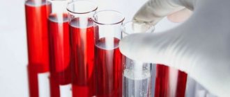
- For the study, use the second and subsequent drops of blood, which are collected using a glass adapter, then placed in test tubes and signed.
- After the blood has been taken, the injection site is treated with alcohol or iodine and clamped with a cotton swab until the blood stops completely.
The algorithm for collecting capillary blood from a child is exactly the same as from an adult.
Competition "bio/mol/text"-2019
This work was published in the category “Visually about the beloved” of the “bio/mol/text” competition-2019.
The general sponsor of the competition and partner of the Skoltech nomination is the Skoltech Center for Life Sciences.
Competition sponsor: the largest supplier of equipment, reagents and consumables for biological research and production.
The audience award was sponsored by BioVitrum.
"Book" sponsor of the competition - "Alpina Non-Fiction"
Why from the ring finger?
Perhaps someone is interested in which finger the blood is taken from and why. The sampling occurs from the ring finger, although it is allowed from the middle or index finger. A puncture, like any violation of the integrity of the skin, can lead to infection. The ring, index and middle fingers have an isolated inner membrane, so if penetration occurs, the infection will first be localized, which means there is time to eliminate it. The thumb and little finger are directly connected to the lining of the hand, and when infected, the infection spreads to the entire hand. The choice of the ring finger is explained by the fact that it bears the least physical load.
2.General blood test
A general blood test is a very important and informative examination. Based on its results, without resorting to other types of examination, doctors of many specialties can already make a preliminary diagnosis, which is then only confirmed by subsequent diagnostics.
To donate blood for a general analysis, no special preparation is required, however, the most accurate readings will be the blood test that the patient took in the morning on an empty stomach. Blood is most often taken from the capillaries of a finger or from a vein in the bend of the elbow.
The blood taken for analysis is subjected to several studies, then the doctor must decipher the blood test (compare the data obtained with normal values).
What does the analysis show?
Taking blood from a finger is taken for preventive purposes, for diagnosis and treatment monitoring. This is a basic examination, and the main, most necessary characteristics for doctors that blood shows are the following:
- hemoglobin level;
- red blood cell level;
- ESR;
- leukocyte level;
- relative content of lymphocytes, monocytes, neutrophils, basophils, eosinophils.
Blood collection using an automatic lancet
Using clinical analysis, doctors can diagnose the following pathological conditions:
- leukemia;
- anemia;
- bleeding disorders;
- the presence of an infectious or inflammatory process in the body.
1.Blood and blood functions
Blood
- This is one of the body’s liquid media, continuously circulating through the system of blood vessels and capillaries. The functions of blood are very important and varied. Blood delivers oxygen and nutrients to all organs and tissues, participates in maintaining immunity to various infections, removes harmful metabolic products from tissues, carries enzymes, hormones, and other substances that give a signal to start certain processes in the body.
Since blood is involved in all processes occurring in different organs and systems of the body, its composition is very variable under the influence of substances received or released into the blood. Healthy blood, “serving” the diseased organ, receives from it a kind of information about the quality and degree of disorders. During positive events in the human body (for example, the production of sex hormones, endorphins), the resulting substances enter the brain with the blood, which commands the organs and systems to most effectively participate in processes beneficial to the body.
Interpretation of results
Deciphering should be carried out only by the attending physician. You should not try to do this yourself based on tables that indicate the norm for each indicator. The doctor evaluates the main parameters not only individually, but also in aggregate.
- Hemoglobin level. The norm for women is 120-140 g/liter, for men – 130-160 g/liter. If the content is higher than normal, dehydration, intestinal infections, and congenital heart disease are possible. A low level indicates anemia.
- CPU (color index). The norm is from 0.85 to 1.15%. Low values indicate anemia; elevated values are observed with folic acid deficiency and stomach cancer.
- Red blood cells. The norm for men is 4-5 g/l, for women – 3.7-4.7 g/l. An increase in level indicates renal pathologies, tumors, Cushing's syndrome. A slight excess of the norm can be observed with diarrhea, taking diuretics, and burns. Low levels indicate anemia, overhydration, and blood loss.
- ESR. The red cell sedimentation rate is an indicator of the level of plasma proteins. Normally, in women – up to 20 mm/hour, in men – up to 15 mm/hour. A high level is typical for inflammatory processes, infections, autoimmune diseases, intoxication, endocrine, renal and hepatic pathologies, and oncology. The reasons for the decrease are circulatory failure, hyperbilirubinemia, erythremia.
- Leukocytes. The norm of white cells is 4-9X10⁹/liter. The reasons for the decrease are cancer with secondary tumors in the brain, diffuse connective tissue diseases, typhoid fever, viral hepatitis, leukemia. Increased levels are observed in bacterial and fungal infections, acute inflammation, purulent infections, pneumonia, otitis media, pancreatitis, bronchitis, meningitis, and so on.
- Platelets. The normal content of blood platelets responsible for blood clotting is 180-320X10⁹/liter. High platelet counts indicate the development of rheumatoid arthritis, polycythemia, tuberculosis, and myeloid leukemia. A reduced content accompanies thrombocytopenic purpura, aplastic and hemolytic anemia, hemolytic disease, and lupus erythematosus.