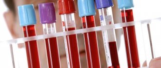
How is blood plasma different from serum?
Plasma is a yellowish, cloudy substance that is part of the blood.
It contains basic information about the individual's health status. It helps to identify hormonal imbalances and problems in the functioning of individual organs and systems. Among the disadvantages of plasma, experts note its short shelf life, after which it becomes unsuitable for study and use.
Serum is plasma without fibrinogen, which increases its lifespan. The serum can be used to obtain various drugs that have medicinal properties.
It helps conduct large-scale studies of the capabilities of the human body, testing the reaction of blood cells to various types of pathogenic microorganisms.
The difference between plasma and serum is as follows:
- Plasma is the entire component of blood, while serum is only a part.
- Plasma contains fibrinogen, a protein responsible for blood clotting.
- Plasma is always yellowish, and serum may have a reddish tint due to damaged red blood cells.
- Plasma coagulates under the influence of the enzyme coagulase, and serum is resistant to this process.
The differences between these two components of blood are so enormous that it is impossible to consider them identical.
Laboratory tests of serum can determine the amount of proteins, carbohydrates and minerals in the blood. The results are used to draw conclusions about the coherence of the internal organs.
If a decrease in total serum protein is detected, prolonged fasting or a low-protein diet may be suspected.
When a person has not limited his diet, and the indicators are significantly below the norm, they speak of the following violations:
- Serious pathologies of the liver, kidneys, endocrine system.
- Burns or major blood loss.
- Presence of neoplasms.
- Problems with protein production under the influence of medications.
Exceeding the norm leads to:
In such cases, additional diagnostics are often required. If the problems are caused by dehydration, the patient is recommended to adjust the drinking regime. In other situations, special treatment is necessary, which is prescribed by an appropriate specialist.
Special serums with markers are used for scientific and research purposes.
Serum is the most informative reagent when performing blood biochemistry, which allows you to diagnose pathologies:
- Pancreas.
- Liver.
- Kidney.
- Prostate gland.
- Bone tissue.
- Muscle fibers.
When studying human serum, a decrease in the amount of ferritin, which is responsible for transporting iron in the body, may be detected.
If its levels are reduced, problems begin with the level of iron in the blood. Neopterin reflects the speed of the immune response to adverse conditions.
Each protein is responsible for its own area, so the likelihood of error when making a diagnosis is minimal.
Technique for obtaining blood, serum and plasma.
Receiving blood. Small amounts of blood from animals are usually taken from the auricle; for this purpose, the collection site is pre-treated, clipped or shaved. Wipe with a swab moistened with 70% alcohol, after which either an incision is made in the vessel or the vein is pierced with a needle. The first drop that appears is removed with a swab, since after treatment with alcohol the red blood cells in it are destroyed. Then the blood is collected drop by drop into a watch glass and immediately pipetted for testing. Blood is taken from the person's middle or ring finger of the left hand, after wiping it with 70% alcohol. A large amount of blood from a horse, cattle, or small cattle is taken from the jugular vein at the border of the upper and middle third of the neck. To do this, a tourniquet is placed on the animal’s neck below the puncture site to fill the vein with blood, wipe it with alcohol or tincture of iodine, then a sterile sharp needle is inserted into the vein at an angle of 45 degrees against the blood flow towards the head. Blood is collected in test tubes or flasks. In pigs, large quantities of blood are obtained from the tail. To do this, cut off about 1 cm of the tail with a scalpel, collect the blood in test tubes, then compress the tip of the tail with an elastic band or bandage for 1-2 days, and thoroughly disinfect the wound. In dogs, large quantities of blood are obtained from the saphen vein (outer thigh). In rabbits from the ear vein, in guinea pigs from the heart, in chickens from the crest or crane vein.
Obtaining blood serum. To obtain blood serum, it must clot; for this, the blood is collected in clean, dry test tubes, a stream of blood is directed along the wall of the test tube to prevent foam from forming, then the test tubes with blood are placed in a thermostat for several hours for complete coagulation, then the resulting clot is separated from the walls of the test tube with a glass rod: circling the clots around, then the blood is settled or centrifuged, while the clot becomes compacted - retraction and a straw-yellow serum is released from it.
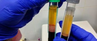
Obtaining blood plasma. The blood must be protected from clotting, that is, stabilized. Anticoagulants are used to stabilize the blood. These include Na citrate, Na oxalate, and heparin. An anticoagulant is placed in a test tube, then blood is drawn. After taking, the test tube is carefully inverted several times to mix. When settling or centrifuging, the blood is divided into 2 layers: On top there is a yellowish cloudy liquid-plasma, below there is a dark cherry-colored layer of red blood cells, and on it there is a small white coating of leukocytes.
Blood serum differs from plasma in that it does not contain the protein fibrinogen, since undissolved fibrin is formed from it, which became the basis for a blood clot.
In order to obtain serum from plasma, the fibrinogen protein must be precipitated from it. Then centrifuge and sediment the serum into a separate tube. TCA-trichloroacetic acid.
Determining the volume ratio between plasma and blood cells is the hematocrit indicator.
The blood contains 55-60% plasma of 40-45 formed elements. To determine the hematocrit, stabilized blood (with the addition of an anticoagulant) is used. A little stabilized blood is placed on a watch glass. Take 2 capillary tubes. Both capillaries are filled with blood. Then both ends of the capillary are plugged with plasticine, then placed in a special centrifuge and centrifuged at 3-4 thousand rpm for 8-10 minutes. The capillaries are removed. The blood in them is divided into 2 layers. From the center there is plasma, from the periphery there are formed elements. Then the capillaries are placed in a special frame. The amount of plasma in both capillaries is determined and the average value is taken.
Formed elements of blood.
1. Red blood cells. Red blood cells in higher animals are round, nuclear-free, 5-6 macromeres in diameter, covered on top with a protein-serous membrane, inside the stroma it is represented by hemoglobin. Hemoglobin is a pigment, which in structure is a complex protein consisting of a simple globin protein and a heme coloring substance, which is a prosthetic part. Hemoglobin consists of 4 peroid rings in the center of which is divalent iron. In cross-section, the red blood cell has a biconcave disk to increase its surface area.
2. Leukocytes are white blood cells, larger than red blood cells, contain a nucleus, and are capable of independent movement both in the bloodstream and beyond. Based on their structure and color reproduction, they are divided into 2 groups: granular (granulocytes) and non-granular (agranulocytes). Granular cells include basophils, eosinophils, and neutrophils. Based on the degree of maturity, neutrophils are divided into young, band and segmented.
Agranulocytes-lymphocytes (small, medium, large). Monocytes. In all animals and humans, segmented neutrophils and lymphocytes predominate with different variations.
Hemoglobin is a complex protein that is found inside red blood cells and makes up its stroma. Hemoglobin, due to divalent iron in the composition of heme, can attach oxygen-oxyhemoglobin, this is not a strong compound, it is formed in the vessels of the lungs, releases oxygen into the tissue fluid and is restored. The compound of hemoglobin with carbon dioxide is carbhemoglobin, it is formed in tissues and transported in the lungs. Carbohemoglobin easily releases carbon dioxide and attaches oxygen.
Cito! - Fast. A triad of express blood tests is the hemoglobin level, the number of leukocytes in the blood and the erythrocyte sedimentation rate. In order to determine the level of hemoglobin, it is necessary to destroy red blood cells and release the hemoglobin into solution.
Physiological compounds of hemoglobin are oxyhemoglobin, carbhemoglobin, reduced. There are more than 50 pathological types of hemoglobin. Of these, carboxyhemoglobin is a compound of hemoglobin with carbon monoxide, a very strong compound, methemoglobin is a compound in which iron changes its valence and becomes trivalent and this also leads to the formation of very strong compounds.
ESR (erythrocyte sedimentation rate).
If you take blood stabilized with sodium citrate and draw it into a capillary, place the capillary vertically, the red blood cells will begin to settle, and plasma will remain on top. The erythrocyte sedimentation rate may vary. It depends on several factors, on the type of animal. The highest erythrocyte sedimentation rate in a horse is 64 mm/h. In cattle and small cattle, rabbits 0.5-1 mm/h, in dogs 2.5 mm/h, in pigs 34 mm/h, in humans 4-8 mm/h. In addition, the ESR may change for each individual throughout his life. Under physiological conditions, that is, normally, the ESR does not change significantly, but in a female individual in the 2nd half of pregnancy, the ESR increases significantly. Most often, ESR accelerates or slows down due to pathologies.
For example, ESR slows down when the blood thickens, accelerates when there are inflammatory processes in the body during which many protective globulin proteins are formed, they are adsorbed on red blood cells, lowering their surface charge and red blood cells will settle faster in the blood column. ESR is an important diagnostic indicator and is one of the three express blood tests.
Hemolysis of red blood cells. This is the destruction of the membrane of red blood cells and the release of hemoglobin into solution. There are several types of hemolysis:
1. Osmotic.
2. Chemical.
3. Temperature.
4. Mechanical.
Osmotic hemolysis is based on the fact that in hypotonic solutions red blood cells are destroyed depending on their concentration, and the red blood cells of different individuals are destroyed differently in hypotonic solutions - this means that red blood cells have osmotic resistance - this is the ability of red blood cells to withstand low osmotic pressure.
Chemical is based on the destruction of red blood cells under the influence of chemicals (ammonia, distilled water, acids).
Temperature is based on the destruction of red blood cells after thawing, previously frozen red blood cells. Mechanical when stirring.
Blood clotting.
Blood clotting is a protective reaction of the body that protects it from blood loss. Blood clotting is triggered when the integrity of the vascular wall is violated, due to mechanical or other damage, or when roughness appears on the vascular wall. There are 3 categories of substances in the blood that are involved in blood clotting:
1. Substances that promote coagulation. They combine to form the blood coagulation system.
2. Substances that prevent blood clotting. They combined into an anti-coagulation system.
3. The system of substances that cause the liquefaction of already coagulated blood is the fibrinolytic system.
Blood coagulation consists of the interaction of their components:
1. Hemostasis - stopping blood flow.
2. Reflex narrowing of damaged blood vessels up to spasm.
3. Humoral narrowing under the influence of hormones and mediators adrenaline, serotonin, norepinephrine.
4. Aggregation (sticking) of platelets to each other and their damage.
Blood coagulation system.
There are 13 coagulation factors:
I. Fibrinogen is a blood plasma protein that, when exposed to an unusual environment, turns into an insoluble phase—fibrin.
II. Prothrombin is an inactive form of an enzyme that, under certain conditions, is converted into active thrombin.
III. Thromboplastin is an enzyme released from platelets that activates prothrombin.
IV. Ionized calcium participates in all phases of coagulation and activates all enzymes.
V. Accelerin (Ac) is an enzyme that accelerates the activation of thromboplastin.
VI.
VII. Proconvertin is similar to factor 5 and is involved in the formation of thromboplastin.
VIII. Antihemophilic globulin A. Participate in education
IX. Antihemophilic globulin B. Thromboplastin .
X. Plasma thrombotropin – participates in the formation of a blood clot.
XI. Plasma prothromboplastin.
XII. Hageman factor. It activates prothromboplastin.
XIII. Fibrin stabilizing factor (FSF).
Blood coagulation occurs in 3 phases and is a chain of enzymatic reactions. The result of each enzymatic reaction becomes the trigger for the next enzymatic reaction.
A chain of enzymatic reactions precedes the next reaction. This is a reflex spasm of the injured vessel and the formation of a “platelet nail”. Its formation begins with a change in the charge of the vascular wall at the site of damage. A charge change occurs. This leads to the fact that the formed elements begin to crowd around the damaged area. A special role belongs to platelets. They crowd together and stick together (aggregation), which leads to primary hemostasis. Platelet aggregation leads to their disruption. BAS come out of them, which trigger enzymatic coagulation (coagulation hemostasis). There are three phases in it:
1. When platelets are destroyed, thromboplastin is released from them, which is called blood thromboplastin. Tissue thromboplastin is released from the damaged vascular wall. Normally, they are absent in the body and are released only when the wall is damaged. With their participation, the enzyme blood and tissue prothrombinase is formed. This occurs under the influence of 5 8 9 10 and 11 factors with the participation of calcium ions.
2. Consists in the activation of prothrombin and its conversion into active thrombin under the influence of 5 7 factors and in the presence of calcium ions.
3. Consists in the formation of fibrin. Goes in 3 stages:
a. Proteolytic stage. Thrombin acts on fibrinogen, splits off individual monomers from it, and profibrin is formed.
b. Polymerization. With the participation of calcium, profibrin molecules stick together. Fibrin polymer is formed.
c. Under the influence of factor 8, fibrin polymer molecules are cemented. The final fibrin is formed. It falls out in the form of threads, the formed elements become entangled in them, and a clot is formed.
This is not the final stage, since after some time the fibrin filaments begin to shorten, which causes the clot to thicken and the liquid-serum to be squeezed out of it. Compaction of a clot by retraction is a complex biological process that leads to a tight closure of the vessel with a plug, thus bringing the edges of the wound closer together. The fibrin plug dissolves over time. This fibrinolysis process is carried out under the action of the fibrinolytic system.
How does BLOOD SERUM differ from PLASMA?
The cells of our body are washed by a certain amount of bodily fluids, or humors. Due to the fact that these fluids occupy an intermediate position between human cells and the external environment, they ensure the survival of cells and play the role of a so-called shock absorber during sudden external changes, in addition, they are an effective means of transporting nutrients and waste products in the body.
An important role in the human metabolic process is played by blood, which consists of the liquid part of blood plasma and formed elements suspended in it:
- leukocytes - white blood cells that perform protective functions;
- erythrocytes - red blood cells containing hemoglobin (red respiratory pigment);
- platelets - blood platelets necessary for blood clotting.
Formed elements make up 40–45%, plasma – 55–60% of the total blood volume. This ratio is called the hematocrit ratio, or hematocrit number. In some cases, the hematocrit number includes only the volume of blood that accounts for the formed elements.
Blood plasma is a solution that consists of:
- water (90-92%) and dry residue (10-8%);
- organic and inorganic substances;
- formed elements (blood cells and plates);
- dissolved substances: proteins (albumin, globulins and fibrinogen); inorganic salts that are dissolved in the form of anions (sulfate, chlorine ions, phosphate, bicarbonate) and cations (potassium, magnesium, sodium and calcium); transport substances derived from digestion (amino acids, glucose) or respiration (oxygen and nitrogen), metabolic products (urea, carbon dioxide, uric acid) or substances absorbed by the lungs, skin and mucous membranes.
Plasma constantly contains all microelements, vitamins and intermediate metabolic products (pyruvic and lactic acids).
Lymph, blood, tissue, pleural, spinal, joint and other fluids form the internal environment of the human body. They originate from blood plasma and are formed through the process of plasma filtration by passing through the capillary vessels of the human circulatory system.
Plasma protein contains fibrinogen, which appears due to changes in the physicochemical state during blood clotting. Fibrinogen has the ability to pass from a soluble to an insoluble form, converting into fibrin and forming a clot.
Blood serum is a clear, yellowish (or light yellow) liquid separated from a blood clot after blood has coagulated outside a living body. From the blood serum of animals and people immunized with certain antigens, it is possible to obtain immune sera used in the diagnosis, treatment and prevention of various diseases.
The serum can be either red due to hemolysis - this is the process of destruction of red blood cells with the release of hemoglobin into the environment surrounding the red blood cells, or icteric - due to increased values of bilirubin (a pigment that is contained in the blood and excreted with bile, due to which it is called bile pigment).
Blood serum is used for preventive, diagnostic or therapeutic purposes. To obtain it, it is necessary to place sterilely collected blood in a thermostat for 30–60 minutes, remove the clot from the wall of the test tube with a Pasteur pipette and place it in the refrigerator for several hours (preferably for a day). The settled blood serum is aspirated or drained using a sterile Pasteur pipette into a sterile test tube.
Conclusions:
- Blood plasma is the liquid part of the blood that remains after the removal of formed elements. In a suspended state, it contains formed elements - blood cells and platelets (or blood cells).
- Blood plasma in its composition is a very complex liquid biological medium, which includes vitamins, carbohydrates, proteins, various salts, lipids, hormones, dissolved gases and intermediate metabolic products.
- Blood serum (or blood serum) is the liquid fraction of clotted blood.
- Blood plasma is obtained by precipitation of formed elements, and serum is obtained by introducing coagulants (substances that promote blood clotting) into the blood plasma.
- Blood serum differs from plasma in the absence of a number of proteins of the coagulation system, such as fibrinogen and antihemophilic globulin, therefore it does not coagulate in the presence of coagulase, incl. microbial
Collection, storage conditions and delivery of venous blood for ELISA and PCR
Preparation of subjects
Venous blood is taken on an empty stomach, in the morning. When taking venous blood, it is necessary to take into account a number of factors that can affect the result of hematological studies: physical stress (running, fast walking, climbing stairs), emotional arousal, eating on the eve of the test, bathing, drinking alcohol, etc. To exclude these factors, the following conditions for preparing patients should be observed: • venous blood is taken after a 15-minute rest of the subject; • the patient sits during collection; in severely ill patients, blood collection can be performed lying down. • smoking, drinking alcohol and eating immediately before the study are excluded; The main method of collecting venous blood for laboratory testing is vein puncture. Venous blood is usually collected from the cubital vein. If necessary, it can be obtained from any vein (wrist, back of the hand, above the thumb, etc.). In newborns and infants, blood is usually taken from the frontal, temporal or jugular vein. When taking blood from a vein, it is necessary to avoid: places of scars, hematomas; veins used for transfusion of solutions; leg veins (in patients with diabetes, with peripheral blood flow disorders, angiopathy).
Equipment
For venipuncture, you can use three options for puncture systems: • disposable plastic systems (vacutainers), consisting of a container with a disposable needle screwed onto it and a test tube with a tight-fitting stopper and a vacuum inside; • disposable syringes with a suitable needle diameter; • needles with an internal diameter of 0.55-0.65 mm. Conditions for transporting venous blood
Properly collected venous blood must be delivered to the laboratory in a timely manner. At room temperature, delivery time should not exceed 60 minutes after blood collection. If blood is delivered to the laboratory during the day, it is stored at a temperature of +40C-+60C (in the refrigerator) and then delivered to the laboratory in special transport containers in an ice bath. During transportation, blood tubes and containers must be adequately protected from harmful environmental influences and weather conditions. When transporting venous blood, safety rules, asepsis and antiseptics must be strictly observed. The tubes must be labeled, packaged and tightly closed. The packaging should be convenient for transportation. Storage periods depend on the indicator being studied, storage temperature and the anticoagulant used to draw blood.
Method for obtaining blood serum (without the use of separation or centrifugation aids)
Equipment 1. Centrifuge glass tubes with a total volume of 10-12 ml. 2. Glass rods or Pasteur pipettes with capillaries sealed at the end (to separate the clot). 3. Laboratory centrifuge (up to 3000 rpm). Preparation of whey
Venous blood obtained without anticoagulants into a glass centrifuge tube is left in it at room temperature (15-200C) for 30 minutes until a clot is completely formed. Once the clot has formed, the tubes are opened and a thin glass rod or a sealed capillary Pasteur pipette is carefully passed along the inner walls of the tube along the circumference in the upper layer of blood to separate the clot column from the walls of the tube. The serum is poured into another centrifuge tube, holding the clot with a glass rod, and centrifuged, or centrifuged in the same primary tubes.
Centrifugation
After clot retraction, samples are centrifuged at an RCF of 1000 to 1200 xg (maximum 1500 xg) for 10 minutes. In the case of using microtubes and a centrifuge for them, centrifugation is carried out at 6000-15000 xg for 1.5 minutes. After centrifugation, the serum is poured into secondary (transport) tubes. The serum should not be hemolyzed. Plasma is obtained from blood by separating blood cells. It is a cell-free supernatant obtained by centrifugation of blood whose clotting is inhibited by the addition of an anticoagulant immediately after collection. Plasma contains blood clotting factors. Due to the fact that plasma and serum contain about 93% water, in contrast to whole blood, which contains about 81% water, the concentration of components in plasma is 12% higher than in whole blood. This may be of fundamental diagnostic importance when studying activity, for example LDH, which has a higher concentration in blood serum than in plasma. Commercial plasma production systems are widely used. They are tubes or syringe-type devices (“vacutainers”) with a vacuum inside, containing various anticoagulants and/or glycolysis inhibitors. As with serum devices, these plasma tubes come in a variety of options containing separation gels and polystyrene granules for faster plasma production, easier transportation and storage. They already contain anticoagulants and marks to which blood should be drawn.
Plasma production method
Plasma preparation
Venous blood obtained with an anticoagulant immediately after collection is mixed by inverting the capped blood tubes at least 5 times. Mixing should be carried out without shaking or foaming. The time between starting to apply the tourniquet and mixing the blood with the anticoagulant should not exceed 2 minutes. After equilibrating the tubes with blood, they are centrifuged at an RCF of 1000-1200 xg, but not more than 1500 xg, for 10-15 minutes. The plasma is immediately drained into a transport centrifuge or chemical tube. The test tube is closed with a lid. Conditions for transporting blood plasma Properly obtained and collected blood plasma must be delivered to the laboratory in a timely manner. At room temperature, delivery time should not exceed 24 hours. If plasma is delivered to the laboratory during the day, it is stored at a temperature of +4...+80C (in the refrigerator) and then delivered to the laboratory in special transport containers in an ice bath. For longer storage, plasma can be frozen at –200C. The rules for transporting plasma are the same as for venous blood.
What should be the biochemical blood test?
Using a biochemical blood test, a specialist can evaluate the functioning of a certain human organ and identify the lack of vitamins and microelements in the body.
Let's consider what a blood test should be based on this research method.
- Total protein is the total content of all blood serum proteins. The norm for total protein in adults is 64-82 g/l, in children under one year of age - 46-72 g/l, up to 6 years old - 52-77 g/l, up to 12 years old - 58-78 g/l.
- Albumin is the main blood protein that is produced in the liver. The amount of albumin in the blood should be 35-50 g/l in adults, 38-53 g/l in children under 14 years of age.
- C-reactive protein is a sensitive element in the blood that reacts faster than other elements to tissue damage. The normal level of its content in blood serum is less than 0.5 mg/l.
- Glycated (glycosylated) hemoglobin is a hemoglobin protein to which glucose is attached. The norm of glycated hemoglobin is 4.0-6.5% of the amount of free hemoglobin in the blood.
- Alanine aminotransferase (ALAT) is a liver enzyme that is involved in the metabolism of amino acids. The normal value of ALT in the blood in women is less than 31 U/L, in men – less than 41 U/L.
- Aspartate aminotransferase (AST) is a cellular enzyme that takes part in the metabolism of amino acids. What should be the value of this indicator in a biochemical blood test? The AST norm for women is less than 31 U/l, for men – less than 41 U/l.
- Gamma glutamyl transpeptidase (GGT) is an enzyme that takes part in the metabolism of amino acids. Its normal content in the blood of women is less than 32 U/l, in men - less than 49 U/l. For newborn children, the value of this indicator is 2-4 times higher than the normal value for adults.
- Total cholesterol is an organic compound that is an important component of fat metabolism in the human body. The normal level of total cholesterol in the blood in adults is 3.0-6.0 mmol/l.
- Triglycerides are neutral fats, which are derivatives of higher fatty acids and glycerol. What should be the triglyceride value in a child’s blood test? For children under 10 years of age, the norm for this indicator is 0.34-1.24 mmol/l, for children under 15 years of age – 0.36-1.48 mmol/l. In women, the normal amount of triglycerides in the blood is 0.44-2.70 mmol/l, in men – 0.52-3.29 mmol/l.
- Glucose is the main indicator of carbohydrate metabolism in the body. The normal level of blood glucose in adults is 3.89-5.82 mmol/l, after 60 years - up to 6.38 mmol/l. In children under 14 years of age, this indicator has a normal value of 3.33-5.55 mmol/l.
- Total bilirubin is a bile pigment that is a breakdown product of hemoglobin and some other blood components. The normal content of total bilirubin in the blood is 3.4-17.0 µmol/l.
- Creatinine is a substance that is formed as a result of protein metabolism in the body. The normal level of creatinine in the blood in men is 60-115 µmol/l, in women - 53-96 µmol/l, in children under one year of age - 18-35 µmol/l, up to 14 years - 26-62 µmol/l.
Blood glucose
Blood glucose determination is one of the most widely used tests in clinical laboratory diagnostics. Glucose is determined in plasma, serum, and whole blood. According to the Laboratory Manual for the Diagnosis of Diabetes presented by the American Diabetes Association (2011), it is not recommended to measure serum glucose when diagnosing diabetes because the use of plasma allows samples to be quickly centrifuged to prevent glycolysis without waiting for clot formation.
Differences in glucose concentrations between whole blood and plasma require special attention when interpreting results. The concentration of glucose in plasma is higher than in whole blood, with the difference depending on the hematocrit value, therefore, using a constant factor to compare blood and plasma glucose levels may lead to erroneous results. According to WHO recommendations (2006), the standard method for determining glucose concentration should be the method for determining glucose in venous blood plasma. The concentration of glucose in the plasma of venous and capillary blood does not differ on an empty stomach, but 2 hours after a glucose load the differences are significant (Table).
| Glucose concentration, mmol/l | ||||
| Whole blood | Plasma | |||
| venous | capillary | venous | capillary | |
| Norm | ||||
| On an empty stomach | 3,3–5,5 | 3,3–5,5 | 4,0–6,1 | 4,0–6,1 |
| 2 hours after OGTT | <6,7 | <7,8 | <7,8 | <7,8 |
| Impaired glucose tolerance | ||||
| On an empty stomach | <6,1 | <6,1 | <7,0 | <7,0 |
| 2 hours after OGTT | >6,7<10,0 | >7,8<11,1 | >7,8<11,1 | >8,9<12,2 |
| SD | ||||
| On an empty stomach | >6,1 | >6,1 | >7,0 | >7,0 |
| 2 hours after OGTT | >10,0 | >11,1 | >11,1 | >12,2 |
The glucose level in a biological sample is significantly affected by its storage. When storing samples at room temperature, glycolysis results in a significant decrease in glucose content. To inhibit glycolysis processes and stabilize glucose levels, sodium fluoride (NaF) is added to the blood sample. When collecting a blood sample, according to a WHO expert report (2006), if immediate separation of plasma is not possible, the whole blood sample should be placed in a tube containing a glycolytic inhibitor, which should be kept on ice until plasma is isolated or analyzed.
Indications for the study
- Diagnostics and monitoring of diabetes;
- diseases of the endocrine system (pathology of the thyroid gland, adrenal glands, pituitary gland);
- liver diseases;
- obesity;
- pregnancy.
Features of sample collection and storage.
Before the study, it is necessary to exclude increased psycho-emotional and physical stress.
Preferably, venous blood plasma. The sample should be separated from the formed elements no later than 30 minutes after blood collection; hemolysis should be avoided.
Samples are stable for no more than 24 hours at 2–8 °C.
Research method.
Currently, enzymatic methods for determining glucose concentration - hexokinase and glucose oxidase - are most widely used in laboratory practice.
Increased values
- DM type 1 or 2;
- pregnancy diabetes;
- diseases of the endocrine system (acromegaly, pheochromocytoma, Cushing's syndrome, thyrotoxicosis, glucoganoma);
- hemachromatosis;
- acute and chronic pancreatitis;
- cardiogenic shock;
- chronic liver and kidney diseases;
- physical exercise, strong emotional stress, stress.
Reduced values
- Overdose of insulin or hypoglycemic drugs in patients with diabetes;
- diseases of the pancreas (hyperplasia, tumors) causing disruption of insulin synthesis;
- deficiency of hormones with counter-insular effects;
- glycogenosis;
- oncological diseases;
- severe liver failure, liver damage caused by poisoning;
- gastrointestinal diseases that interfere with the absorption of carbohydrates.
- alcoholism;
- intense physical activity, feverish conditions.
How is blood plasma different from serum?
Plasma is a yellowish, cloudy substance that is part of the blood. It contains basic information about the individual's health status. It helps to identify hormonal imbalances and problems in the functioning of individual organs and systems.
Serum is plasma without fibrinogen, which increases its lifespan. The serum can be used to obtain various drugs that have medicinal properties.
It helps conduct large-scale studies of the capabilities of the human body, testing the reaction of blood cells to various types of pathogenic microorganisms.
The difference between plasma and serum is as follows:
- Plasma is the entire component of blood, while serum is only a part.
- Plasma contains fibrinogen, a protein responsible for blood clotting.
- Plasma is always yellowish, and serum may have a reddish tint due to damaged red blood cells.
- Plasma coagulates under the influence of the enzyme coagulase, and serum is resistant to this process.
Blood in a test tube
conclusions
After the comparison, it becomes clear that there is a difference between serum and blood plasma. It depends on various factors and indicators. Sampling is carried out using ordinary test tubes. Plasma and serum are different in composition. The serum promotes rapid blood clotting - this property allows the body to resist severe bleeding. Plasma is responsible for transporting various substances through the bloodstream throughout the body. If you conduct research, the results of diagnosing plasma and serum will be almost the same. This is due to the similar composition of these two blood components.
How does BLOOD SERUM differ from PLASMA?
The cells of our body are washed by a certain amount of bodily fluids, or humors. Due to the fact that these fluids occupy an intermediate position between human cells and the external environment, they ensure the survival of cells and play the role of a so-called shock absorber during sudden external changes, in addition, they are an effective means of transporting nutrients and waste products in the body.
An important role in the human metabolic process is played by blood, which consists of the liquid part of blood plasma and formed elements suspended in it:
- leukocytes - white blood cells that perform protective functions;
- erythrocytes - red blood cells containing hemoglobin (red respiratory pigment);
- platelets - blood platelets necessary for blood clotting.
Formed elements make up 40–45%, plasma – 55–60% of the total blood volume. This ratio is called the hematocrit ratio, or hematocrit number. In some cases, the hematocrit number includes only the volume of blood that accounts for the formed elements.
Blood plasma is a solution that consists of:
- water (90-92%) and dry residue (10-8%);
- organic and inorganic substances;
- formed elements (blood cells and plates);
- dissolved substances: proteins (albumin, globulins and fibrinogen); inorganic salts that are dissolved in the form of anions (sulfate, chlorine ions, phosphate, bicarbonate) and cations (potassium, magnesium, sodium and calcium); transport substances derived from digestion (amino acids, glucose) or respiration (oxygen and nitrogen), metabolic products (urea, carbon dioxide, uric acid) or substances absorbed by the lungs, skin and mucous membranes.
Plasma constantly contains all microelements, vitamins and intermediate metabolic products (pyruvic and lactic acids).
Lymph, blood, tissue, pleural, spinal, joint and other fluids form the internal environment of the human body. They originate from blood plasma and are formed through the process of plasma filtration by passing through the capillary vessels of the human circulatory system.
Plasma protein contains fibrinogen, which appears due to changes in the physicochemical state during blood clotting. Fibrinogen has the ability to pass from a soluble to an insoluble form, converting into fibrin and forming a clot.
Blood serum is a clear, yellowish (or light yellow) liquid separated from a blood clot after blood has coagulated outside a living body. From the blood serum of animals and people immunized with certain antigens, it is possible to obtain immune sera used in the diagnosis, treatment and prevention of various diseases.
The serum can be either red due to hemolysis - this is the process of destruction of red blood cells with the release of hemoglobin into the environment surrounding the red blood cells, or icteric - due to increased values of bilirubin (a pigment that is contained in the blood and excreted with bile, due to which it is called bile pigment).
Blood serum is used for preventive, diagnostic or therapeutic purposes. To obtain it, it is necessary to place sterilely collected blood in a thermostat for 30–60 minutes, remove the clot from the wall of the test tube with a Pasteur pipette and place it in the refrigerator for several hours (preferably for a day). The settled blood serum is aspirated or drained using a sterile Pasteur pipette into a sterile test tube.
Conclusions:
- Blood plasma is the liquid part of the blood that remains after the removal of formed elements. In a suspended state, it contains formed elements - blood cells and platelets (or blood cells).
- Blood plasma in its composition is a very complex liquid biological medium, which includes vitamins, carbohydrates, proteins, various salts, lipids, hormones, dissolved gases and intermediate metabolic products.
- Blood serum (or blood serum) is the liquid fraction of clotted blood.
- Blood plasma is obtained by precipitation of formed elements, and serum is obtained by introducing coagulants (substances that promote blood clotting) into the blood plasma.
- Blood serum differs from plasma in the absence of a number of proteins of the coagulation system, such as fibrinogen and antihemophilic globulin, therefore it does not coagulate in the presence of coagulase, incl. microbial
Comparison
The comparison should begin with the main difference, according to experts, between the components of the blood - the plasma contains an agent that allows the blood to gradually clot. There is no this agent in the serum. Accordingly, only plasma takes part in the blood clotting process. In appearance, it is a clear or yellowish liquid part of the blood. Serum is also a liquid upon visual inspection, but it is formed after the coagulation process.
Despite the fact that many consider these two elements to be the same, the differences in them are already noticeable. Terms cannot be used as synonyms for the same concept or property of blood. You also need to take into account the fact that whey has a limited shelf life. Shelf life is limited to only a few months. Blood serum is used when health problems caused by cholesterol, blood sugar or blood pressure are identified.
You may also be interested: Soul and spirit - what are they and the differences
The main function of plasma is the transfer of proteins, hormones, nutrients, and antibodies through the bloodstream throughout the body. The peculiarity is that the cells release their waste products into the plasma. This ensures high-quality cleansing and renewal of the cellular composition.
In addition to water, plasma contains:
- Hormones.
- Albumen.
- Amino acids.
- Nutrients.
- Globulin.
- Fibrinogen.
Nitrogen waste is also present. In most cases, plasma helps regulate body temperature and blood pressure. Plasma has a long shelf life and can be stored for 12 months, which is 2-3 times longer than plasma.
Serum is formed after the removal of clotting factors. This is achieved through a process called centrifugation. As a result, a protein called fibrinogen is converted into fibrin. It belongs to the insoluble types of proteins. The substance is used to restore damaged tissue. This is achieved by forming a characteristic clot on the wound. It is this that prevents strong blood flow.
To better understand the functions and differences, it is recommended to pay attention to the work that its main protein, albumin, does in plasma. It transports various substances, including hormones, fatty acids, ions, bilirubin, and drugs. It is albumin that takes part in the metabolic process and carries out protein synthesis. This protein controls plasma pressure and blood clotting parameters. It makes sure that a certain level of amino acids is maintained. If, under the influence of a number of circumstances, the level of albumin in plasma changes, this indicator becomes an additional sign of a pathological condition during diagnosis. Protein concentration helps determine the condition of the liver, since its decrease is a characteristic sign of chronic diseases.
Separating blood plasma and serum is a common process as these components are used to treat various diseases. The process takes place in a centrifuge. The components can be extracted because they have different weights and densities. It is possible to extract, for example, leukocytes and erythrocytes from plasma.
You may also be interested: Drama and tragedy - what are they and the differences
Blood composition
The material is a red liquid that, when moving, delivers nutrients to organs and tissues
Another important property of blood is the cleansing of cells from decay products. This is what protects the body from self-poisoning
Oxygen saturation, regulation of body temperature and protection from pathogenic bacteria are also the merits of the red liquid.
The composition of the liquid consists of the following components:
- 55% plasma;
- 45% red blood cells;
- less than 1% of leukocytes.
Plasma, in turn, consists of:
- albumins that carry out transportation;
- globulins that protect the blood;
- fibrinogen - a protein that coagulates the material.
Important!
90% of the total plasma volume is water, and only 10% are important protein components.
Production
Fetal bovine serum is a by-product of the dairy industry. Fetal bovine serum, like the vast majority of animal serum used in cell culture, is produced from blood collected in commercial slaughterhouses from dairy cattle that also supply meat intended for human consumption.
The first step in the fetal bovine serum manufacturing process is the collection of blood from the bovine fetus after the fetus has been removed from the slaughtered cow. The blood is collected under sterile conditions in a sterile container or blood bag and then clotted. The usual collection method is cardiac puncture, in which a needle is inserted into the heart. This minimizes “the risk of contamination of the serum by microorganisms from the fetus itself and the environment.” It is then centrifuged to remove the fibrin clot and remaining blood cells from the clear yellow (straw) serum. The serum is frozen before further processing, which is necessary to make it suitable for cell culture.
The second stage of processing involves filtration, typically using a filtration chain, with the final filtration being triple sterile 0.1 micrometer membrane filters. When processed by a trusted commercial serum supplier, sterilized fetal bovine serum is subject to strict quality control and is supplied with a detailed Certificate of Analysis. The certificate provides full test results and information about the origin of the whey. Certificates of Analysis vary between commercial providers, but each typically includes the following details: filtration statement, country of origin, cell growth performance testing, microbial sterility testing, mycoplasma and virus screening, endotoxin, hemoglobin, IgG, and total protein assays.
Blood determination
Serum for blood group recognition must be standard, that is, a certain group prepared from human blood. For the test, you need to prepare a dry glass slide, standard serum of the three blood groups, sodium chloride solution, cotton wool, glass rods and pipettes.
Blood serum is not just a complex mixture that can tell about the state of the body, but also an important element of most antiviral drugs.
All materials are published under the authorship or editorship of medical professionals (about the authors), but are not a prescription for treatment. Contact the specialists!
When using materials, a link or indication of the source name is required.
The thymol test (thymoloveronal test, thymol turbidity test, Maclagan test) is not one of the particularly popular biochemical methods of blood testing, however, it is not discounted when identifying certain diseases and is still used in clinical laboratory diagnostics.
A nonspecific reaction, based on the interaction with thymol in the veronal buffer of individual plasma proteins (gamma globulins and beta globulins associated with lipids - low density lipoproteins), and the turbidity of the solution, does not give a clear answer in relation to certain diseases, but often significantly helps in combination with other tests, and in some cases even outperforms them. This occurs in the initial stages of the disease (hapatitis A in children, for example), when other laboratory tests are still within normal limits. In addition, it has other advantages that do not allow laboratory diagnostic doctors to consign this analysis to oblivion.
Serum iron norm
When assessing the serum concentration of a microelement, the nutritional-dependent nature of this indicator should be taken into account. Iron enters the body with food, so a moderate decrease in the concentration of bound transferrin during a non-strict diet or taking drugs that interfere with the absorption of Fe is considered a physiological phenomenon that can easily be eliminated by correcting the diet.
If severe iron deficiency is detected, appropriate drug treatment is prescribed. It is worth considering that in the morning the serum contains slightly more of this microelement than in the evening. With all this, serum Fe may vary in patients belonging to different age categories.
Among women
In the body of representatives of the fairer sex, iron metabolism occurs under the influence of constantly changing hormonal levels, therefore the norm of serum iron in the blood of women is slightly underestimated and is about 10.7-21.5 µmol/l, which is mainly due to menstruation. During pregnancy, plasma Fe levels can also decrease significantly. So, during gestation, this indicator should not fall below 10.0 µmol/l.
In men
Subject to a balanced diet and adherence to a daily routine, iron reserves in the stronger sex are consumed optimally. A decrease in ferritin inside cells in men occurs as a result of liver disease, which often occurs against the background of abuse (or even poisoning) of alcoholic beverages and their surrogates. The normal level of serum iron in men ranges from 14.0 to 30.4 µmol/l.
In children
The Fe content in the blood of young patients varies depending on their age, weight and height. Children under one year old who are exclusively breastfed are susceptible to a slight decrease in hemoglobin. This fact is due to the limited content of so-called heme iron in the body of babies, which is not a cause for concern. The norm of serum Fe in children under one year of age is 7-18 µmol/l, and in older children this figure can reach 9-21 µmol/l.
