ESR or erythrocyte sedimentation rate indicates an imbalance of blood proteins, which can result from cell destruction as a result of many pathological conditions. In case of cancer, a change in ESR is not at all necessary, but in some clinical situations, an acceleration or deceleration of the rate of fall of red blood cells may occur.
- ESR: what is it?
- ESR norm
- ESR indicators in oncology
- Table of ESR indicators for oncology in men and women
- ESR analysis
- Preparation for analysis
- How to get tested
ESR: what is it?
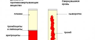
ESR and ROE - erythrocyte sedimentation reaction are the same, but the latter abbreviation is considered outdated. ESR shows the intensity of red blood cell aggregation followed by their sedimentation to the bottom.
The clumping of red blood cells is enhanced in the presence of the protein fibrinogen, which is involved in the formation of blood clots and is normally practically absent in plasma. A decrease in the largest fraction of proteins - albumin and an increase in numerous protein fractions of globulins - gamma, beta and alpha changes the ESR towards an increase.
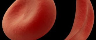
The sedimentation rate will certainly change if the erythrocyte has an abnormal shape and non-standard size, which is typical for anemia of various etiologies, including those caused by complications of chemotherapy.
Any pathological process - infection, inflammation, tumor, especially a malignant blood disease, affects the ratio of blood proteins and, of course, can shift the indicator in one direction or another.
Classification
There are no clear digital gradations for dividing the increase in ESR by degree. Conventionally, moderate and high degrees are distinguished. According to the mechanism of occurrence they distinguish:
- True increase in ESR
. The cause is various inflammations, infectious, and oncological pathologies. Develops as a result of dysproteinemia, which increases erythrocyte aggregation. - False increase in ESR
. False acceleration of ESR is observed in anemia, azotemia, alkalosis, and high blood cholesterol levels. The cause of this phenomenon is various pathological processes in which the ESR increases due to changes in the number or shape of red blood cells, the protein-lipid composition of plasma, a shift in blood pH, and the presence of other chemical compounds.
ESR norm
Normal interval rates of subsidence rates have been determined for all age groups of children; for adults there is only a gender norm without taking into account age, but it is known that over the years of life the rate slightly accelerates. In women, normal values are higher than in men: from 2 to 15 mm per hour versus an interval of 1 to 10 mm.
The higher value of female ESR is explained by the lower density of red blood cells per milliliter of blood, which allows them to fall faster, similar to the higher speed of a car on an open highway and the inevitable decline in heavy traffic.
During pregnancy, or rather from the second half, ESR increases, reaching 50-60 mm per hour. After childbirth, the balance of protein fractions and sex hormones, which are proteins, as well as plasma volume gradually return to normal and the rate of fall of blood cells stabilizes.
The erythrocyte sedimentation rate responds to food intake, so blood tests are taken on an empty stomach. The rate of red blood cell sediment formation is sensitive to some medications and physical activity, even to the duration of the summer heat period, so normal values are given with a spread, where the maximum value is several times higher than the minimum value.
Every twentieth healthy adult has his own ESR standard, deviating from the generally accepted standard.
If any abnormalities from the norm appear in the blood test of a cancer patient, and not just changes in ESR, you need to consult with an oncologist; an examination may be required. Our Clinic specialists are ready to help and find the root cause.
Book a consultation 24 hours a day
+7+7+78
Important to remember
- Inflammation tests—ESR, CRP, and procalcitonin test—help determine whether there is underlying inflammation in the body
- The most sensitive test of this trio, the procalcitonin test, allows you to distinguish a viral infection from a bacterial one, but does not allow you to find out which bacteria caused the infection. Typically, it is used in a hospital to monitor the condition of critically ill patients.
- Less sensitive tests - ESR and CRP - allow you to understand whether there is inflammation or not. CRP does this more accurately than ESR, but is more expensive, so it is rarely prescribed “just in case.” However, both CRP and ESR are often prescribed to patients who are recovering at home
- There is no point in doing inflammation tests as part of a check-up: in healthy people they will not provide any interesting information. And even if they do, it will be very difficult to figure out the results on your own. It is better to leave the appointment of tests for inflammation to the doctor - he will determine which test is needed, help you understand the results and prescribe competent treatment.
ESR indicators in oncology
Erythrocyte sedimentation rate is a nonspecific marker, since it changes with any pathology. ESR is only evidence of trouble and the release of “extra” proteins into the blood during tissue breakdown, insufficient synthesis of albumin due to impaired liver or hemoglobin function, as well as changes in the structure of the blood cell itself during anemia.
For a long time, in the majority of those suffering from a malignant neoplasm, including those at the metastatic stage, the blood picture can be absolutely normal. The exception is blood diseases accompanied by the synthesis of pathological proteins - paraproteins, which occurs in myeloma and some malignant lymphomas.
ESR may increase in the following clinical situations:
- disintegration of a cancer tumor, when its central part is deprived of nutrition, forming an area of necrosis, and the destroyed cells enter the bloodstream;
- the addition of inflammation along the periphery of the cancer node, which often accompanies metastases in the lung or primary lung cancer;
- disturbances in protein synthesis in the liver affected by metastases;
- loss of proteins with frequent removal of ascitic or pleural fluid;
- malabsorption disorders due to a malignant tumor of the gastrointestinal tract or removal of a large volume of the stomach and intestines;
- disturbances in the synthesis of the hormone erythropoietin by the kidneys due to the toxic effects of chemotherapy drugs;
- myocardial damage due to certain cytostatics;
- development of anorexia-cachexia syndrome in the terminal stage of the disease.
A decrease in ESR in a cancer patient is possible:
- when red blood cells are deformed as a result of the use of platinum derivatives;
- with jaundice - a complication of pancreatic cancer;
- dehydration as a result of severe vomiting, especially during hot periods;
- progression of failure due to tumor damage to the liver;
- severe pulmonary heart failure with cancerous pleurisy or advanced lung cancer.
A change in ESR in a cancer patient is not a mandatory characteristic of cancer and does not reflect the course of the malignant process, but always indicates clinical distress. In each case of a change in the analysis, it is necessary to seriously understand; it is not at all necessary that this change is a sign of cancer progression.
Diagnostics
Any, even asymptomatic, increase in ESR requires contacting a doctor to find out the cause. The doctor asks the patient in detail whether there was an increase in body temperature, whether the patient experienced pain in the joints, muscles, fatigue, etc. This can help in the diagnostic search. An additional examination is prescribed, depending on what nosology is suspected:
- Blood tests
. The concentration of hemoglobin and blood cells (erythrocytes, platelets, leukocytes) is measured. Very often an increase in the level of fibrinogen and C-reactive protein is detected. The blood is checked for the presence of autoaggressive antibodies (aCCP, antineutrophil cytoplasmic antibodies, antibodies to double-stranded DNA). In severe bacterial infections, high levels of presepsin and procalcitonin are observed in the blood. - Identification of the infectious agent
. Antibodies to antigens of viruses, bacteria, parasites are determined using ELISA methods and serological tests. PCR detects DNA and RNA of microorganisms. Microscopy, bacteriological culture of urine, sputum, and blood are performed. A stool test is performed for worm eggs. - Radiography
. With tuberculosis, an X-ray of the lungs shows an increase in mediastinal lymph nodes and infiltration in the upper lobes of the lungs. In paraproteinemic hemoblastoses, x-rays of the bones reveal numerous foci of bone tissue destruction. Multiple myeloma is characterized by a “puncture sign” on a skull x-ray. - Ultrasound
. An ultrasound scan of the abdominal cavity reveals thickening of the walls of the gallbladder in cholecystitis, enlargement and diffuse changes in the pancreatic parenchyma in pancreatitis, and enlargement of the liver and spleen in hemoblastosis. - Angiography.
In case of infarctions of various organs caused by thrombosis, radiography or computed tomography with contrast reveals a filling defect at the site of vessel occlusion. With systemic vasculitis (Horton's, Takayasu's arteritis), areas of vascular stenosis are visible. - Histological studies
. If the cause of increased ESR is oncological pathology, a biopsy is necessarily performed. A common symptom is the detection of a large number of atypical cells. In patients with malignant blood diseases, a predominance of blast cells is noted in the bone marrow aspirate, and atypical lymphoid proliferation is noted in the lymph node biopsy.
Table of ESR indicators for oncology in men and women
On the Internet you can find tables of changes in ESR for various types of malignant processes separately for women and men. This information could be trusted if the ESR indicator were included in the mandatory criteria for cancer, as we know, most tumor markers produced by cancer cells do not have this honor.
The erythrocyte sedimentation rate in most cancer patients does not deviate from the norm over the period of cancer development. On the eve of death from the progression of a malignant disease, when the volume of the tumor and its aggression are practically incompatible with life, and the function of the most important body systems is reduced to a minimum, the ESR can demonstrate “full health.” A banal respiratory infection, which occurs in the average Russian six times a year, in a radically treated patient without any signs of a malignant process can increase the rate of red blood cell precipitation to 40-60 mm per hour for a month.
With a very small range of oncohematological diseases, ESR may coincide with the progression of the process, but this is not necessary, nor is a decrease in the indicator considered a victory over the tumor.
The interpretation of test changes is individual, and tables of ESR correspondence to certain cancer diseases can be considered a marketing ploy of the site that does not reflect the objective oncological reality.
Correction
Conservative therapy
A transient increase in ESR after exercise, food intake or medications does not require correction. Elderly people with high ESR without other clinical and laboratory signs of any pathology also do not need any intervention. To normalize the pathological increase in ESR, it is necessary to treat the nosology that caused its development.
- Fighting infection
. For bacterial infections, antibiotics are prescribed according to sensitivity. If the patient's condition is serious and requires immediate treatment, broad-spectrum antibiotics (penicillins, cephalosporins, fluoroquinolones) are used. For influenza, oseltamivir is used; for other acute viral infections, symptomatic therapy is used. For the treatment of chronic viral hepatitis, combinations of peligated interferon with ribavirin and entecavir are effective. - Anti-inflammatory therapy
. For diseases accompanied by immune inflammation, anti-inflammatory drugs are needed - glucocorticoids (prednisolone), cytostatics (azathioprine, methotrexate), 5-aminosalicylic acid derivatives (sulfasalazine). If they are ineffective, tumor necrosis factor inhibitors - monoclonal antibodies (infliximab) are used. - Blood thinning
. If the cause of the heart attack is thrombosis or embolism, antiplatelet agents (clopidogrel, acetylsalicylic acid) and anticoagulants (heparin, warfarin, dabigatran) are prescribed. Thrombolytics (streptokinase) are used to dissolve a blood clot. - Chemotherapy.
Antitumor drugs (alkylating agents, antimetabolites) are used to treat cancer. Malignant blood diseases require a combination of several chemotherapy drugs.
Surgery
In case of acute abdominal pathology (cholecystitis, pancreatitis), it is necessary to perform a surgical (sometimes emergency) operation - pancreatic resection, laparoscopic cholecystectomy. Endovascular thrombectomy is performed to remove the thrombus. In case of myocardial infarction, percutaneous coronary intervention (stent placement) is performed. Unsuccessful conservative therapy for oncohematological diseases is considered an indication for bone marrow transplantation.
How to get tested
You need to take the test calmly, it’s too late to get nervous - everything has already happened in the body and all that’s left is to get the information. It would be more correct to pose the question differently: where to take it. It is advisable for an oncology patient to undergo tests in a specialized institution, where not only analysis on the machine is available, but also monitoring of the results obtained “in manual mode”. With specific treatment, especially after the use of medications, the conventional boundary of normality of indicators may temporarily shift. Often, haste in interpreting the result results in excessive examination and unnecessary treatment.
If you want an optimal and unconditionally professional approach, accuracy and objectivity, come for a consultation at our clinic.
Book a consultation 24 hours a day
+7+7+78
Bibliography
- Lugovskaya S.A., Morozova V.T., Pochtar M.E., Dolgov V.V. /Laboratory hematology// M.: Publishing house. Unimed-press; 2002.
- Pervushin Yu.V., Lugovskaya S.A., Marchenko L.A., Kovalevich N.I. / Hematological studies in clinical laboratory diagnostics//Stavropol; 2006
- Nazarenko G.I., Kishkun A.A. / Clinical assessment of laboratory research results // M., 2000.
- Oganesyan A.A., Garbatsevich M.S., Kostyukovich D.I. and others /ESR: old test, new opportunities // Lab. diagnostics. Eastern Europe; 2012; No. 4.
- Chizhevsky A. L. /Biophysical mechanisms of erythrocyte sedimentation reaction// Novosibirsk: Science, 1986.
Popular promotions
View all promotions
Biorevitalization and contour plastic surgery
Do you want to maintain a youthful face, improve skin condition and preserve natural beauty?
Read more…
Dermatoscopy free of charge
Dermatoscopy is a non-invasive diagnostic method for the visual assessment of skin lesions, which allows a more thorough examination of both the surface of the skin and its deeper layers.
Read more…
SMAS lifting using the UCOS HIFU device
The procedure for exposure to highly focused ultrasound on the latest HIFU UCOS device is a triple rejuvenating effect on the deep ligaments of the face, subcutaneous fat packets and skin!
Read more…
Chronic tonsillitis
Chronic tonsillitis is not only a sore throat, but also general weakness, fatigue, low-grade fever, etc. Only in November, treatment with the Tonsillor device instead of 450 rubles . per session you will pay 350 rubles !
Read more…