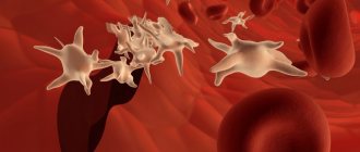
In blood analysis, MID denotes the combination of three formed elements included in the cellular composition of biological fluid: monocytes, eosinophils, basophils. The study and calculation of the number of MIDs is carried out as part of clinical hematology (general blood test).
CCA (general clinical analysis) is a laboratory method for determining the chemical composition and physical properties of blood. This is the most common study that allows us to identify violations of microbiological processes in the body. Clinical hematology is prescribed to adults and children of all ages:
- for medical reasons (diagnosis of diseases and monitoring of treatment);
- for preventive purposes (medical examinations, routine medical examination).
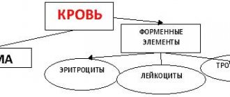
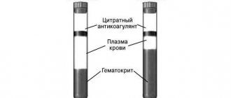
For the study, capillary blood is taken (from a finger). A transcript of the results is transferred to the attending physician, or the patient collects the results independently. The analysis takes into account the quantitative characteristics of various blood cells:
- ESR – erythrocyte sedimentation rate (ESR), indicating the ratio of plasma protein fractions;
- RBC – erythrocytes or red blood cells;
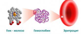
- Hb – hemoglobin (a complex protein component of blood);
- Hct – hematocrit (red blood cell volume);
- PLT – platelets (bone marrow blood platelets);
- RET – reticulocytes (young anucleate red blood cells).
Separately, the results of the leukogram (leukocyte formula) are displayed, which includes WBC - total leukocytes (colorless or white blood cells), and the components:
- NEU or NEUT – neutrophils (band and segmented)
- LYM – lymphocytes;
- MID triad, including: MON - monocytes, EOS - eosinophils, BAS - basophils.
The microscopy process is automated. The results are assessed using a comparative method of the patient’s indicators obtained and the accepted reference values. The advantages of general analysis lie in its accessibility, simplicity, information content, and efficiency of implementation in a laboratory setting.
Important! A complete blood count is an important diagnostic test that cannot be ignored. Based on its results, the doctor may suspect the presence of a serious pathology that requires immediate treatment.
What is MID?
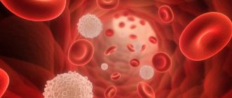
Leukocytes are white blood cells that are produced in the bone marrow and lymph nodes. These blood components play a large role in protecting the body from infections. Leukocytes are divided into several types:
- eosinophils;
- neutrophils;
- basophils;
- lymphocytes;
- monocytes.
The relative or absolute content of a mixture of eosinophils, basophils and monocytes shows the MID in a blood test. What it is? Relative content is measured as a percentage of the total number of leukocytes. The absolute indicator is calculated in the number of cells per 1 liter of blood. Nowadays, the MID percentage is more commonly used. Otherwise, this indicator is called MXD.
Components of MID
MID in the leukocyte formula combines the smallest types of white blood cells: monocytes, eosinophils, basophils.
Monocytes (MON)
Agranulocytic leukocytes of this type are large in size and have a red-violet nucleus. The cells are formed in the bone marrow and then move into the systemic circulation, where they live on average for about three days. Monocytes then transform into macrophages and move to the tissues of the liver, spleen and lymph nodes.
Macrophages are active phagocytes of the immune system that cleanse the body of cellular debris (dead cells) and bacterial microorganisms. The hallmark of MON is its survival. Neutrophilic granulocytes (neutrophils) are initially programmed to die after meeting foreign agents and performing their function, while monocytes are not destroyed, but continue their security activities.
In addition to phagocytosis, the responsibilities of monocytes include the production of the protective protein interferon, which inhibits the activity of viruses and is involved in building specific immunity, tissue regeneration, participation in the process of hematopoiesis, suppression of the activity of cancer cells, and protection of the body from cancer.
The monocyte variety of white cells copes more effectively with viral agents than with bacteria and parasites. An increased content of monocytes in the blood is defined by the term “monocytosis”. A reduced amount is called monocytopenia.
Eosinophils (EOS)
Of all MID representatives, eosinophils remain in the blood for the shortest amount of time. After moving from the bone marrow, they remain in the systemic circulation for several hours and are then transported to the tissues of the digestive system, lungs and epidermis (skin). Eosinophilic leukocytes are intended for the eradication of helminthic infestations through phagocytosis of pathogens, the formation of antiparasitic immunity, and the metabolism of histamine (a bioactive indicator of allergic reactions).
Along with basophils, eosinophil cells take part in the initiation of immediate hypersensitivity (the system's response to the introduction of allergens into the body). Eosinophilia (high concentration of eosinophils), first of all, means the presence of parasites or allergic antigens in the body. Eosinopenia (low cell count) has no particular diagnostic value.
Basophils (BAS)
The smallest, but very important type of granulocyte. The phagocytic properties of the cells are less developed than those of other leukocytes, but their membranes contain receptors for immunoglobulin E (IgE). When allergenic agents enter the body, IgE activates the allergic reaction through the release of histamine.
An increased concentration of basophils in the blood (basophilia) is a clinical sign of the development of allergies. In addition, these colorless cells contain heparin, which maintains stable blood flow in the capillaries and prevents increased blood clotting.
This effect helps maintain optimal blood circulation in small vessels, liver and lungs. Basophils do not have accumulative properties in tissues, like other leukocytes. They move to the inflamed area as needed, eliminate the foreign invasion and die. A decrease in BAS levels is called basopenia.
How is the test done?
Blood for a general clinical analysis (CCA) is usually taken from a finger, in rare cases it is taken from a vein. An area of skin is treated with a disinfectant solution, a small puncture is made and the material is collected into a test tube. This type of research does not require special preparation. It is advisable to donate blood in the morning on an empty stomach. A general analysis is taken at any clinic. In addition to MID, such an examination reveals other important hematological data: hemoglobin, ESR, red blood cell and platelet counts.
Abbreviated and extended blood test
In the abbreviated version of the study, the MID is necessarily determined in a blood test. What it is? If a person does not have any complaints, and OAC is carried out for the purpose of prevention, then a shortened analysis is done. In addition to MID, the following indicators are calculated:
- hemoglobin;
- ESR;
- platelets;
- red blood cells;
- total number of leukocytes.
If abnormalities were detected during the abbreviated CBC, then a more detailed study is carried out. For example, if the MID norm in a blood test is exceeded, decoding must be carried out for each type of cell separately. For this purpose, a detailed examination is prescribed to determine the leukocyte formula.
MID norms in blood tests
The relative MID in a general blood test is 5-10%. This is considered the norm. The research is quite accurate and errors in the results are extremely rare. The percentage of leukocyte cells is calculated automatically.
The absolute MID should be 0.2 - 0.8x10⁹/l. It should be noted that the MID standards for deciphering a blood test are the same for women and men. Minor fluctuations in these data are possible only during the menstrual period due to hormonal imbalance.
Norms and deviations of cell analysis
Each group of leukocytes included in the MID has its own laboratory standards. Parameters are measured using absolute (numerical) and relative (percentage) values.
Standard indicators of monocytes
The absolute content of monocytes for adults is 0.09–0.6*10^9/l. A slight excess of the norm is allowed for women in the third trimester of the perinatal period and before childbirth. The children's indicator is 0.05–1.1*10^9/l. The percentage in adults and adolescents (over 15 years old) is from 3 to 11%. In children, the MON range depends on age.
| Age | Up to 1 year | 1-5 years | 5–15 years |
| Norm in % | 4–10 | 3–10 | 3–9 |
Deviation of indicators from the norm in one direction or another indicates the development of infectious viral, oncological or hematological pathologies. Causes of monocytosis characteristic of childhood: acute infection provoked by the Epstein-Barr virus (infectious mononucleosis), specific “childhood” infectious diseases (whooping cough, rubella, measles, mumps).
Infections affecting adults and children: systemic disease of the lung tissue (sarcoidosis), infection with Koch's bacillus (tuberculosis), zoonotic infection transmitted from animals (brucellosis), venereal disease syphilis, typhoid and typhus, helminthic infestations (ascariasis, enterobiasis, opisthorchiasis, teniarinhoz, etc.).
An increase in the number of monocytes in the blood can be a sign of systemic autoimmune diseases (lupus erythematosus, type 1 diabetes mellitus, scleroderma), oncohematological diseases (blood cancer). In rare cases, monocytosis occurs when the body is poisoned with chemicals. Monocytopenia develops in the presence of purulent bacterial infections caused by streptococci and staphylococci. The most dangerous is a generalized lesion of the circulatory system - sepsis.
Eosinophil norms
In adult men and women, the absolute reference values of eosinophils are 0.2–0.4*10^9/l, the relative values are 0.5–5%. Children's indicators (up to 8 years) are graded by age:
The reason for the increased content of eosinophils in children, in comparison with adult norms, is the imperfection of the immune system. Eosinophilia occurs due to:
What do eosinophils show in the blood?
- penetration into the body of helminths (ascaris, pinworms, toxocara, whipworms, bovine tapeworm) and protozoan single-celled parasites (giardia, trichomonas);
- rapidly developing allergic reaction (anaphylactic shock, Quincke's edema, acute allergic rhinitis, etc.);
- lesions of the respiratory system (bronchial asthma, pleurisy);
- relapse of tuberculosis or development of an acute form of the disease;
- diseases of the gastrointestinal tract (eosinophilic gastritis and colitis)
- development of malignant blood pathologies.
Eosinopenia is recorded in purulent-inflammatory processes of a protracted or chronic nature or acute purulent conditions (phlegmonous appendicitis, peritonitis). In addition, a decrease in the level of EOS is associated with the development of a pre-infarction state, pain shock, symptoms of heavy metal intoxication, and distress (constant neuropsychological tension). With progressive blood cancer, the eosinophil count drops to zero.
Reference values for basophils
Normal BAS (BASO) indicators are:
- from 0.01*10^9/l to 0.065*10^9/l – absolute values;
- 1% is a relative value.
The change in standards is provided only for infants up to one year old.
| Newborns | 1 day | 4 days | A week | 2 weeks | 14 days – 1 year |
| 0,75% | 0,25% | 0,4% | 0,5% | 0,5% | up to 0.9% |
Exceeding the norm in women during the ovulation phase of the menstrual cycle does not pose a danger, since it is a physiological feature. In addition, physiological reasons for an increase in BAS concentration include short periods after infectious diseases, X-ray examinations, and taking oral contraceptives.
The pathological causes of basophilia are:
- malignant change in lymphatic tissue (lymphogranulomatosis);
- receiving large doses of ionizing radiation (radiation sickness);
- oncohematology and cancer of other organs;
- the presence of an allergenic antigen in the body (the results of OKA are usually supported by corresponding symptoms: itching, cough, respiratory failure, swelling, rash);
- parasite infestations and infectious diseases of the digestive system;
- disruption of the endocrine system (diabetes, decreased synthesis of thyroid hormones);
- autoimmune pathologies.
In childhood, basophilia most often indicates an allergic reaction of the body. If the results of the OKA reveal basopenia, but there are no pronounced symptoms of changes in well-being, then the reason lies in the patient’s unhealthy eating behavior or unstable psycho-emotional state.
Deviation from norm MID
If the MID concentration in a blood test is increased or decreased, this usually indicates pathology. This indicator is not affected by random reasons, and the survey results are rarely distorted. But it is impossible to make a diagnosis using the abbreviated CBC alone. Therefore, in such cases, a study on the leukocyte formula is prescribed.
If the MID in a blood test is elevated, what does this mean? Such indicators indicate that the body has to fight pathology. And for this reason, leukocyte cells are produced in large numbers. To guess the nature of the disease, it is necessary to do a more detailed analysis.
Pathologies in which the MID in the blood test is increased are more common. Low levels of this indicator are observed less frequently. This may be due to hematopoietic disorders, taking certain medications, intoxication, anemia, or decreased immunity. In these cases, an additional detailed study is also prescribed for eosinophils, basophils and monocytes.
What to do if your monocyte count is high
First of all, diagnostics are prescribed, which will allow us to establish the exact cause of the pathology.
Types of research:
- Blood analysis. The leukocyte formula is studied, the presence of autoantibodies and cancer markers is determined.
- Microbiological examination (PCR test, ELISA). Diagnostics is aimed at studying the composition of sputum and allows you to determine the specific pathogen.
- X-ray. The study detects pathologies in the lung tissue.
- Sonographic examination. Diagnosis is an ultrasound examination of the abdominal organs and helps determine the presence of brucellosis or mononucleosis.
- Histology. The procedure involves the collection (puncture) of biological material and its subsequent laboratory study. Most often used for suspected parasitic infestations.
After all examinations and diagnosis are made, treatment is prescribed. In this case, the treatment regimen is developed individually and depends on the form and severity of the disease.
All actions are aimed at eliminating the underlying pathology, which provoked an increase in the concentration of formed elements. For previous infectious diseases, treatment is not carried out.
The MID in the blood test is elevated and requires mandatory conservative therapy. In this case, one or another method is selected depending on the form of the disorder and the age of the patient, since childhood and adult diseases require different therapeutic approaches.
Conservative methods:
- Anti-infective therapy. In most cases, treatment is focused on bed rest, drinking plenty of fluids, and taking nonsteroidal anti-inflammatory drugs (NSAIDs). In addition, medications are prescribed that eliminate concomitant symptoms - analgesics, antiseptics, nasal drops. In case of bacterial infection, antibacterial drugs are used. If tuberculosis is present, anti-tuberculosis drugs are used (for example, Isoniazid, Rifampicin, Ethambutol).
- Treatment of inflammatory processes. When inflammation occurs, glucocorticosteroids are used. The most commonly prescribed drug is Prednisolone. In more complex and severe cases, immunosuppressants (for example, Methotrexate) are prescribed.
- Chemotherapy. Similar drugs are used in the presence of malignant tumors. In this case, all medications are injected into a specific body cavity (depending on the location of the tumor).
In addition to the main therapy, dietary nutrition is prescribed, which must be followed until complete recovery.
In severe conditions, surgical intervention is performed. This method of treating monocytosis is used in the presence of tumor-like processes, as well as in the case of congenital forms of neutropenia. The technique allows you to completely get rid of the disease, but has a high risk of death.
The prognosis for complete clinical cure directly depends on the pathology that led to an increase in monocytes in the blood. Thus, some childhood diseases caused by natural physiological causes do not require therapy and go away on their own. At the same time, there is no threat to the child’s life.
However, in other cases (with congenital forms of neutropenia or blood cancer), the risks of mortality are quite high, so mandatory therapy and observation by a doctor are required.
An increase in the concentration of formed elements in the blood may be asymptomatic, so it is necessary to periodically undergo such tests as a preventive measure. MID analysis will allow you to detect pathology in time and select appropriate and timely treatment.
Eosinophils
Eosinophils are cells produced by the bone marrow. When an infection enters the body, the immune system produces antibodies. Complex complexes are formed from antigens of microorganisms and cells that fight foreign proteins. Eosinophils neutralize these accumulations and cleanse the blood.
The normal percentage of eosinophils in the leukocyte formula is from 1 to 5%. If these indicators are exceeded, then doctors talk about eosinophilia. This may indicate the following diseases:
- helminthic infestation;
- allergy;
- malaria;
- bronchial asthma;
- skin diseases of non-allergic origin (pemphigus, epidermolysis bullosa);
- rheumatic pathologies;
- myocardial infarction;
- blood diseases;
- malignant tumors;
- pneumonia;
- lack of immunoglobulins;
- cirrhosis of the liver.
In addition, eosinophilia can be triggered by taking medications: antibiotics, sulfonamides, hormones, nootropics. The reasons for such a deviation in the blood test for leukocyte formula can be varied. Additional examinations are required to clarify the diagnosis.
If eosinophils are reduced, doctors call this condition eosinopenia. This suggests that cell production is suppressed due to the depletion of the body's defenses. The following reasons for the decrease in eosinophils are possible:
- severe infections;
- sepsis;
- appendicitis complicated by peritonitis;
- infectious-toxic shock;
- emotional stress;
- injuries;
- burns;
- operations;
- lack of sleep.
Test results may be affected by recent childbirth, surgery, or medications.
Basophils
If the patient has complaints of allergic reactions, then testing for basophils plays an important role in case of increased MID in the blood test. What it is? Basophils fight allergens that enter the body. This releases histamine, prostaglandins and other substances that cause inflammation.
Normally, the relative amount of basophils in the blood in adults is 0.5-1%, and in children 0.4-0.9%.
An increased content of these cells is called basophilia. This is quite a rare occurrence. It is usually observed in allergic reactions and hematological pathologies such as leukemia and lymphogranulomatosis. Basophils can also be elevated in the following pathologies:
- gastrointestinal diseases;
- diabetes;
- chickenpox;
- early stages of respiratory tumors;
- hypothyroidism;
- iron deficiency;
- taking thyroid hormones, estrogens and corticosteroids.
Sometimes basophils can be slightly elevated in minor chronic inflammations. Slightly increased levels of these cells are observed in women at the beginning of menstruation and during ovulation.
If, with a reduced MID, the decoding of the blood test for basophils shows results less than normal, then this indicates a depletion of the leukocyte supply. The reasons for this analysis result may be different:
- physical and emotional stress;
- increased activity of the thyroid or adrenal glands;
- acute infections;
- exhaustion.
It must be remembered that false test results are possible in women during pregnancy. This is due to an increase in blood volume, which causes the relative number of basophils to decrease.
Reasons for the increase in monocytes, what symptoms accompany monocytosis
The MID in a blood test can be elevated due to various internal diseases. In this case, the level of formed elements increases to 1 thousand per 1 μl of blood and depends on the severity of the disorder and other physiological factors.
Symptoms for violations are practically absent. The diagnosis is made based on the manifestations characteristic of a particular disease.
The development of a disorder can be suspected based on the following indirect signs:
- weight loss (in the absence of changes in lifestyle;
- complete or partial loss of appetite;
- general weakness and fatigue;
- psycho-emotional disorders – anxiety, panic attacks;
- dislike of meat foods;
- disorders of the nervous system - insomnia, irritation, apathy;
- changes in stool - presence of impurities, irregularity, presence of foamy discharge;
- pain syndrome developing in the epigastric region;
- cough (especially dry and with the release of viscous secretions or blood);
- pain in muscles and joints;
- rash on the skin or mucous membranes, which has a specific character (in some cases, rashes can form in the genital area);
- discomfort and development of pain during sexual intercourse.
These symptoms can develop either individually or in combination, depending on the pathology that provoked a decrease in the number of blood cells.
Convalescence
The condition is otherwise called recovery - the period after the disease has been suffered and cured. In most cases, the process develops in children after an infection. In this case, the condition indicates the complete removal of the pathogen that provoked the development of the disease.
There are no manifestations, the form of the flow is insignificant. The process lasts up to 2 weeks and does not require any therapy.
Viral infections
MID in blood tests is slightly increased in viral infections and does not require special therapeutic measures. When infected with a virus, monocytes act as the body's first defense reaction. Heading to the lesions, they transform into macrophages and absorb microorganisms. In this case, the elements produce antigens on their surface to a specific infectious agent.
Macrophages are also responsible for the production of various mediators and cytokines and guide neutrophils to the affected areas.
In some cases, infectious viral diseases can additionally lead to enlarged lymph nodes.
Classification of violations:
- Acute viral infections. The most common diseases are influenza or ARVI. The increase in the number of monocytes is insignificant and gradually returns to normal 2 weeks after the condition improves.
- Infectious mononucleosis. Pathology develops in children when infected with the Epstein-Barr virus. The condition is accompanied by characteristic symptoms and can persist from several months to several years. In this case, a large number of lymphocytes are formed, which have properties similar to monocytes.
The exact form of the disease can be established only after complex diagnostic procedures.
Bacterial infections
An increase in the level of monocytes can be observed during chronic bacterial infections. Most often, the condition develops against the background of tuberculosis, brucellosis or syphilis. Pathology can also be observed with endocarditis and rickettsiosis. At the same time, the level of blood cells differs slightly from the number of monocytes during the development of viral infections.
The main cause of the disorder is disruptions in the processes of phagocytosis. When capturing pathogenic cells, macrophages are not able to completely destroy them, as a result of which the microorganisms remain on the surface of monocytes and are protected from the influence of other immune cells.
Against the background of the process, a chronic course of the disease occurs, which can persist for several months or years. The condition stabilizes only after appropriate therapy aimed at eliminating the underlying cause of the disease. The concentration of monocytes is moderate. The main and only disorder in children leading to pathology is scarlet fever.
Systemic granulomatous processes
The pathology is a chronic non-infectious disease of a systemic nature that develops against the background of the formation of granulomas from macrophages, lymphocytes and mast cells. These formations accumulate in tissues.
Disorders occur in children as a result of the development of ulcerative colitis and Crohn's disease.
In adult patients, the main causes of pathology are the following diseases:
- sarcoidosis;
- histiocytosis;
- Wegener's granulomatosis.
Monocyte levels are slightly elevated compared to bacterial or viral infections.
Diffuse connective tissue diseases
An increase in MID in the blood can also occur when diffuse connective tissue diseases occur. However, the disease is rare and requires additional tests to accurately determine its presence, stage of development and form.
At the moment, the exact reasons for the development of pathology have not been established. According to one of the most popular theories, against the background of the production of autoantibodies, monocytes are formed in the bone marrow tissue. The inflammatory process is directly related to the production of monocytes.
The main causes of the disease are the following disorders:
- systemic lupus erythematosus;
- systemic scleroderma;
- dermatomyositis;
- polymyositis.
The number of formed elements is associated with the stage of the disease. During remission, their levels remain within normal limits.
Malignant blood diseases
Oncological pathologies of the circulatory system can also lead to an increase in the level of monocytes. The process is associated with the transformation of bone marrow stem cells. At the same time, the concentration of formed elements is high and can reach half the level of the total number of leukocytes. The increased number of monocytes persists for a long time and decreases only after chemotherapy or surgical interventions associated with bone marrow transplantation.
Possible causes (pathologies):
- myeloid leukemia, occurring in a chronic form (observed in adults);
- Hodgkin's lymphoma (a disorder typical of children);
- acute course of monocytic leukemia (in children).
In acute forms of the disease, a so-called leukemic failure may occur.
Neutropenia
A group of diseases characterized by decreased production of neutrophils, which are produced by the bone marrow. Most often, the pathology develops in children and is caused by a congenital (genetic) predisposition.
Causes:
- childhood agranulocytosis (CAA);
- neutropenia (cyclic form of the disorder);
- chronic neutropenia (myelocachexia).
In most cases, this form of monocytosis is accompanied by an increase in the level of eosinophils in the blood.
Rare causes
MID in a blood test can also be elevated for other rarer reasons, which occur in isolated cases and are most often observed in young children.
Thus, the disorder develops against the background of the following factors:
- Parasitic infestations. These pathologies include malaria, Borovsky's disease (including the visceral form of the disorder).
- Intoxication (poisoning). In most cases, it occurs due to poisoning with tetrachloroethane or phosphorus.
- Use of medications. Monocytosis can develop as a result of prolonged use of glucocorticosteroids (injection).
The condition may also be caused by the use of chemotherapy drugs (treatment of myelosuppression).
Monocytes
Monocytes are blood cells that fight primarily against viral infection. They are able to digest not only foreign proteins, but also dead leukocytes and damaged cells. It is precisely because of the work of monocytes that suppuration never occurs during viral inflammation. These cells do not die when fighting infection.
The normal percentage of monocytes in the blood is 3-10%. In infants up to 2 weeks, the norm is from 5 to 15%, and in children under 12 years old - from 2 to 12%. Exceeding this indicator is observed in the following conditions:
- viral infections;
- helminthic infestation;
- diseases caused by fungi and protozoan microorganisms;
- tuberculosis;
- syphilis;
- brucellosis;
- autoimmune pathologies (systemic lupus erythematosus, rheumatoid arthritis);
- monocytic leukemia and other malignant blood diseases;
- bone marrow diseases;
- tetrachloroethane intoxication.
In childhood, the most common cause of increased monocytes is infectious mononucleosis. This is how the immune system reacts to the Epstein-Barr virus entering the body.
In women during menstruation, a slight increase in the monocyte count to the upper limits of normal is possible. In the first months of pregnancy, moderate monocytosis is possible, as the immune system reacts to the embryo.
Sometimes monocytes deviate from the norm to a lesser extent with a reduced MID in a blood test. What does this data mean? Monocytopenia can be observed in the following pathologies:
- states of shock;
- purulent-inflammatory diseases;
- general exhaustion of the body and immune system;
- excessive intake of hormones;
- blood diseases.
When is the test prescribed?
https://www.youtube.com/watch?v=iMcScN4zKeI
The study is prescribed during preventive examinations, as well as during the treatment of the disease to monitor the dynamics.
Indications for the procedure:
- inflammatory diseases;
- diseases of the hematopoietic system;
- allergy;
- infectious and viral damage to the body;
- anemic conditions, especially during pregnancy and childhood.
Also, the study is mandatory during pregnancy. If any negative signs develop, diagnosis is prescribed to patients during menstruation.
Please note that the accuracy of the results depends on preliminary preparation, which includes the following:
- Eating should be no later than 12 hours before the test. In this case, you should avoid eating fried, spicy and flour foods.
- The day before the procedure, you must stop drinking alcohol and coffee. During this period, it is recommended to drink more clean water.
- You should also completely stop smoking 12 hours before the test.
- Before diagnosis, it is necessary to avoid stressful situations and give up physical activity the day before.
- 1-3 days before the test, you should stop taking medications, especially those that affect blood function.
- The study should not be carried out during menstruation (only according to doctor's indications).
- The procedure is not performed after some other examinations have been previously performed - ECG, X-ray, fluorography.
If these rules are followed, the risks of incorrect data display are almost completely absent.
Lymphocytes and neutrophils
The MID blood test shows the content of monocytes, eosinophils and basophils. However, during a detailed examination, you need to pay attention to other types of leukocyte cells: lymphocytes and neutrophils.
Lymphocytes play a major role in the formation of immunity against infections. Normally their content ranges from 20 to 40%.
Lymphocytosis is observed in serious infectious diseases such as HIV, whooping cough, hepatitis and others. The number of these cells can be increased in case of blood diseases and poisoning with lead, arsenic, and carbon disulfide.
Lymphocytopenia (decreased lymphocytes) can occur with the following diseases:
- immunodeficiency states;
- acute infectious pathologies;
- tuberculosis;
- autoimmune processes;
- anemia.
Neutrophils are divided into band-nuclear (normal 1-6%) and segmented (normal 47-72%). These cells have bactericidal properties; they rush to the site of inflammation and destroy microorganisms.
An increased count of neutrophils is called neutrophilic leukocytosis. This may be due to the following reasons:
- any inflammatory processes;
- malignant diseases of the blood and bone marrow;
- diabetes;
- gestosis and eclampsia;
- the first 24 hours after surgery;
- blood transfusion.
A decrease in the number of neutrophils is observed in the following conditions:
- acute viral infections (measles, rubella, chickenpox, mumps);
- severe bacterial diseases;
- intoxication with chemicals;
- radiation exposure (including radiation therapy);
- anemia;
- high body temperature (from 38.5 degrees);
- taking cytostatics, antidepressants, non-steroidal anti-inflammatory drugs;
- blood diseases.
What to do if MID deviates from the norm?
If there is a deviation from the norm in the blood test for MID, it is necessary to undergo additional diagnostics. It is impossible to detect the disease only by CBC and leukocyte count. Treatment will depend on the type of pathology.
If deviations from the norm are caused by infectious diseases, then antibiotics and antiviral drugs will be required. When basophils increase due to allergies, antihistamines are prescribed. If changes in the leukocyte composition are associated with blood diseases, then such pathologies are treated for a long time with complex methods.
Sometimes abnormalities in the analysis do not require special therapy. To improve blood composition, changes in the patient’s lifestyle may be sufficient. But this is only possible in the absence of serious diseases.
The results of the blood test must be shown to the doctor. Only a specialist will be able to prescribe further diagnostics and determine treatment tactics.