Photo: UGC How does the human heart work, how does it work, what are its functions? All this is studied in a school biology course, but is forgotten over the years. Attention to this small but powerful organ appears later, especially in connection with various diseases. What is unique about the heart - a creation of nature that does not stop throughout a person’s life? Let's talk about this today.
The role of the heart in the body
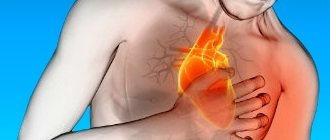
The heart has a very important task - ensuring blood circulation. Why is this so important? The thing is that every organ in the body, to one degree or another, needs a constant supply of oxygen and nutrients. In addition, “waste” material in the form of carbon dioxide and various metabolites must be removed from the organs. Both provide blood, the movement of which is associated with the work of the heart. Contracting with a certain force and frequency, it maintains constant blood circulation through the vessels. Thus, cardiac arrest or disruption of its functioning leads to oxygen starvation of the entire body.
Is the heart an ordinary muscle?
The myocardium, the main functional component of the heart wall, does consist of muscle fibers, but their structure is significantly different from skeletal muscles. Structurally and biochemically, the myocardium is designed in such a way that fatigue practically does not occur. The heart “rests” only in the periods between contractions. This mode of operation requires a constant supply of oxygen to the myocardium. This explains the fact that if the blood supply to the myocardium is disrupted, a heart attack develops relatively quickly, that is, cell death due to lack of oxygen. Skeletal muscles can remain viable for a long time in the absence of oxygen.
What happens if the heart stops beating?
For clarity, it is enough to imagine a situation where the heart no longer performs its function. In this case, the brain will be the first to be affected. In humans, this organ has reached its highest development and requires an appropriate amount of resources for its work. Therefore, as soon as the blood supply stops, the person almost instantly loses consciousness, which is a sign of a severe violation of brain function. Next, the death of neurons occurs. This process begins from the first minute after cardiac arrest, steadily progressing. After 3 minutes, irreversible changes in brain tissue become possible. Even after successful resuscitation measures, the patient will most likely remain disabled. After 5 minutes, in most cases, the death of the cerebral cortex occurs, which results in the death of the patient. Successful resuscitation at this stage can save life. But can a person’s state in which he is unable to speak, move, breathe or think be called “life”? A longer cessation of blood circulation inevitably leads to death.
Of course, the given time frames are arbitrary. A younger and healthier person will last longer, an older and sick person - less. But one thing is clear: stopping the work of any other organ can hardly cause such a sudden death.
Figure 1. What's good for heart health. Image: MedPortal
Properties of the myocardium
The main property of the myocardium is contractility. As the heart chambers contract, they perform rhythmic movements. So, they move blood along the vascular bed. During their work, the atria and ventricles constantly repeat two stages: contraction and relaxation. In medicine they are called systole and diastole. Systolic contraction of the myocardium occurs due to the interaction of two substances. These are myosin and actin. Systole is also provided by the energy of adenosine triphosphoric acid and potassium ions that leave the cells. Thin muscle fibers slide along thick ones, and the bundles become smaller in size when contracted.
In the phase of myocardial relaxation, it is customary to speak of diastole. Myosin and actin stop interacting. The energy spent during the cycle is restored by the heart with enzymes, vitamins and hormones. It receives them constantly, in the process of blood flow. The strength of heart contractions depends on how much calcium the myocyte cells contain.
After systole and diastole there is a short pause, after which the entire cardiac cycle repeats again. If the rhythm is normal, the cycle duration is 0.8 seconds. During systolic contraction of the atria, the ventricles fill with blood. After pushing blood into the ventricles, the atria relax and the ventricles begin to contract.
During the research, cardiologists were able to calculate how long the heart remains in a state of contraction and relaxation. Over a period of 24 hours, the following figures were obtained:
- contraction phase – 9 hours 24 minutes;
- relaxation phase – 14 hours 36 minutes.
Total diastole lasts almost one and a half times longer. This is important for the functioning of the myocardium: it does not wear out so quickly and “rests” longer than it contracts.
In addition to contraction, the myocardium has a number of properties on which the work of its cycle directly depends:
- automaticity - there is always an electrical impulse in the organ, giving rise to its contraction. This is the constant potential of the myocardium. Thanks to it, the heart is able to contract even if it is removed from the human body. This property is very valuable and is decisive in organ transplantation: it continues its work outside the body, maintaining blood circulation within itself. Myocardial automatism is no less valuable when carrying out resuscitation measures - with general collapse of the vascular bed and depression of respiratory and heartbeat functions. When the heart operates autonomously, special pacemaker cells are activated, providing control and regulation of rhythm;
- conductivity - a certain group of cells is also responsible for it. They transmit electrical impulses to all parts of the heart. Under a microscope, the processes of these myocytes resemble neurons - brain nerve cells;
- excitability - myocytes always respond to external stimuli. The concept of absolute and relative refractoriness is closely related to excitability. In the phase of absolute refractoriness, there is no response from the myocardium even if the incoming stimulus is very strong (period of 200-300 milliseconds). When a muscle is in a relative refractory phase, it reacts to strong stimuli.
The refractory state, or temporary loss of the ability to excite, protects the heart from signals above its threshold of perception.
The unique abilities of the heart ensure constant contraction of its muscle fibers. They regulate the rhythm so that the cavities are filled with blood in a timely manner and it is sent further through both circles of blood circulation. If a person finds himself in an extreme situation, compensatory mechanisms ensure the movement of blood and can save his life.
Where is the heart
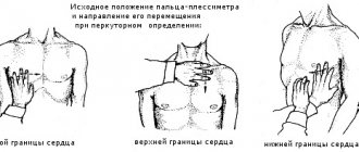
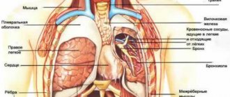
The heart is located in the chest cavity directly behind the sternum. Most of it is to the left in relation to the sternum, in the middle and to the right of it is the smaller part. However, it is extremely rare that the heart may, on the contrary, be located more to the right of the sternum. This anatomical feature is called dextrocardia.
The heart is enveloped by the lungs on both sides, in the front it touches the chest in a small area, and in the back there are the spine, blood vessels, nerves, digestive and respiratory tracts. Thus, the heart is, as it were, “walled up” with organs and bone formations on all sides. The space between the lungs, sternum and spine, where the heart is located, is called the mediastinum.
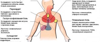
How to distinguish heart pain from others?
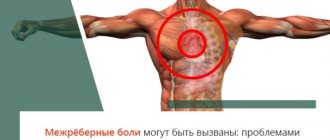
Pain due to intercostal neuralgia, pathology of the respiratory system and musculoskeletal system is very similar to pain in the heart. If you do not know some of the features of this type of pain, you can easily mistake the existing sensations for cardiac pathology. Heart problems are unlikely if:
- You can easily indicate the pain point, that is, the source of pain is small.
- Pain occurs when turning the body, sudden movements.
- Taking a full breath increases the intensity of pain.
- Feeling the painful area on the chest increases the pain.
- Taking analgesics such as Nurofen reduces pain.
Despite these seemingly clear differences in pain syndromes, pain due to cardiac pathology can sometimes be atypical. In this case, even a doctor cannot always make a correct diagnosis based only on the characteristics of the pain. For example, there are cases when a patient goes to the dentist about pain in the lower jaw, without even suspecting that in this case it is a symptom of angina pectoris. Other variants of atypical symptoms are also known. Therefore, all newly detected pain in the heart area is a reason to consult a doctor.
A doctor can determine some diseases simply by listening to the heartbeat. Photo: branin / freepik.com
Location anomalies
In typical clinical cases, which are the vast majority, the heart is on the left side. But there are at least two known conditions when this is not the case.
Dextrocardia
It is extremely rare. It is a congenital anomaly. The essence of the deviation (it is not considered a disease in the full sense of the word) is the reverse, mirror arrangement of the heart.
Normally it is on the left, in patients with a similar disorder it is on the right. Further options are possible regarding the localization of other organs.
With isolated dextrocardia, unpaired organs remain in their proper places. Only the heart moves. With systemic, complex transposition is observed. All organs mirror move in the opposite direction.
As a rule, such a deviation does not cause discomfort to the patient. This is normal and physiological in 85-90% of cases. Unfortunately, in the remaining 10-15% of situations, pronounced problems are found.
Usually these are congenital defects of cardiac structures that require surgical care.
In the absence of this, a typical clinical picture of physical and mental retardation in development is revealed.
Possible symptoms include:
- Insufficiently rapid weight gain in early periods of life.
- Drowsiness. The child is not active enough and has little interest in the world around him. Almost does not cry and is not capricious even in the presence of an irritating factor.
- Paleness of the skin.
- Cyanosis (blue discoloration) of the nasolabial triangle, nails, limbs, mucous membranes (the change is especially clearly visible in the gums).
Structure and functions of the parts of the heart
The heart is a cone-shaped muscular organ, the apex of which faces downwards. The upper part of the organ, from which the large vessels emerge, is called the base of the heart.
The heart consists of four chambers: two atria, located above, at the base, and two ventricles, located below, forming the apex. To better imagine the structure of the organ and the function of each of its sections, it is convenient to divide the heart into 2 halves, right and left, and consider them separately.
Right side of the heart
On the right, the heart is represented by the right atrium and the right ventricle. The atrium is significantly smaller than the ventricle in size. These formations are delimited from each other by the tricuspid valve.
Mainly the right half of the heart (right heart) is characterized by its connection with the lungs. The right heart transports venous blood from the organs to the lungs, where gas exchange occurs, as a result of which the blood becomes arterial. This happens as follows. Venous blood from all organs and systems of the body flows through the superior and inferior vena cava into the right atrium. Having collected a sufficient volume of blood, the atrium contracts and blood flows into the right ventricle. As soon as the blood pressure in the ventricle reaches the required value, the tricuspid valve leaflets collapse and the walls of the ventricle contract. Venous blood flows from the ventricle into the pulmonary trunk, which divides into two pulmonary arteries and carries blood further to the lungs.
Figure 2. Right half of the heart. Red arrows indicate the course of venous blood. The lungs are shown schematically at the top. Illustration by Danila Melnikov
As can be seen in Figure 1, there is also a valve at the exit of the right ventricle called the pulmonary valve. Both valves are designed to prevent blood from returning to previous parts of the heart during the next contraction of the myocardium.
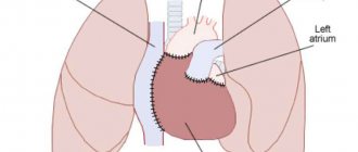
Left half of the heart
The left half of the heart (left heart) is responsible for transporting arterial blood enriched with oxygen in the lungs to all organs and systems. The left heart is more developed than the right, since in order to move blood into every capillary of the human body, it is necessary to create high blood pressure. Also, unlike the right side, between the left atrium and the left ventricle there is a bicuspid (mitral) valve. Otherwise, the structure of the left parts of the heart is similar to the structure of the right (see Figure 3).
Figure 3. Left half of the heart. Red arrows indicate arterial blood flow. The lungs are shown schematically at the top. Blue vessels are pulmonary veins. Illustration by Danila Melnikov
When describing the direction of blood flow, we focused on the lungs. What happens next? After gas exchange occurs, blood flows through the pulmonary veins into the left atrium. The atrium then contracts, the mitral valve opens, and the left ventricle fills with blood. Having collected the required volume of blood, the ventricle contracts, the aortic valve, located at the base of the aorta, opens, and blood enters the aorta. The aorta, in turn, giving off numerous branches, supplies blood to all organs and systems.
Circulation circles
Having collected the two halves of the heart together, we will see the following picture (see Figure 4). Two circles of blood circulation are formed. The first, small circle, begins in the right ventricle and ends in the left atrium. It is necessary for oxygenation of blood in the lungs. The second, large circle, begins in the left ventricle and ends in the right atrium. Its purpose is to distribute oxygenated blood to the organs, and take venous blood from them to the heart in order to circulate it again in a small circle.
Figure 4. Circulation circles. Designations: R/R - right atrium; R/F - right ventricle; L/R - left atrium; L/F - left ventricle. The liver at the bottom of the picture is an example of a blood-supplying organ. Illustration by Danila Melnikov
As you can see, the heart is inextricably linked by blood circulation circles with all human organs, but first of all with the lungs, because there is a separate circle for them. That is why, with heart pathology, the lungs often suffer.
What is cardio muscle tissue
A muscle is a fibrous tissue that contracts to produce movement. There are three types of muscle tissue in the body: skeletal, smooth and cardiac. Cardiac muscle is highly organized and contains many cell types, including fibroblasts, smooth muscle cells, and cardiomyocytes.
Cardiac muscle exists only in the heart. It contains heart muscle cells that perform highly coordinated actions that support the heart pump and blood circulating throughout the body.
Unlike skeletal muscle tissue, such as that found in the arms and legs, the movements produced by cardiac muscle tissue are involuntary. This means that they are automatic and cannot be controlled by humans.
How does a person's heart beat?
Like any muscle, the heart is innervated by peripheral nerves, which allows you to regulate the strength and frequency of its contractions. However, the following fact is known. If you denervate the heart, that is, cut the nerves going to it, it will not stop beating. If you do the same with any other muscle, it will no longer be able to contract. Why is this happening?
The thing is that the heart has its own analogue of the nervous system, which is called the cardiac conduction system. It is represented by two nodes and several branches of cells that are somewhat different from ordinary myocardial cells. They are able to continuously generate electrical impulses at a specific frequency that trigger muscle contraction. When doctors install a pacemaker in a patient, they are thereby trying to restore the disrupted functioning of our “congenital pacemaker” - the conduction system of the heart.
But the peripheral innervation, which we “got rid of” in the experiment above, also has an important function. When we are lying down in a calm environment, it is hardly possible to feel our own pulse - we do not feel the heartbeat. But after a short run, you can feel your heartbeat literally throughout your entire body. This phenomenon can be explained by the fact that during physical activity the tone of the sympathetic nerves going to the heart increases. As a result, the strength and frequency of heart contractions increases to meet the increased oxygen demand of working muscles. In a quiet stop or during sleep, the work of parasympathetic innervation prevails, and everything happens the other way around.
Why does the heart beat?
By placing your ear to a person's chest, you can hear a characteristic rhythmic knocking sound.
Normally, it consists of two tones: the first - strong, ringing and the second - quieter. Tones are created by valves when their valves close. When a doctor listens to a patient's heart with a phonendoscope, he can evaluate these sounds and give a preliminary conclusion on the operation of the valve apparatus. Since the sounds of the valves correspond to the next heart contractions, they can be used to characterize the heart rhythm. Various pathological phenomena can change the rhythm, make it irregular, frequent, rare, dull, etc. These characteristics allow the doctor to diagnose some heart diseases by barely hearing the heartbeat.
Compensatory mechanisms
Our body protects the myocardium from overload through the complex functions of the neuroendocrine system. It transmits signals to the muscle about the need to increase heart rate (heart rate per minute). Compensatory mechanisms can be activated under the following conditions:
- diseases and malfunctions of other organs;
- changes in environmental conditions and climate;
- the appearance of strong irritants (including during neuropsychic stress).
In such conditions, two strong hormones are released into the blood - adrenaline and norepinephrine. To compensate, the body needs more oxygen. The heart begins to work at an accelerated rate, and the heart rate increases. The faster the heart rate, the more oxygen the body receives. This compensatory reaction is normal, for example, during high physical activity in athletes, during pleasant excitement, and positive emotions. If the heart is in this state too often, it can lead to the development of a serious illness.
How does your heart hurt?
Heart pain is most often caused by coronary heart disease (CHD). In this condition, blood circulation in the heart itself is disrupted, and pain occurs due to lack of oxygen. This pain has its own characteristics:
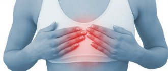
- Localized behind the sternum.
- It is impossible to pinpoint the pain point.
- Accompanied by a feeling of pressure and burning behind the sternum.
- May spread to the left shoulder and upper arm, left arm, lower jaw.
- Often appears due to stress or physical activity.
- The intensity varies: from a feeling of discomfort to unbearable pain.
- With angina, the pain lasts up to 10 minutes.
- With myocardial infarction, it can last for hours, accompanied by suffocation and a feeling of lightheadedness.
There are also pains not associated with coronary artery disease, they are called cardialgia. They may indicate completely different diseases and are not always associated with pathology in the heart. Cardialgia varies in nature, but is rarely pressing and burning, as with ischemic heart disease. Their intensity is also lower.
Important!
If you detect any pain in the heart area, you should contact a cardiologist. If the pain is intense and lasts longer than 10 minutes, this is a reason to call an ambulance. Ignoring heart pain can be life-threatening.
Other symptoms of heart disease
Heart disease makes itself felt not only by pain, but also by other symptoms:
- Shortness of breath occurs not only when the respiratory system is damaged, but also the heart. This is an unfavorable symptom that indicates the neglect of the pathological process. It can be caused by valve defects, arrhythmias, cardiomyopathies and a number of other diseases. Shortness of breath may also indicate chronic heart failure, which can be the outcome of other heart diseases (myocardial infarction, hypertension, myocarditis, etc.)
- The feeling of interruptions in the work of the heart occurs most often with various arrhythmias. It may seem as if the heart is periodically tumbling in the chest, and sometimes there is a feeling that the heart is beating irregularly. The culmination of such symptoms is loss of consciousness due to arrhythmia.
- A rare pulse often indicates a disruption in the conduction system of the heart. Normally, the heart contracts at a rate of 60-89 beats per minute. If, after feeling the pulse on the wrist, it is found that this value is below 55 beats, the presence of the disease is quite likely. The exception is professional athletes, whose rare pulse is normal.
During physical activity, the strength and frequency of heart contractions increases, and we feel our own pulse well. Photo: axesor/Depositphotos
How to eat to keep your heart healthy?
A healthy diet allows you to achieve prevention against two of the most common cardiovascular pathologies: hypertension and coronary heart disease. The most effective measures are the following³:
- Reduce your intake of saturated fatty acids. They are found in large quantities in many cheap confectionery products. It is better to choose products that do not contain palm, palm kernel and coconut oil.
- Olive oil, nuts, fatty fish, and seafood are preferred sources of fat. These foods contain mono- and polyunsaturated fatty acids, the consumption of which reduces the risk of cardiovascular disease (CVD).
- Minimize foods fried in oil in your diet, especially fast food. The trans fats contained in the latter increase the risk of CVD diseases.
- It is not recommended to regularly consume sugar-containing drinks such as lemonade, soda and juice. They do not satisfy thirst, which leads to uncontrolled consumption. Simple sugars in such drinks increase the risk of both diabetes and coronary heart disease.
- Increase the amount of dietary fiber in your diet. Grains, fruits and vegetables will help with this.
- Reduce table salt consumption. Recommended figures are up to 5 g/day, but according to new standards, the maximum reduction in CVD risk is achieved by consuming 3 grams of salt per day. This aspect of diet plays a large role in the context of hypertension.
- Foods high in potassium are beneficial. This element is found in large quantities in fresh fruits and greens and helps lower blood pressure. It is recommended to eat 400-600 g of vegetables, fruits and berries daily.
Prevention of heart disease
In the absence of severe chronic diseases, you have every chance of maintaining the health of your heart muscle for a long time. For this you need:
- healthy and proper nutrition;
- strengthening the immune system (exercise, light exercise);
- monitoring the state of the endocrine system;
- to give up smoking;
- abstaining from alcohol or drinking it in moderation.
If you want to improve your health at the fitness center, you need to understand the differences between cardio exercise and regular gymnastics. Cardio exercise is for those with a healthy heart. They cannot be used in case of hypertension and serious heart diseases.
In people actively involved in sports, moderate enlargement of the left or right ventricles of the heart is often detected.
If you have been diagnosed with myocardial disease, you can engage in therapeutic exercises. You must first consult with your doctor. Exercise therapy is prescribed:
- to accelerate the healing of muscle tissue after heart attacks;
- to strengthen the spinal apparatus;
- to improve immunity;
- to restore neuroendocrine functions;
- to improve the functioning of the vascular bed.
The heart is a unique organ on which the blood supply to all parts of our body depends. The myocardium is able to withstand heavy loads, but, most importantly, do not overdo it and take care of your health. Moderate physical exercise, a healthy lifestyle and a favorable psychological microclimate in the family and at work will help strengthen the heart muscle.
The correct physiological location of the heart in humans involves its location in the chest asymmetrically, slightly to the left of the center.
This is a generally accepted calculation that is used by both theorists and practitioners as part of echocardiography (ultrasound) or MRI diagnostics.
Preventing proper heart function
When it comes to preventing cardiovascular diseases, physical activity is the first thing to think about. Research figures unanimously confirm this.
Regular physical activity, compared with a sedentary lifestyle, is associated with a 22–36%² reduction in all-cause mortality and a 25–35%² reduction in cardiovascular mortality.
In people already diagnosed with coronary heart disease, exercise training reduced the risk of overall mortality by 19–58%² and cardiovascular mortality by 20–62%². Even 1 hour of walking per week already reduces the above risks². What if you walk for an hour every day? A correlation was also found with walking speed: the faster the step, the higher the result of prevention. Thus, in order to significantly reduce the risk of heart disease, it is enough to spend half an hour to an hour of intense walking every day.
Physical activity is a good preventive measure for heart disease. The faster the step, the better the result. Photo: Kzenon/Depositphotos
Is the heart really that vulnerable?
In fact, the heart has a large number of adaptive reactions. They can maintain its normal activity for a long time even in the face of a large number of negative factors such as poor diet, inactive lifestyle and obesity. Under conditions of increased load, the myocardium increases in size and compensates for it. If the conduction system is disrupted, the heart does not stop contracting—second-order pacemakers come into play and continue to send impulses, albeit at a lower frequency. With valve defects, due to disturbances in hemodynamic parameters, the heart contracts more strongly and also increases in size. There are many such examples.
The problem is that these adaptive responses only help for a while. Over the course of several years, they turn against the heart and irreversible damage begins. Moreover, the symptoms, unfortunately, appear just at a time when it is already too late - the changes that have arisen are irreversible, and the disease becomes chronic.
The importance of biochemical processes in the myocardium
The viability of cardiomyocytes is ensured by the supply of nutrients, oxygen and the synthesis of energy in the form of adenosine triphosphoric acid.
All biochemical reactions occur maximally during systole. The processes are called aerobic because they are possible only with a sufficient amount of oxygen. The left ventricle consumes 2 ml of oxygen per minute per 100 g of mass.
To produce energy, the following are used in the blood:
- glucose,
- lactic acid,
- ketone bodies,
- fatty acid,
- pyruvic and amino acids,
- enzymes,
- B vitamins,
- hormones.
If the heart rate increases (physical activity, anxiety), the need for oxygen increases 40–50 times, and the consumption of biochemical components also increases significantly.
Sources
- Prives M. G., Lysenkov N. K., Bushkovich V. I. / Human Anatomy. — 12th ed., revised. and additional - St. Petersburg: Publishing house SPbMAPO, 2006. - 720 p., ill.
- Krivoshapova Kristina Evgenievna, Tsygankova Daria Pavlovna, Barbarash Olga Leonidovna Low physical activity as a risk factor for cardiovascular morbidity and mortality // Systemic hypertension. 2021. No. 3.
- Smetneva Natalya Sergeevna, Pogozheva Alla Vladimirovna, Vasiliev Yuri Leonidovich, Dydykin Sergey Sergeevich, Dydykina Irina Stepanovna, Kovalenko Alexey Anatolyevich The role of optimal nutrition in the prevention of cardiovascular diseases // Nutrition Issues. 2020. No. 3.
- Yakushin S.S., Filippov E.V. Physical activity and its importance for the prevention of cardiovascular diseases // Clinician. 2015. No. 3.
What compensatory mechanisms does the heart muscle have?
A person does not develop pathology as long as the compensation mechanisms work well. Regulation is carried out by the neuroendocrine system.
The sympathetic nerve delivers signals to the myocardium about the need for increased contractions. This is achieved by more intense metabolism and increased ATP synthesis.
A similar effect occurs with increased synthesis of catecholamines (adrenaline, norepinephrine). In such cases, increased work of the myocardium requires an increased supply of oxygen.
The vagus nerve helps reduce the frequency of contractions during sleep, during rest periods, and preserve oxygen reserves.
It is important to consider reflex adaptation mechanisms.
Tachycardia is caused by congestive stretching of the mouths of the vena cava.
Reflex slowing of the rhythm is possible with aortic stenosis. In this case, increased pressure in the cavity of the left ventricle irritates the endings of the vagus nerve, contributing to bradycardia and hypotension.
The duration of diastole increases. Favorable conditions are created for the functioning of the heart. Therefore, aortic stenosis is considered a well-compensated defect. It allows patients to live to an old age.
Types of violation
Cardiac dextrocardia is divided into 3 types:
- Simple. It occurs quite rarely and consists of a displacement to the right side of only the heart and its vessels.
- Mirror. Characterized by incorrect location of parts of the digestive and respiratory organs.
- Full transposition. With it, all the organs of the chest are positioned incorrectly.
There is also a type of dextrocardia called dextroversion. It is characterized by the fact that the apex of the heart is turned to the right side. In this case, the anatomical structure of the organ does not suffer.
How myocardial pathology occurs, mechanisms of clinical manifestations
Myocardial diseases develop under the influence of various causes, but appear only when adaptation mechanisms fail.
Long-term loss of muscle energy, the impossibility of independent synthesis in the absence of components (especially oxygen, vitamins, glucose, amino acids) lead to a thinning of the actomyosin layer, breaking the connections between myofibrils, replacing them with fibrous tissue.
This disease is called dystrophy. It accompanies:
- anemia,
- avitaminosis,
- endocrine disorders,
- intoxications.
Arises as a consequence:
- hypertension,
- coronary atherosclerosis,
- myocarditis.
Patients experience the following symptoms:
- weakness,
- arrhythmia,
- shortness of breath during physical exertion,
- heartbeat.
At a young age, the most common cause may be thyrotoxicosis and diabetes mellitus. In this case, there are no obvious symptoms of an enlarged thyroid gland.
The inflammation of the heart muscle is called myocarditis. It accompanies both infectious diseases of children and adults, as well as those unrelated to infection (allergic, idiopathic).
It develops in focal and diffuse forms. Proliferation of inflammatory elements affects myofibrils, interrupts pathways, and changes the activity of nodes and individual cells.
As a result, the patient develops heart failure (usually right ventricular failure). Clinical manifestations consist of:
- pain in the heart area;
- rhythm interruptions;
- shortness of breath;
- dilation and pulsation of the neck veins.
Atrioventricular blocks of varying degrees are recorded on the ECG.
The most well-known disease caused by impaired blood flow to the heart muscle is myocardial ischemia. It proceeds in the form:
- angina attacks,
- acute heart attack,
- chronic coronary insufficiency,
- sudden death.
All forms of ischemia are accompanied by paroxysmal pain. They are figuratively called “the cry of the starving myocardium.” The course and outcome of the disease depends on:
- speed of assistance;
- restoration of blood circulation due to collaterals;
- the ability of muscle cells to adapt to hypoxia;
- formation of a strong scar.
Ventricular tachycardia (VT)
VT refers to an abnormally rapid heartbeat. The source of such a rhythm may be an ectopic focus in the myocardium of the right or left ventricle. Typically, the cause of ventricular abnormal pacemakers is diseases of the heart muscle (coronary heart disease, arrhythmogenic dysplasia of the right ventricle, etc.). In VT, the heart does not pump blood as efficiently as it does in normal sinus rhythm. The rapid rhythm of contractions prevents the ventricles from filling completely between individual heartbeats. As a result, the volume of blood circulation in the body decreases.
With VT, symptoms such as dizziness, fainting, presyncope, and loss of consciousness occur. For most patients, VT is considered a very dangerous rhythm, which can lead to the death of the patient.
Myths and speculation
Naturally, like everything unusual, people with “incorrectly” located organs cause a lot of speculation and gossip. For example, they are often credited with psychic abilities and the ability to read minds.
In addition, it is believed that such babies will get sick more. In fact, there is no scientific basis for this. It’s just that such people must have a note about their particularity in their medical record, and also exercise certain caution when actively engaging in physical education.
How to help the heart muscle?
People involved in sports remain the most prepared for critical impacts. One should clearly distinguish between cardio training offered by fitness centers and therapeutic exercises. Any cardio program is designed for healthy people. Increased training can cause moderate hypertrophy of the left and right ventricles. When the work is done correctly, the person himself monitors the sufficiency of the load using his pulse.
Therapeutic exercise is indicated for people suffering from any diseases. If we talk about the heart, then it has the goal:
- improve tissue regeneration after a heart attack;
- strengthen the spinal ligaments and eliminate the possibility of pinching of the paravertebral vessels;
- “boost” the immune system;
- restore neuroendocrine regulation;
- ensure the functioning of auxiliary vessels.
Treatment with drugs is prescribed in accordance with their mechanism of action.
There is currently a sufficient arsenal of means for therapy:
- relieving arrhythmias;
- improving metabolism in cardiomyocytes;
- enhancing nutrition by dilating the coronary vessels;
- increasing resistance to hypoxia conditions;
- suppressing unnecessary foci of excitability.
You can’t joke with your heart; experimenting on yourself is not recommended. Only a doctor can prescribe and select medications. In order to prevent pathological symptoms for as long as possible, proper prevention is needed. Every person can help their heart by limiting their intake of alcohol, fatty foods, and quitting smoking. Regular exercise can solve many problems.
Why is it so long?
What a lot of trouble a giraffe has with that long neck! Why does he need her at all? Scientists have two main hypotheses. According to one, the long neck is the result of sexual selection; according to another, more widespread, it arose as a result of food competition and helps giraffes to conveniently eat high branches that other herbivorous inhabitants of the savannah, which are very numerous, cannot reach. For years people took this version for granted. To those who have observed African wild grasslands filled with herbivores of all sizes, nothing else could have occurred to them. However, scientists conducted the first experiments confirming the benefits of long-necks at meals only at the beginning of the 21st century.
Rice. 4.
The acacias, surrounded by a high fence, are accessible only to giraffes and are eaten evenly.
Public trees are gnawed like an umbrella. Light rectangles
are fenced acacias,
dark ones
are unfenced. Image: Chemistry and Life
South African researchers Elissa Cameron and Johan du Toit performed a very simple experiment. Having chosen a savannah area in the Kruger National Park where giraffes grazed, they surrounded several acacia trees in this area with a 2.2 m high fence. It was 1 m from the crown boundary. For adult giraffes, such a fence is not a hindrance; they can gnaw branches at a height of four meters and easily lean over the fence. The rest of the animals could not eat the fenced trees (the fence was protected from elephants). In the immediate vicinity of the experimental acacias, the scientists designated control trees of approximately the same height, so that each fenced tree had a proportionate pair. After two wet seasons, the researchers compared leaf biomass on shoots located at different heights. (The scientists used averaged data for ten randomly selected branches.) In the experimental acacias, it was almost the same on all branches, but in the control trees, the lower branches turned out to be much more eaten than the upper ones. And only at a height of four meters the number of leaves on fenced and unfenced acacias was practically the same. Looking at these results, scientists concluded that giraffes evolved a long neck to help them avoid competition for food with other herbivores. And although this experiment has been criticized in the scientific press for not taking many factors into account, it certainly deserves mention as an attempt to scientifically substantiate an obvious fact.
Diagnosis of heart position
You can confirm the doctor’s assumption about the abnormal location of organs using instrumental research methods:
- X-rays help determine the degree of movement of the heart and neighboring organs;
- The ECG has a mirror direction of the waves, the voltage is reduced, the QRS complexes are increased in leads V1–V3 and decreased in V4–V6;
- Ultrasound helps to assess the structure of the heart, the presence of valve defects and great vessels; when examining the abdominal cavity, one can assess the correct location of the main organs in it;
- CT and MRI are prescribed for detailed diagnosis and the need for surgery.
Treatment methods
Treatment depends on whether dextrocardia is accompanied by other cardiac pathologies. If not, then therapy is not required. The patient can lead a full active life. Only when deciding to engage in sports is it recommended to undergo an ECG.
If the incorrect location of the heart occurs along with other diseases, treatment tactics will depend on the specific diagnosis. Typically, heart defects are eliminated through surgery, during which the existing defects are removed.
Before the operation, the patient must be prepared by prescribing medications. To maintain the patient's condition, diuretics, antihypertension agents, and drugs that support the myocardium are used.
Special attention is paid to antibacterial treatment. It is carried out both before surgery and during the period of rehabilitation of the body after it. But taking antibiotics for a long time is not recommended, as they negatively affect the condition of the organs.
Any medications are prescribed exclusively by the attending doctor, taking into account the results of the patient’s examination. The specific list of medications depends on the type of disease and the severity of its course.
Forecast
Patients with dextrocardia, which does not manifest itself, do not have to worry about their health if they follow recommendations regarding nutrition, giving up bad habits and maintaining physical activity. Their risk of heart failure is the same as that of other people. In such cases, the pathology is considered just an anatomical defect.
In case of multiple deviations in the structure and formation of organs, the prognosis will depend on the type of malformation and the timeliness of its treatment, and the possibilities for radical surgery.