Currently, the most common methods for studying the human cardiovascular system are ultrasound (ultrasound) with Doppler techniques:
- Duplex scanning.
- Doppler ultrasound (USDG).
- Echocardiography (Echo-CG).
The main advantages of these methods are their absolute non-invasiveness (no trauma to the skin and mucous membranes), safety for the patient, high information content, sensitivity and specificity of the data obtained, the possibility of conducting studies in dynamics with registration of both background blood flow parameters in real time and induced parameters when applying a variety of functional load tests.
Ultrasound Dopplerography (USDG)
In Doppler ultrasound, ultrasound responds to moving red blood cells in the blood, sending signals to a monitor and giving an idea of the speed and pattern of blood flow. Doppler ultrasound mainly gives an idea of the patency of the veins and the ability to identify their damage and the causes that provoked vascular changes. As a result of the ultrasound examination procedure, a one-dimensional image is obtained.
Advantages of ultrasound scanning
The main advantage of Doppler ultrasound is the availability of the method, since scanning in Doppler mode is much cheaper. Doppler ultrasound also allows us to identify stenoses and their degree, atherosclerotic plaques and blood clots.
When is routine Doppler sonography necessary?
Doppler of both extra- and intracranial arteries and veins should be performed at least once a year as a routine study (even before the appearance of any complaints):
- all women over 45 years old
- all men over 40 years old
- those who have close relatives suffering from hypertension, diabetes, coronary artery disease
- for diabetes
- smoking
- antiphospholipid syndrome
- for osteochondrosis of the cervical spine
- metabolic syndrome
- arterial hypertension
- if you have had a stroke or transient cerebrovascular accident
- if a person suffers from rhythm disturbances (the likelihood of cerebral thromboembolism with subsequent stroke is increased)
- increased levels of cholesterol, triglycerides, low-density lipoproteins in the blood (signs of atherosclerosis)
- had surgery on the spinal cord or brain
- before planned heart surgery.
Ultrasound duplex scanning (USDS)
Unlike Doppler Doppler Doppler, Doppler Doppler Doppler Doppler Ultrasound
includes not only Doppler ultrasound, but also B-mode, in which veins and arteries are visualized, as well as the condition of nearby tissues. Ultrasound scanning is more informative than Doppler ultrasound, since when it is performed, damage to blood vessels and nearby tissues can be seen, rather than assuming their presence. Based on the results of ultrasound scanning, a two-dimensional color cross-sectional image of the vessels is obtained.
Advantages of ultrasonic testing
The main advantage of ultrasound scanning is its high information content, which allows one to obtain information not only about the patency, but also about the condition of any vessels. This makes it possible to diagnose a wide range of vascular diseases, including parietal thrombotic formations, aneurysms and inflammatory changes in the walls.
Types of ultrasound examination
Doppler ultrasound (CDS - color duplex scanning of the brachiocephalic arteries) is indicated for patients with a wide variety of complaints. The method is used for different organs of the human body:
- Ultrasound Doppler Doppler (CDS) of neck vessels. They scan those arterial and venous branches of vessels that are located outside the cranium and are responsible for normal nutrition of the brain and the outflow of blood from it. The condition of the carotid, subclavian, vertebral arteries, and brachiocephalic trunk is assessed.
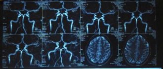
- Ultrasound Doppler Doppler (CDS) of the brain. Provides information about the condition of extracranial blood vessels (located outside the skull), and intracranial vessels (located inside the skull).
- Ultrasound Doppler Doppler (CDS) of the arteries of the lower extremities. During the procedure, it is possible to identify pathologies of the vessels of the extremities and other disorders in the blood flow system.
- Ultrasound Doppler Doppler (CDS) of the veins of the lower extremities. Used to assess the level of patency of deep and superficial veins, determine the functional state of venous valves, and diagnose typically located valves of perforating veins.
- Neurosonography. It is used in children from birth to one year to determine the condition of the brain, possible pathological changes, the size of some of its parts, as well as formations - cysts, hematomas.
The procedure is carried out in real time, the conclusion is immediately issued upon completion.
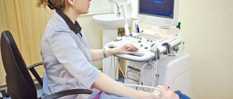
How are both studies conducted?
These two studies are carried out in specially equipped ultrasound diagnostic rooms. Both procedures are performed with the patient in a supine position; in some cases, functional tests may be necessary. No special preparation is required for duplex scanning or Doppler ultrasound, but you should refrain from drugs and drinks that can change vascular tone. When choosing an ultrasound scan or duplex scanning, it is worth knowing what the difference is in the process of carrying out these procedures.
USDG
The duration of the procedure depends on which organ is being examined. For example, the neck requires 15-20 minutes, while an ultrasound scan of the brain will require about 30 minutes. The peculiarity of performing an ultrasound scan is that this procedure requires the high competence of a sonologist: he applies a Doppler sensor to areas of the body standard for this study to check whether the vessels perform their functions. The sensor moves according to the flow of blood.
UZDS
What is the difference between duplex scanning and ultrasound scanning of blood vessels
, takes this type of research to a different level: in the process, the vessels are clearly visible, which means that the sonologist moves the sensor along a clearly visible trajectory, and not blindly. This allows you to make the research more detailed and informative. The duration of the procedure is no more than half an hour.
Vein scanning: duplex and triplex - what is the difference
This is how the doctor sometimes wraps up such a word, and in a tongue twister, and the patient stands in thought, what kind of study is this?.. duplex and triplex scanning refer to just such terms. The main thing is that it is not clear what to expect - either they will pierce the leg, or this is a type of x-ray.
Now we will decipher it for you, translate it from “medical to human.” In general, in medicine, scanning (from the English scan - to consider) is the controlled movement of a beam of radiation across the body, in our case - ultrasonic.
When pulses are reflected, they are recorded on the monitor screen. This is the essence of an ultrasound scan; its purpose is to obtain a picture of the internal organs or veins in order to determine the extent of the disease.
So we found out that this is all a regular ultrasound examination, without damaging the skin. The skin is smeared with a special gel, the doctor slides a sensor over the body, and a picture of the insides is obtained on the screen in real time. Not only the lumen of the vein and the condition of its walls are visible, but also the speed of blood flow and other characteristics of the blood.
But it was not always so. Previously, vessels were examined using X-rays - they injected a radiopaque substance into the vessel and watched how it filled the vein. The most noticeable violations, of course, could be seen, but, firstly, the method was invasive (that is, puncture of the skin and injection of a substance), and secondly, it did not show everything.
As for the names, when duplex scanning
The device operates in 2 modes at once (normal and Doppler).
The usual (traditional) ultrasound scanning mode allows you to see the vein in a two-dimensional image, its structure, and developmental anomalies.
The Doppler mode allows you to determine the “fluidity” of blood, the direction of its movement and the speed of blood flow.
The Doppler mode allows you to determine the “fluidity” of blood, the direction of its movement and the speed of blood flow.
Duplex scanning is an improved diagnostic method that combines the already familiar ultrasound and Doppler ultrasound.
It has an advantage over the last two, which is expressed in the ability to diagnose vascular pathologies at the earliest stage of development. This is achieved due to the fact that with the help of duplex it is possible to evaluate not only external characteristics, but also to examine the internal structure of blood vessels and identify their internal pathology.
Duplex ultrasound examination is a three-dimensional echography method that has a high degree of information content, which is important in determining the causes of diseases and making a diagnosis.
With all its research capabilities, this procedure is characterized by absolute safety and painlessness and the absence of contraindications and side effects. To carry out the diagnostic procedure, the patient does not need special preparation. However, it requires a highly qualified doctor to obtain reliable results.
This research provides an opportunity to:
- reliable determination of any pathology of the vascular bed;
- determination of blood flow speed;
- identifying the causes of changes and disturbances in blood flow.
Triplex scanning
is not any other research method. The same ultrasound machine already works in 3 modes - color Doppler is added to the first two.
Triplex scanning of blood vessels allows you to examine:
- the anatomy of the vessel, the degree of its tortuosity, which is very important to know during operations;
- structure of the vascular wall;
- speed and direction of blood flow;
- vein patency in color mode (presence of narrowings or blood clots).
Color scanning today provides doctors with the most accurate information about vascular diseases (which is very important not only for conservative treatment, but also before surgery).
Color scanning is performed in the following cases:
- Thrombophlebitis, its early diagnosis is especially valuable.
- Varicose veins, especially in the moderate and severe stages.
- Thrombosis.
- The method helps to accurately determine whether surgery is necessary for these diseases and where exactly the greatest destruction of blood vessels is.
- If deep veins are affected, triplex scanning is a must!
- You will be surprised, but when preparing operations in the abdominal cavity, doctors often scan blood vessels. This helps reduce the risk of postoperative complications (thrombosis).
In what cases is ultrasound examination performed?
Ultrasound diagnostics with the Doppler effect are more often used to diagnose large vessels (carotid, vertebral and cerebral arteries, as well as leg veins).
The main indications for ultrasound scanning are:
- constant migraines combined with dizziness;
- frequent fainting;
- noise in ears;
- used in patients who have had a stroke;
- diseases of the spinal column;
- persistent increase in blood pressure;
- decreased visual function;
- diabetes;
- neurological diseases;
- swelling of the legs, accompanied by a feeling of heaviness and pain;
- the appearance of a vascular network, change in skin color on the legs;
- excess weight.
How is the examination carried out?
When examining the head and neck, ultrasound examination is performed in a sitting position.
All other types of examination are performed lying down. In some cases, when examining the veins of the lower extremities, the patient will be asked to stand or place the leg in a certain position. A gel is applied to the area under study to facilitate better penetration of ultrasound into the vessels. Next, a sensor is passed over this place several times in different directions, which transmits data to the monitor.
Based on the data obtained, a conclusion is formed. Based on it, a neurologist, phlebologist, cardiologist or other specialized specialist makes a diagnosis and prescribes treatment
Where can I do an ultrasound scan?
Duplex scanning is carried out on the direction of the attending physician in neurological departments in hospitals.
If the outpatient department at your place of residence has the necessary equipment, then you can be examined there.
In addition, there are many medical and diagnostic centers offering a whole range of diagnostic procedures.
A preventive examination will help prevent serious illnesses. Thus, it is possible to promptly detect and prevent arterial embolism, stroke, and complications after operations.
Preparation
No special preparation methods are required for ultrasound scanning. But there are some restrictions.
First of all, on the eve of the procedure, the patient is not recommended to take drinks that increase vascular tone.
These include:
- coffee;
- tea;
- energetic drinks.
It is not advisable to smoke a lot before the examination.
After consulting with your doctor, you may temporarily stop taking certain medications that may interfere with your results. Such drugs include Betaserc, Cinnarizine and others.
Immediately before starting the procedure, you will have to remove metal jewelry and objects.
Indications and contraindications
Vascular ultrasound using Doppler has no contraindications; it is allowed for children, bedridden patients and even those who are unconscious.
They perform ultrasound examination at home or in a hospital. Doppler ultrasound is recommended for all people over 40 years of age, as well as those suffering from:
- osteochondrosis;
- overweight;
- vegetative-vascular dystonia;
- ischemia;
- hypertension.
Doppler ultrasound of the head is prescribed for:
- chronic headaches;
- dizziness;
- fainting;
- frequent nausea and vomiting not associated with gastrointestinal disorders;
- visual impairment;
- tinnitus.
Doppler ultrasound of the lower extremities is indicated for:
- the appearance of a pronounced vascular network and bulging veins;
- changes in skin color on the legs and feet;
- the occurrence of itching;
- the formation of edema, which increases with stress and overheating;
- repeated seizures.
It is recommended to postpone the study only if:
- inflammatory disease in the acute stage;
- in case of damage to the skin or the presence of a dermatological disease (eczema, psoriasis) in the area under study until they are cured or reach the stage of remission.
You can undergo an ultrasound scan in our clinic on an outpatient basis or call a specialist at home.
For diagnostics, we use modern high-precision equipment, which allows us to identify complex pathologies at the very beginning of their development. Medical specialists have extensive experience in the field of ultrasound examination of both adults and very young newborns. Make an appointment at Novomed by calling the phone number listed on the website!
Procedure for conducting the examination
The ultrasound examination technique is simple.
It is carried out according to the following algorithm:
- the patient takes a horizontal position;
- a cushion is placed under the head, the head is turned to the side (if the vessels of the head are examined);
- gel is applied to the place where the sensor will be placed;
- the doctor then begins to move the scanning device along the surface of the skin in the area of interest.
The operating time of the scanner takes on average no more than 40 minutes.
The doctor can provide diagnostic results immediately after the process is completed.