Brain cancer is a process of pathological transformation of brain cells or the surrounding meningeal, glandular and bone tissues. As a result of transformation, abnormal cells begin the process of uncontrolled division, which causes the formation of a tumor that compresses healthy tissue and increases intracranial pressure. In conditions of limited space in the cranium, even a benign neoplasm, if left untreated, can lead to disability or even death.
Kinds
Brain tumors are divided into:
- primary – developing directly from intracranial tissues;
- secondary – developing from metastases spread by a tumor located in another organ.
Tumors are classified according to the location of their formation:
- neuroepithelial, or gliomas – appearing directly in the brain tissue;
- meningiomas - developing in the tissues of the membranes of the brain;
- neuromas – originating in the cells of the nerve fibers of the skull;
- pituitary adenomas – rarely showing malignant signs;
- metastases from other organs;
- dysembryogenetic neoplasms that develop during the embryonic period of a child’s development.
What is a brain tumor?
A brain tumor is a collection of abnormal cells that divide and grow rapidly.
They rarely spread to other parts of the body, but most can spread through brain tissue. The danger of this type of tumor lies in the disruption of basic brain functions. There are more than 130 types of brain cancer. They are classified depending on how quickly they grow and the likelihood of relapse after treatment.
There are two main types of brain tumors - benign and malignant.
Benign brain tumors, grade 1 or 2, are characterized by relatively slow growth and are less likely to return after treatment. Benign tumors in the brain, which are rarely life-threatening when they develop in other parts of the body, can press on and destroy healthy brain tissue as they grow, which can lead to serious and sometimes life-threatening damage.
Benign ones include: chordomas (most common in people aged 50 to 60 years), craniophagiomas, gangliocytomas, gangliomas, angioplastic gangliomas (occur mainly in young people), meningiomas, pineocytomas, pituitary adenomas (usually affecting people over the age of 30 up to 40 years) and schwanoma.
Malignant brain tumors, grade 3 or 4, can develop directly in the brain (primary tumors) or metastasize there from a tumor in another part of the body (secondary tumors), and also have an increased likelihood of recurrence. The most common tumors that metastasize to the brain are lung tumors, breast tumors, colorectal cancer, kidney cancer, and skin melanoma.
The most common type of malignant brain tumor in adults is gliomas, these include: astrocytomas (most often found in middle-aged adult men), ependyomas, multihormonal gliolastoma (more common in people aged 50 to 70 years and in men than in women), medulloblastomas and oligodendrogliomas.
Symptoms
When brain cancer appears, symptoms in the early stages are local and depend on the location of the tumor. The disease manifests itself:
- impaired sensitivity in certain parts of the body, deterioration of orientation in space;
- memory impairment;
- decreased muscle activity up to complete paralysis;
- convulsive seizures of an epileptic nature;
- hearing damage and decreased speech recognition function;
- partial or complete visual dysfunction;
- partial or complete loss of the ability to write and speak;
- weakness, fatigue;
- pathological changes in the production of hormones from the pituitary gland or hypothalamus;
- changes in character, emotional imbalance;
- decrease in intellectual level;
- the appearance of hallucinations.
The first signs of brain cancer appear as minor changes in one or more of these areas, but the deterioration progresses over time. With a significant increase in pressure inside the skull, general cerebral symptoms appear:
- persistent headaches of high intensity that cannot be relieved with analgesics;
- dizziness caused by compression of the cerebellum or decreased blood supply to brain structures;
- vomiting independent of food intake.
Symptoms of the disease
In the early stages, it is almost impossible to detect a brain tumor. The fact is that the main symptoms are very similar to other pathologies, which complicates diagnostic measures.
Most often observed:
- nausea, unlike poisoning or rotavirus disease, relief does not occur after the stomach is emptied;
- severe pain in the head, in an upright position, but without movement, the person feels relief, but the spasm worsens if he begins to move;
- epileptic seizures or convulsions;
- memory weakens and concentration decreases.
Such symptoms are rarely taken seriously, and in the meantime, it is much easier to cure a tumor in the head in the early stages of the disease. All of the listed symptoms will indicate the disease if they are present simultaneously in the patient. At the second stage, when the tumor grows and the meninges are excited, cerebral changes appear. These include:
- the sensitivity of the skin is lost in certain areas of the body;
- sudden dizziness accompanies the patient;
- muscle tissue weakens on one or both sides of the body;
- severe fatigue and drowsiness become a constant companion of a person;
- there is double vision in the eyes.
At this stage of a brain tumor, treatment may still be effective. However, therapeutic procedures are more difficult and longer than in the early stages of the pathology. At the same time, the risk of relapse increases significantly.
The symptoms of the second stage of a brain tumor are similar to several diseases - epilepsy, hypotension or neuropathy. Therefore, you should not panic and make terrible diagnoses in advance. It is necessary to visit a specialized doctor and undergo appropriate examinations. Any of the listed conditions requires doctor's supervision and timely treatment.
Important!
If you suspect something is wrong, contact your doctor immediately. Once the diagnosis is confirmed, you will be prescribed a course of chemotherapy. It is recommended to take the course in trusted clinics. They provide high-quality diagnostics and also use the latest treatment technologies.
Causes and risk factors
The exact causes of brain cancer are still unclear, but there are certain risk factors that significantly increase the likelihood of developing tumors:
- predisposition inherited from parents or other close relatives;
- work in hazardous production;
- long-term exposure to radiation;
- decreased immune system function;
- infection with Epstein-Barr virus or cytomegalovirus;
- past traumatic brain injury;
- male gender, old age, white race.
The presence of one or even several of the listed signs does not mean that a person will necessarily develop a tumor in the brain, but the risk of developing the disease for these people is increased compared to others.
In children
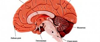
Tumors of the central nervous system in children occupy second place after acute leukemia . In boys, the incidence is higher and its peak occurs at 5-9 years. The location of all tumors is assessed in relation to the tentorium of the cerebellum: above it is a supratentorial location, below it is infratentorial. In 80% of cases, a brain tumor in children is located below the tentorium and affects the anatomical formations of the posterior cranial fossa, in particular the cerebellum. The most common forms include medulloblastoma , astrocytoma , and brainstem glioma , which accounts for 15% of all tumors in children. The brain stem includes the pons, cerebellum, medulla oblongata, midbrain and diencephalon. Tumors in children under 3 years of age are often large and occupy 1/3 of the brain volume. It is this fact that causes an increase in intracranial pressure , which is manifested by headache and vomiting, which occurs in children at the very beginning of the disease and then disappears.
Clinical symptoms depend on age, the severity of hydrocephalus and more on the location of the tumor than on its morphological structure. In young children, symptoms do not appear for a long time or are slightly expressed, which is associated with the elasticity of the bones, the presence of open sutures and fontanelles, so the child remains compensated for a long time. As a result, the tumor is not diagnosed in a timely manner. When examining young children, it is important to measure the head ( macrocephaly if intracranial pressure is increased ). Changes in the fundus are also detected. Signs of increased intracranial pressure develop gradually: fatigue, paroxysmal headaches appear, schoolchildren have decreased academic performance, vision loss, and impaired upward gaze.
Cerebellar tumors are most common in childhood and occupy 60%.
1 - cerebellar hemispheres, 2 - cerebellar vermis, 3 - cerebellar peduncles, 4 - roof of the fourth ventricle, 5 - flocculus, 6 - cerebellar tonsils
In the cerebellum there are medulloblastomas and astrocytomas . Tumors are located only in the cerebellum or spread into the 4th ventricle and brainstem when large, causing hydrocephalus . With a tumor of the cerebellum, disturbances in gait and coordination, dizziness , impaired handwriting, weakness in the legs, and decreased muscle tone appear. With severe cerebellar ataxia, the child cannot walk or sit, nystagmus , and the child loses acquired skills. In most cases, the disease begins with hydrocephalus, as the tumor compresses the cerebrospinal fluid flow paths. In addition to lack of coordination, the child complains of a headache, nausea and vomiting. With a large tumor, compression of the brain stem occurs.
CT or MRI is used to diagnose cerebellar tumors in children. Children under 1 year of age can undergo an ultrasound scan of the brain. The method of choice is surgery to remove the tumor. It must be completely removed, since the result and prognosis for life depend on it. If the tumor is malignant, a course of radiation therapy or chemotherapy is given if the child is under 3 years old.
Brainstem glioma.
The brain stem connects the structures of the central nervous system and is involved in organizing human behavior. Glioma is a diffuse tumor that does not have a specific shape. It spreads through finger-like projections into healthy tissue. Due to this nature of growth and localization, removal of pontine gliomas is surgically impossible, and there is no cure for these tumors. In this regard, the survival rate of children is very low - 10% live 2 years after diagnosis. Trunk gliomas grow rapidly and symptoms develop within one month and progress rapidly.
Symptoms of diffuse pontine glioma include:
- Difficulty swallowing, drooling.
- Weakness in the arms and legs (usually one-sided).
- Loss of balance, impaired walking.
- Jerky movements.
- Double vision, blurred vision, drooping eyelids, squint, eye movements.
- Facial muscle weakness or paralysis.
- Developmental delay.
- Abnormal reflexes.
- Behavioral changes, learning difficulties.
Craniopharyngioma is often diagnosed between the ages of 5 and 14 years. The tumor is benign - it grows slowly, rarely spreads to healthy tissue, very rarely malignizes, but recurs quite often. However, if vital structures of the brain are compressed, the tumor can become dangerous. Often localized in the pituitary gland and hypothalamus. This arrangement affects endocrine functions. When the tumor is located near the optic nerve, vision is impaired.
Craniopharyngioma is diagnosed at a late stage (many years after the first symptoms appear). At the onset of the disease, intracranial hypertension appears, then visual and hormonal disturbances appear (the secretion of gonadotropins, growth hormone, and thyroid-stimulating hormone is impaired). Patients also have diabetes insipidus . Impaired circulation of cerebral fluid as the tumor enlarges causes hydrocephalus .
Stages
Since signs of brain cancer do not appear immediately after the onset of the disease and remain subtle for a long time, accurately determining the stage of tumor development is a rather difficult task. Oncologists distinguish four stages of the disease.
- Pathological changes affect a small number of cells localized in a limited area of the brain. The patient experiences periodic headaches, weakness, constant drowsiness, and dizziness often occurs.
- The tumor grows, penetrates nearby tissues, and begins to compress individual centers of the brain. This can be manifested by convulsive attacks, periodic vomiting, and digestive disorders.
- Due to the rapid growth of the tumor, surgical removal of malignant tissue becomes almost impossible. Symptoms intensify, cognitive impairment appears, and coordination of movements worsens. A characteristic symptom of the third stage is horizontal nystagmus, or constant horizontal movement of the pupils.
- The tumor grows so much that the patient experiences constant headaches and gradually loses the functions of maintaining vital processes. In the absence of treatment or its low effectiveness, the patient falls into a coma, followed by death.
Pathogenesis
With any tumor, the defining feature is cell proliferation that gets out of control. The cause of this reproduction is an oncogene. Cell proliferation, invasive growth and the immune response of cells are interrelated, but are independent processes associated with genetic changes. Thus, the TSC-22 gene has been studied, which is important in various malignant brain tumors. In brain gliomas, the metabolism of non-heme iron is important.
Changing the cell genome activates antitumor immunity. However, the immune system also contributes to tumor progression by producing an immunogenic tumor phenotype.
As the tumor grows, the following occurs:
- its invasion into surrounding tissues and their destruction;
- compression of underlying tissues;
- increased intracranial pressure;
- cerebral edema;
- difficulty in the outflow of cerebrospinal fluid;
- the growth of new vessels in the tumor, becoming a source of bleeding;
- disturbance of venous and arterial blood flow.
Diagnostics
It is difficult to determine the presence of a brain tumor based on symptoms, especially in the early stages. Taking a biopsy specimen for histological examination is very difficult, since this requires a full-fledged neurosurgical operation. During the examination, the neurologist prescribes the following types of diagnostics:
- electroencephalography to detect brain disorders;
- Echo-EEG to check intracranial pressure;
- ophthalmoscopy to exclude deterioration of visual function due to ocular pathologies;
- CT scan of the brain is the main study that allows you to detect a tumor, determine its size, the presence of hemorrhages, calcifications and other pathological changes in tissues;
- MRI of the brain is a complementary study to CT, necessary to clarify changes in the soft tissues of the brain;
- MRI of the vessels of the head - to study changes in the structure of the blood supply to the brain and other tissues;
- PET scan of the brain - to determine the level of malignancy of the tumor.
If a decision is made to perform surgery, a histological examination of the removed tumor tissue is required to determine the histological type of tumor and the level of cell differentiation.
Attention!
You can receive free medical care at JSC “Medicine” (clinic of Academician Roitberg) under the program of State guarantees of compulsory medical insurance (Compulsory health insurance) and high-tech medical care.
To find out more, please call +7, or you can read more details here...
Procedures and operations
Brain cancer is a serious disease and tumor removal is important to perform accurately, so a neuronavigation system is used during the operation. Before surgery, the patient’s research data is entered into the navigation system program, and a 3D computer model of the brain with tumors and blood vessels is obtained, which helps virtually plan the operation. Surgical treatment is based on the principle: extracerebral tumors are removed completely, and intracerebral tumors are removed as much as possible, so that brain function does not suffer. Any operation should involve minimal trauma, preservation of brain structures, blood vessels and veins. Some areas of the brain are considered inoperable - these are the motor and speech areas.
Minimally invasive surgery to remove a tumor is performed using laser, stereotactic and endoscopic techniques. Minimally invasive operations are performed through endonasal access (through the nostril), supraorbital (through an incision above the eyebrows), retrosigmoid (incision behind the ear), as well as through any point convenient for removing deep-lying tumors. Minimally invasive approaches are as effective as invasive ones. If the operation is performed on functionally significant areas, then laser technologies are used. This could be laser thermal destruction. For deep-lying tumors, brachytherapy and photodynamic therapy are used to destroy them.
Highly effective technologies allow treatment without surgery. These include radiosurgery methods: linear accelerator, gamma knife. However, they can be used for small lesions.
Gamma Knife is a form of radiation therapy. This ultra-precise method destroys even several formations per session. The method is applicable for multiple lesions (tumors of metastatic origin). The CyberKnife system is equipment based on a linear accelerator that delivers high doses to the tumor and destroys it. The operation does not require open intervention. If the tumor is large, the bulk of the tumor is first surgically removed, and then irradiation with the CyberKnife system is used.
Boron-neutron capture therapy method. With this method of exposure, the destruction of a tumor that has accumulated the boron-10 isotope is carried out by neutron irradiation. Boron accumulated in cells increases sensitivity to radiation. Boron isotopes absorb neutrons and a “nuclear” reaction occurs, during which energy is released that destroys the new formation. Brachytherapy has not become widespread. It is carried out by implanting radioactive sources (iridium-192, iodine-125, palladium-103) into the tumor tissue.
Before the operation, the possibility of its implementation and safety are taken into account. Without these predictions, surgical treatment is considered inappropriate. With any type of intervention, consequences are possible in the form of disturbances in movement, speech, hearing, vision, reading, and mental changes. It all depends on the location of the tumor and the degree of trauma to surrounding tissues. When a pituitary tumor is removed, hormonal function is disrupted. Motor disturbances that occur immediately after surgery are associated with cerebral edema. But with the use of decongestants, patients recover quickly.
Treatment
Regarding the treatment of brain oncology, you should contact a neurologist or neurosurgeon. Based on the stage of development and location of the tumor, the doctor selects the most appropriate treatment methods in each case.
- Neurosurgery. Removal of a malignant neoplasm is the most effective way to guarantee complete relief from the disease. Intervention is possible at the first and partially at the second stage of the disease, but much depends on the location of the tumor and its proximity to vital centers of the brain. The efficiency of surgeons has increased thanks to the use of laser and ultrasound technology.
- Radiation therapy. Removal of malignant cells using ionized radiation is used as an auxiliary means after surgery, and also as a main means in cases where intervention is not possible. The intensity and duration of irradiation in each case is selected individually, in accordance with the size and location of the pathological area, the type of altered cells and other characteristics.
- Chemotherapy. It is difficult to select drugs for the treatment of brain tumors due to the presence of the blood-brain barrier. As a rule, they are prescribed in combination with radiation therapy, and the course consists of several drugs that complement each other. Courses lasting 1-3 weeks are carried out under constant monitoring of blood changes.
- Cryosurgery. The destruction of tumor tissue by freezing is beneficial in cases where traditional surgery is difficult due to the proximity of vital centers or the deep location of the pathological area. Cryosurgery can be used as a primary method or as an adjunct to conventional surgery, laser and chemical treatments.
- Symptomatic treatment. If the tumor is detected too late, and any of the listed methods do not achieve results, the patient is prescribed therapy aimed at reducing symptoms and improving quality of life. These are glucocorticosteroids, sedatives and antiemetics, NSAIDs for pain relief, as well as narcotic analgesics.
Brain tumors: symptoms, risk factors, diagnosis and treatment
Neurosurgery March 23, 2017
A brain tumor is a growth or abnormal growth of cells in or near the brain. There are several types of brain tumors. They can be noncancerous (benign) or cancerous (malignant). Tumors can also develop from brain tissue (primary) or form in other parts of the body and then spread to the brain (secondary or metastatic).
The rate of tumor growth in the brain can vary greatly. The growth rate, as well as the location of the tumor in relation to other brain structures, determines its effect on the functioning of the nervous system.
Treatments for brain tumors depend on the type of tumor, as well as its size and location.
MAIN SYMPTOMS
Signs and symptoms of a brain tumor can vary greatly depending on its size, location, and growth rate. Main signs and symptoms caused by a brain tumor may include:
- The appearance or change of pain in the head
- Headaches that become more frequent and severe
- Nausea or mouth for no apparent reason
- Vision problems such as blurred vision, double vision, or loss of peripheral vision
- Gradual loss of feeling or ability to move arms or legs
- Balance problems
- Speech problems
- Disorientation in time and space
- Personality and behavior changes
- Seizures, especially if they have not happened before
- Hearing problems
When should you see a doctor?
Make an appointment with your doctor if you have persistent signs and symptoms that bother you.
CAUSES OF BRAIN TUMORS
Tumors that arise directly in the brain
Primary tumors arise directly in the brain or in tissues located close to it—the membranes covering the brain (the meninges), the cranial nerves, the pituitary gland, and the pineal gland. Primary tumors form when errors (mutations) appear in the DNA of normal cells. Mutations allow cells to grow, divide quickly, and survive when healthy cells die. The result is a collection of abnormal cells that form a tumor.
Primary brain tumors are less common than secondary tumors, in which cancer appears in other parts of the body and spreads to the brain.
There are several types of primary tumors and each has its own name, depending on the cells that form it. For example:
- Gliomas. These are tumors that form in the brain or spinal cord and include astrocytomas, ependymomas, glioblastomas, oligodendrogliomas, and oligoastrocytomas.
- Meningiomas. A meningioma is a tumor that develops in the membranes surrounding the brain and spinal cord (the meninges). Most meningiomas are cancerous.
- Acoustic neuromas (schwannomas). These are benign tumors that grow on the nerves that control balance and hearing and carry nerve impulses from the inner ear to the brain.
- Pituitary adenomas. These are mostly benign tumors that form in the pituitary gland at the base of the brain. These tumors can affect pituitary hormones, which affect the entire body.
- Medulloblastoma. It is the most common malignant brain tumor in children. Medulloblastoma forms in the lower part of the brain and spreads through the cerebrospinal fluid. These tumors are less common in adults but do occur occasionally.
- Primitive neuroectodermal tumors are rare malignant tumors that develop in embryonic (fetal) cells in the brain. They can appear in any area of the brain.
- Germ cell tumors. Germ cell tumors can occur in childhood, in the area where the reproductive organs form. But sometimes germ cell tumors move to other parts of the body, such as the brain.
- Craniopharyngiomas. These are rare benign tumors that arise near the pituitary gland, which produces hormones and controls many body functions. While a craniopharyngioma slowly grows, it can affect the pituitary gland and other structures located near the brain.
Cancer that starts in other areas of the body and spreads to the brain
Secondary brain tumors (metastases) are tumors that form against the background of a cancerous tumor that has arisen in another organ and spreads (metastasizes) to the brain.
Secondary brain tumors are most common in people with cancer. But, in rare cases, a metastatic brain tumor may be the first sign of cancer in any organ.
Secondary brain tumors are much more common than primary ones. Although any malignant tumor can metastasize to the brain, the most common metastases are:
- Breast cancer
- Colon cancer
- Kidney cancer
- Lungs' cancer
- Melanoma
RISK FACTORS
In most patients with primary brain tumors, their cause is unclear. But doctors have identified a group of factors that increase the risk of developing a brain tumor. Risk factors include:
Age. As you age, your risk of developing brain tumors increases. They are most common in older people. However, the tumor can appear at any age. There are even some types of brain tumors that occur almost exclusively in children. Exposure to radiation. Individuals exposed to ionizing radiation have an increased risk of developing brain tumors. Examples of ionizing radiation are radiation therapy used to treat cancer and radiation exposure from the explosion of atomic bombs. Other types of radiation, such as electromagnetic fields from high-voltage power lines and radiation from cell phones and microwave ovens, have not been scientifically proven to be associated with brain tumors. Family history. A small proportion of brain tumors occur in people with a family history of tumors or genetic syndromes that increase the risk of developing brain tumors.
DIAGNOSTICS
If a brain tumor is suspected, your doctor may recommend a series of tests and procedures, including:
Neurological examination. A neurological examination may include tests of vision, hearing, balance, coordination, stamina and reflexes, among other things. Abnormalities in one or more functions may indicate parts of the brain are being affected by the tumor.
Medical imaging. Magnetic resonance imaging (MRI) is often used to diagnose brain tumors. In some cases, a contrast agent may be injected through a vein in the arm during the examination.
A number of specialized MRI components —including functional MRI, perfusion MRI, and magnetic resonance spectroscopy—can help the doctor evaluate tumors and determine treatment plans.
Other medical imaging studies may include computed tomography (CT) and positron emission tomography (PET).
Tests done to detect cancer in other organs . If it is suspected that a brain tumor may be caused by cancer in other organs, your doctor may order a series of tests and procedures to determine the origin of the cancer. An example would be a CT scan of the chest to look for signs of lung cancer.
Collection and examination of abnormal tissues (biopsy). A biopsy may be part of surgery to remove a brain tumor or may be performed as a needle puncture.
A stereotactic biopsy can be done to examine tumors in hard-to-reach or sensitive areas of the brain that are easily damaged by surgery. The neurosurgeon makes a small hole in the skull and inserts a thin needle through it. The tissue is then removed using a needle, often guided by CT or MRI (neuronavigation).
The collected material is examined under a microscope to determine whether it contains cancerous or benign cells. This is the most important information for making an accurate diagnosis and prognosis, and, most importantly, for determining further treatment.
TREATMENT
Treatment for a brain tumor depends on its type, size and location, as well as the patient's health and preferences. Surgical intervention.
If the location of the brain tumor allows surgery, the neurosurgeon will remove as much of it as possible.
In some cases, tumors may be small and easily separated from surrounding tissue, allowing for complete surgery. In other cases, the tumors cannot be separated from surrounding tissue or are located near sensitive parts of the brain, making surgery very risky. Then the neurosurgeon removes the maximum part of the tumor located in the safe zone.
Even removing part of the tumor helps relieve signs and symptoms of the disease
Surgery to remove a brain tumor carries certain risks, such as infection and bleeding. Other risks depend on the location of the tumor. For example, removing a tumor near the optic nerve risks vision loss.
Radiation therapy
Radiotherapy uses ionizing radiation—X-rays or protons—that destroy the tumor. The beams may come from a machine outside the body (external beam radiation) or, in very rare cases, the radiation source is placed inside the body near a brain tumor (brachytherapy).
The external beam of radiation can be focused only on the tumor localization area or directed to the entire brain. Whole-brain radiation is most often used to treat cancer that has spread from other organs. The side effects of radiation therapy depend on the type and dose of radiation received. Common side effects during or after radiation include fatigue, headache, nausea, and vomiting.
Chemotherapy
Chemotherapy uses drugs that destroy cancer cells. Medicines may be given orally as tablets or may be given into a vein (intravenously). The most commonly used drug to treat brain tumors is temozolomide (Timodal), available in tablet form. Other types of chemotherapy drugs are also available, prescribed depending on the type of tumor.
The side effects of chemotherapy depend on the type and dose of drugs the patient receives. Chemotherapy can cause nausea, vomiting, and hair loss.
RECOVERY AFTER TREATMENT
Because brain tumors can occur in the parts of the brain that control motor skills, speech, vision, and thinking, rehabilitation may be a necessary part of recovery.
A neurosurgeon can use the following rehabilitation methods:
- Physical therapy to restore lost motor skills or muscle strength
- Occupational therapy to help a patient return to normal daily activities and work after treatment for a brain tumor or other illness
- Speech therapy performed by a speech therapist in case of speech impairment (speech pathology).
Author: Danu Adrian, neurosurgeon, highest category
Forecasts
The success of treatment depends on the timely detection of the disease. If therapy is started at the first or second stage of brain cancer, the five-year survival rate is, according to various estimates, 80-85% of patients. If the disease is detected late, a favorable outcome is possible for approximately 31% of patients. However, it should be remembered that cancer is one of the least studied areas of medicine, so you should never give up your chances of life, even in the most advanced cases of the disease.
Treatment methods and prognosis for further survival
Brain tumors are dangerous not only because of their malignancy, but also because of their location. It is quite difficult to remove a formation that develops in the closed space of the skull without affecting the vital centers. But not all tumors need to be considered fatal.
Once the neurosurgeon determines the boundaries, size and exact location of the tumor, it will become clear how the patient should be treated. If there is even the slightest chance of a successful outcome of the operation, then histology of the malignant tissue will be required, if such a procedure is possible and does not complicate the patient’s condition.
Doctor's advice
Brain surgery is not as scary as it seems. If there is a choice to do or not to do, then refusal means a pessimistic forecast, while consent gives a chance, and not a small one. After treatment with modern devices, in most cases, long rehabilitation is not required, the patient returns to his normal life. During this period, he needs to be supported morally, helped with usual household chores and childcare. Students can read the curriculum little by little, this will help them get into a familiar rhythm. If you have panic attacks, neurosis, or depression that develops as a result of an illness, you need to consult a psychologist. All this is very important for recovery.
Victoria Druzhikina Neurologist, Therapist
Treatment is always complex. At an early stage of cancer, all available methods are used: radiosurgery, cryosurgery, radiation and chemotherapy, surgery, symptomatic therapy. The goal is not only to cure cancer; first, it is necessary to relieve pain, alleviate the general condition and prevent swelling of the brain.
The survival prognosis for people with early stage brain cancer depends on two factors: the accuracy and timeliness of diagnosis.
If treatment is started immediately, about 82% of patients survive after five years. With late presentation, the five-year survival rate is only 31%. The prognosis largely depends on the type, aggressiveness and growth rate of the tumor.
Brain tumors are strikingly different from other malignant tumors and occur in approximately 3% of all recorded cases of oncology. They are located so inconveniently that the symptoms of the disease are difficult to differentiate, and this significantly complicates treatment.
The basis for referral to an oncology clinic is sudden development and increasing neurological symptoms. However, modern medicine has hardware diagnostic tools that can not only detect cancer at an early stage, but also identify predisposition to it.
For more information about the symptoms of a brain tumor, watch the video:
Be healthy!
Useful articles on the topic: Why women may feel dizzy: common causes Tension headaches: causes, types of pathology, symptoms and effective treatments
This article has been verified by a current qualified physician, Victoria Druzhikina, and can be considered a reliable source of information for site users.
Rate how helpful this article was
5 2 people voted, average rating 5
Did you like the article? Save it to your wall so you don’t lose it!
Questions and answers
How do you know if you have brain cancer?
In the initial stages of the disease, symptoms of brain cancer can easily be confused with signs of ordinary fatigue, a mild cold, or a neurological or cardiac disease. To be sure that there is no disease, it is necessary to undergo a CT scan, which allows you to detect a tumor in the initial phase of development.
Is there a cure for brain cancer?
With timely detection of a tumor, timely and high-quality surgical treatment, one hundred percent restoration of health is possible.
What causes brain cancer?
A tumor in the brain tissue develops because one or more cells begin the process of abnormal division. In this case, the pathologically altered cells lose their original function and are only capable of rapid division, which is why the size of the tumor grows rapidly. It has not yet been possible to establish what mechanisms trigger this process.
Attention! You can cure this disease for free and receive medical care at JSC "Medicine" (clinic of Academician Roitberg) under the State Guarantees program of Compulsory Medical Insurance (Compulsory Medical Insurance) and High-Tech Medical Care. To find out more, please call +7(495) 775-73-60, or on the VMP page for compulsory medical insurance
Signs in women
The first signs of a brain tumor in women are similar to those observed in men. Therefore, there is no need to talk about specific signs of the development of tumors in women.
Signs often repeat those of other pathological conditions, for example, headache, tinnitus, decreased vision, nausea and others.
However, symptoms that last a long time (several weeks) and are persistent, increasing in nature, indicate the occurrence of tumor processes.
Therefore, it is important to seek medical help as early as possible. This will allow diagnosis and effective treatment methods to be applied.