Article for the "bio/mol/text" competition: The mechanism for stopping bleeding is essential for the survival of the body, however, despite a history of research spanning decades, many details of this system remain unclear. Eight years ago, Mikhail Panteleev told Biomolecule about blood clotting. Since then, a lot of new data has accumulated in this area. In this article we will tell you how a young team of scientists from Moscow State University lifted the veil of secrecy over two mysterious phenomena in the complex system of arterial thrombus formation, showing how dying cells move in it.
Main function of platelets
As is known, these blood cells take an active part in the coagulation process, that is, in hemostasis. This is the main function of platelets. For the human body, this process is one of the most important. It helps prevent significant blood loss during serious injury. Thanks to this function of human platelets, the walls of blood vessels become stronger. The corpuscles clog the injury site in a short time. In essence, these blood cells play the role of the primary vascular plug.
Coagulation occurs as a result of the interaction of enzymes, proteins and about 40 other components. This is a very complex biological mechanism in which platelets, prothrombin and fibrinogen play a major role. The interaction of these elements occurs in the blood plasma.
Concept of platelets
The plates are oval or round in shape. When they reach surfaces on which there is no epithelial layer, the formation of special processes is activated. These branches are intended to stop emerging bleeding and close wounds; their length is 5-9 times greater than the size of a platelet.
The granules of the plates contain platelet enzymes that destroy bacterial membranes and protect the body from harmful microorganisms. The processes serve to accelerate movement in the bloodstream by adhering to foreign bodies; the threads capture elements and digest them. A protective barrier is created by gluing with other platelets, that is, blood clotting occurs.
An important function of platelets is to supply micronutrients to the endothelial layer. Their lifespan ranges from 7 to 12 days, then destruction occurs in the liver, lungs and spleen.
Forms of platelets in the blood:
- degenerative varieties - contained in amounts up to 0.2%;
- stereotypes of irritation - range from 0.8 to 2.3%;
- young cells - from 0 to 0.8%;
- mature forms - from 90.2 to 95%;
- old records - from 2.3 to 5.7%.
Inactivated elements are flattened spherical shapes, in which the semi-axes are located one to the other in a ratio of 2:8. This feature is used in the case of recognizing the optical characteristics of platelets and when regenerating parameters using flow cytometry.
Cell structure
Microscopic studies show that the change in cell shape upon activation is associated with transformation of the tubule ring. This phenomenon is caused by the conversion of the amount of calcium ions.
The structure of platelets is heterogeneous, there is heterogeneity, and the structure contains several layers:
- the outer surface is represented by a three-layer membrane, the thickness of which contains phospholipase A, receptors for adhesion to other elements and tissues;
- the lipid layer is a collection of glycoproteins and helps the plates stay glued together for a long time;
- microtubules in the form of a cellular framework help the structure contract to release the contents of the platelet beyond its boundaries;
- The area of organelles is represented in the structure by various components and factors that promote wound healing.
In the early periods of research, there was no photographic technique and there was no unambiguous terminology, so the time of initial observations of platelets is not precisely established. There is information that the first study was conducted by the inventor of the microscope, the Dutchman van Leeuwenhoek. In England, the cells are now called platelets, a term coined by Bizzocero in 181. He is also credited with the discovery of the relationship of elements with homeostasis and thrombocytosis. In Russian, flat blood cells are called platelets, sometimes the name Bozzocero's plaque is used.
Formation and life cycle
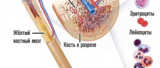
Their formation and maturation begins in the body of the red marrow in the area of the pelvic bones and vertebrae. The spongy substance reproduces stem cells that are incapable of the process of differentiation and are not divided into specific types.
Platelets go through several stages during formation:
- stem cells turn into elements of the megakaryocyte series;
- the formation of a megakaryoblast begins - a large formation of a mesh structure without granules, with 2-3 round nuclei;
- the resulting proplatelet turns into a promegakaryocyte and platelet formation occurs.
Transformations occur as a result of the influence of genetic factors. The lack of cells leads to an increase in the duration of bleeding; the increased volume of elements causes the formation of clots and blockage of blood vessels. As a result of blocked channels, a heart attack, stroke, pulmonary embolism, or vascular plug in other organs of the human body can occur.
Inferiority of platelets or their deficiency is called thrombocytopathy. A low overall indicator of the number of plates in the analysis causes a consequence in the form of thrombotic purpura, in which ischemia of various organs or hemolytic anemia develops, and the number of blockages in the vessels after bleeding increases.
Functions of elements
Platelets form the primary plug to tighten the wound of a damaged vessel and accelerate the plasma coagulation reaction. The peculiarity of the plates is that they play a significant role in the healing and restoration of tissues, while releasing polypeptide molecules for cell growth and division. Platelets are quickly activated when needed and enter a new, irreversible state.
The plates are transformed under the influence of pathogens:
- the main protein of connective tissue - collagen;
- the main protein of the coagulation system - thrombin;
- adenosine monophosphate produced by damaged cells - ADP;
- a second-line activator synthesized by platelets to establish vasoconstriction - thromboxane A2.
The modified flat cells attach to the damaged surface and form a plug as a result of adhesion to the walls and each other. As a result, the shape of the plates is updated, and phosphatidylserine and P-selectin are expressed.
In its normal state, the platelet membrane does not support the blood clotting procedure, while phospholipids are located on its inner layer. Activation of flat elements causes excitation of scramblase protein molecules, which transfer phospholipids to the outer shell. As a result, the concentration of phosphatidylserine in both layers is equalized. In the process, transmembrane proteins of the exogenous layer change, which accelerate coagulation through the characteristic coupling of factors.
Platelet functions
What are platelets needed for? The main function of platelets is timely blood clotting, but their work does not end there. If the body has a normal amount of these elements, then they do not change their structure and size. As soon as pathogenic microorganisms enter the circulatory system, the cells begin to send out up to a dozen processes. The processes are so large that they exceed the size of platelets by 5-10 times. Due to this shield, pathogenic microorganisms stop entering the circulatory system.
Scientists identify four functions of platelets:
- Ensuring the integrity of tissues in the body (angiotrophic function). When platelets interact or are destroyed, active production of biological substances occurs. Due to this function, the release of growth factors begins. These blood elements additionally restore and strengthen the tissues and walls of blood vessels.
- Vasoconstriction. When a vessel is damaged, platelets are the first to come to the rescue. They perform a hemostatic function. The role of platelets is to create a platelet plug that will close the damaged hole in the vessel. Such a plug blocks the flow of blood, narrows the passage of the vessel, where its cells are destroyed.
- Fight against foreign microorganisms. The structure of platelets is so unique that they are able to attach to various bacteria. With their processes they capture the pathogenic pathogen and prevent it from fulfilling its function. Over time, this blood element is excreted along with the captured microorganism due to the active work of the liver and spleen. As a result, the risk of negative impact of external factors is reduced, and resistance to various diseases is increased.
- Restoring the integrity of blood vessels. The lifespan of platelets is short, but they manage to restore damaged vessel walls. A low level of platelets in the blood may indicate an increased risk of internal bleeding, since they are not fully involved in strengthening blood vessels.
Platelet elements are included in the list of main blood test indicators, since their increase or decrease can be caused by a number of diseases.
Competition "bio/mol/text"-2019
This work was published in the “Own Work” category of the “bio/mol/text” competition 2019.
The general sponsor of the competition and partner of the Skoltech nomination is the Skoltech Center for Life Sciences.
The nomination partner is the Russian Science Foundation.
Competition sponsor: the largest supplier of equipment, reagents and consumables for biological research and production.
The audience award was sponsored by BioVitrum.
"Book" sponsor of the competition - "Alpina Non-Fiction"
Specialists and non-specialists are well aware of the ominous word “thrombosis”. The word “thrombus” is traditionally perceived as something dangerous. However, not all blood clots are dangerous. If the vascular wall is damaged, the body must quickly form something like a plug that will prevent blood from flowing out of the artery or vein. Thus, the formation of plugs, or, in scientific terms, hemostatic blood clots , is a key task that is solved by the human hemostasis system (and, of course, not only humans). However, sometimes this system fails, and damage to the vessel leads to the formation of a massive intravascular thrombus, which almost completely blocks blood flow. If this process occurs in a large artery that supplies blood to a vital organ, such a blood clot can cause serious complications and even death. Myocardial infarction and ischemic stroke are perhaps the most well-known and common complications caused by arterial thrombosis, which are today the most common cause of death and disability in people in developed countries [1].
Why in some cases, in response to damage, the hemostasis system works excessively and forms a deadly plug in the vessel? Despite many decades of research, this question remains unanswered. The lack of understanding of the mechanisms that regulate the formation of a blood clot leads to the fact that today there is no reliable way to prevent thrombosis: taking existing antithrombotic drugs is associated with a fairly high risk of bleeding, including life-threatening ones.
Platelets are...
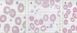
A thrombus is a blood clot (translated from Greek), the basis of which is platelets.
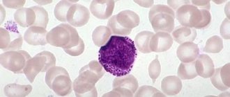
A platelet is a small, colorless, spherical platelet of blood (Bizzocero plaque). Plates are formed from the plasma structure (megakaryocytes - plasma cells) of the bone marrow. Platelets do not have a nucleus, but are equipped with an abundant number of granules (more than 200).
The value of granulations is due to the high content of special components of platelet growth (thromboxane, thrombin, adenosine diphosphorate and other factors), which ensure the formation of amino acids and enzymes (what are they?) that destroy the membranes of bacterial cells, preventing the penetration of pathogens into the blood.
The size of platelet plates varies within their age (young, middle, mature), from 2 to 5 microns.
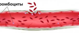
However, when a platelet comes into contact with a surface that does not correspond to the internal vascular endothelium or the cavity of the cardiac pericardium, the plates are activated, releasing up to 10 pseudopods (processes) tens of times larger than the size of the plate itself.
This feature allows platelets to serve as a kind of “patch” that, if necessary, covers the wound surfaces of blood vessels, preventing bleeding.
- High platelets with RA - why worry - Arthritis - 2021
It is the pseudopods that ensure the movement of the plates through the circulatory system. Platelets have the ability to adhere to foreign agents, capture them and destroy them, form a blood clot by aggregation (gluing plates together) to prevent hemorrhagic processes (bleeding).
» alt=»»>
The main role of platelets is to actively participate in the process of blood clotting (hemostasis).
They provide a transport function, delivering nutritional components to the tissues (endothelium) lining the internal cavity of the vascular walls. They remain viable for up to 10 days, after which they are destroyed in various organs (spleen, lungs, or liver).
Recent developments by Japanese scientists have proven that megakaryocytes are not the only sources of formation of Bitsocerro plaques (platelets). They were able to obtain platelets from the patient's own stem cells.
These studies increase the success of transplantation, since the presence of such platelets does not cause the body to reject donor organs.
How platelets participate in the process of hemostasis can be seen in the diagram:
Having appreciated the basic functions of platelets, you can understand what their imbalance in the body entails and what the negative consequences may be.
Any imbalance in platelet activity and number is medically called thrombocytopathy.
- A decrease in the concentration of platelet platelets is called thrombocytopenia.
- And an increased concentration is thrombocytosis.
- A dysfunction of their activity is diagnosed as thrombosthenia.
Blood tests
In the category of qualitative transformations of platelets, there is a deficiency or blockade of membrane receptors or the absence of compact granules. Symptoms of hemorrhagic diathesis arise due to a shift in the release of spherosomes, when the production of thromboxane and prostaglandins is impaired. An anomaly and deficiency of von Willebrand factor, as well as a violation of the interchange of nucleotides and calcium movement, are important.
In modern medicine, the following types of blood tests are used:
- general clinical;
- biochemical research;
- determination of the degree of coagulation;
- Sukharev's test.
Blood testing is recommended as the first step in making a diagnosis. As a result of laboratory tests, pathology is revealed and the true state of the human body is reflected.
General clinical
Based on the results of the study, the level of hemoglobin, the number of leukocytes, lymphocytes are checked, the color coefficient, the degree of erythrocyte sedimentation (ESR) are determined, and the volume of platelets present is shown in the overall picture. Based on the research, the degree of functioning of the body is determined and deviations from the norm are identified.
A general analysis is prescribed by a doctor to confirm or refute:
- the appearance of inflammation;
- development of diseases of hematopoietic organs and systems;
- the occurrence of immune failures;
- allergic reactions.
The analysis is recommended for pregnant women, patients with varicose veins, heart and vascular diseases. The study is necessary for organ pathologies and autoimmune diseases. The analysis does not require complex preparation; the morning time before breakfast is more suitable for taking blood.
Biochemical research
The analysis informs the doctor and provides a detailed table of indicators, so a large volume of blood is required, which is taken from a vein. Biochemical indicators reflect the functioning of most organs and the degree of development of the disease.
Check shows:
- inflammatory processes;
- indicators of the state of the blood system;
- position of water-salt exchange;
- volumes of microelements important for life.
As a result, the indicator of proteins and carbohydrates is determined, the level of blood enzymes, and the concentration of bilirubin are checked. An advanced biochemical analysis shows whether the content of trace elements is normal or not. As a result, nitrogen metabolism is examined, the presence of urea and creatinine is established.
Determination of clotting
The study reveals the rate of blood clot formation and platelet aggregation. Extension of the indicator leads to excess blood loss, and low activity of flat bodies causes blockages of blood vessels. Pregnant women are tested twice, since timely clotting is very important during childbirth. The occurrence of traffic jams is dangerous with varicose veins because it causes the appearance of blood clots. A coagulogram is prescribed before surgical treatment or extensive blood transfusion.
The analysis is carried out before the morning meal or 8 hours after a meal; alcohol intake is not recommended. 1 ml of blood is taken from a vein and divided into two tubes. The samples are kept at a temperature of +37˚C, and the time from blood collection to the start of the clotting stage is determined.
Diagnostics according to Sukharev
In the process, the time of blood thickening to the time of its complete immobility is studied. The development of the procedure should be limited to a period of 35-120 seconds, and the end of the process is extended to 3.5-5.0 minutes.
Decreased indicators indicate changes:
- anemia of various origins;
- pregnancy;
- improper functioning of platelets;
- excessive use of blood thinning medications.
Accelerated clotting indicates hormonal imbalances, the development of atherosclerosis, and infectious lesions in the body. For analysis, a Panchenkov tube is used, blood is taken from a finger, the first drop is removed with cotton wool. The capillary is filled to the control level and placed horizontally. After 30 seconds, the laboratory technician turns the tube over, changing sides, and uses a stopwatch while working.
What do elevated platelets mean?
An increase in the concentration of Bizzocero plaques can warn of the presence of various pathological processes in the body - the development of tuberculosis infection, blood cancer (leukemia), cancer of various organs, Hodgkin's lymphoma.
- Platelet counting according to Fonio - is the method accurate?
Thrombocytosis (increased platelets) is a constant companion:
- erythrocytosis;
- inflammatory intestinal and joint pathologies;
- chronic form of myeloid leukemia;
- acute course of infectious diseases;
- intravascular hemolysis and anemia.
An increase in the number of platelets is caused by severe stress, general intoxication of the body and blood loss, which can ultimately lead to the development of thrombocytemia and failure of the functions of stem cells in the bone marrow.
Oddly enough, significant blood loss does not reduce the concentration of platelet platelets, but, on the contrary, increases it. This is due to the presence of compensated properties of the body that can replenish platelet losses.
It doesn’t matter for what reasons platelets go beyond the normative values. In such a situation, a medical assessment with appropriate therapy is necessary.
Auxiliary functions of platelets
In addition to their protective properties, these red plates have another useful ability. It consists in feeding the endothelium of the human circulatory system. Thanks to this function of platelets, blood vessels receive vital microelements that help normalize the flow of red cells and the general functioning of internal organs. The degree of protection of the body (immunity) largely depends on this property. Blood elements are also actively involved in regeneration, that is, in the healing of tissue after damage. This effect is achieved by the accelerated process of division and release from the vessels. In other words, platelets perform the function of clogging the entire damaged area. Moreover, they promote accelerated growth of affected cells. This process refers to the division of polypeptide molecules.
During platelet activation, fibroblast growth also occurs. Also at this moment, microcomponents are produced that are responsible for cell transformation and restoration of insulin levels.