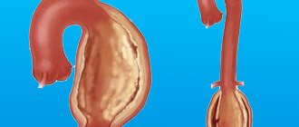
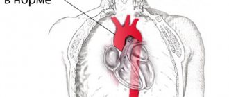
Calcium is an essential trace element necessary for the functioning of the body. But its excess, caused by metabolic disorders, can provoke a disease such as calcification of the aortic walls. Excess calcium enters the blood, and then begins to be deposited on the vascular walls, growing and gradually clogging the vessels. It is especially dangerous if it occurs in the largest and most important vessel, the aorta and aortic valve. In this case, they speak of aortic calcification, and the rapid development of the disease requires immediate diagnosis and treatment.
The formation of calcium deposits can damage the valve.
Calcification classification
Calcification is a pathological process characterized by calcium deposition. Depending on the location, the macroelement can accumulate and affect:
- vascular system;
- heart muscle;
- brain;
- joints and tendons;
- soft tissues and be diagnosed in the mammary glands, muscles and ligaments, fat deposits;
- liver and gall bladder;
- organs of the urinary system, most often the kidneys and urinary reservoir.
Depending on the etiology, calcification is of 3 types:
- dystrophic – the most common type of pathological process that develops as a response to any damage to soft tissues and internal organs, including after implantation of various medical devices;
- the metastatic type of the disease develops as a result of an imbalance of calcium, phosphorus and magnesium in the body against the background of renal failure, dyscalcemia and other severe pathologies;
- the tumor type of calcification is associated with the formation of spherical neoplasms around the joints; its etiology is not fully understood.
Calcification can also be systemic, affecting all human organs, or local, localized in one organ or system.
Symptoms of limescale formation
Despite the fact that calcification is quite common, its intravital diagnosis is difficult. There are no specific signs for this pathology, so it is mistaken for rheumatism, post-infarction cardiosclerosis, the consequences of the inflammatory process and hypertension.
Clinical symptoms that may occur when heart valves are desiccated include:
- the formation of an acquired defect of the mitral or aortic valve - calcification is likely after excluding rheumatism, endocarditis, and autoimmune diseases;
- murmur when listening to the heart in the absence of a defect, occurs during systole or diastole, rough;
- impaired conduction of impulses and myocardial excitability in the form of atrial fibrillation or blockade of the legs of His; with mitral calcinosis, complete atrioventricular block against the background of atrial flutter (Frederick's syndrome) often occurs;
- extrasystole, paroxysmal ventricular tachycardia;
- valve prolapse;
- heart failure (shortness of breath, tachycardia, swelling in the legs, severe weakness) in the absence of underlying diseases;
- formation of intracardiac blood clots with subsequent blockage of blood vessels.
- Aortic heart defects: causes, diagnosis and treatment
Changes in myocardial conductivity are explained by the fact that the valve leaflets pass into the septum between the ventricles, in which the cells of the conduction system are located. Valve calcification may also be accompanied by an inflammatory reaction of tissues, which spreads to neighboring areas of the heart.
The most common clinical form of aortic calcification is progressive valve stenosis. It is accompanied by the following symptoms:
- fast fatiguability;
- shortness of breath with slight exertion;
- feeling of strong and rapid heartbeat;
- dizziness;
- fainting conditions;
- heartache;
- attacks of suffocation at night;
- swelling;
- pain in the liver area.
Complications of stenosis include acute circulatory failure (cardiac asthma, cardiogenic shock, pulmonary edema), endocarditis, cerebral ischemia (transient attacks or stroke), complete blockade of impulse conduction, severe angina and myocardial infarction.
Causes of calcification
Calcium deposition in soft tissues and internal organs occurs as a result of metabolic disorders, which leads to impaired absorption of an important macronutrient for the human body. Most often, disruptions in metabolic processes are caused by endocrine pathologies, infectious and autoimmune kidney diseases, impaired enzyme production due to liver pathologies and pancreatic diseases.
Calcium metabolism disorders can be caused by insufficient intake of magnesium and excess vitamin D, which are directly involved in the body’s absorption of the macronutrient.
Calcification of an individual organ can develop with the formation of cysts, benign and malignant tumors, and tissue degeneration.
The process of formation of calcium conglomerates also affects connective and cartilage tissue, atherosclerotic plaques, dead parasitic microorganisms, and implants.
Signs of calcification
At the initial stages, it is extremely difficult to recognize the pathology due to its asymptomatic course. However, some types have a fairly pronounced clinical picture.
In case of systemic calcification or damage to the skin, joints, the epidermis becomes covered with small blisters, no changes in structure or color are observed. As the pathology progresses, calcium conglomerates grow and become denser to the touch and change color. Fistula formation is possible.
During routine examinations by specialists or during instrumental examination, lime deposits can be detected on teeth, bones, blood vessels, muscle and nerve fibers. The accumulation of macroelements on organ tissues leads to disruption of their functioning.
When the heart muscle and vascular system are damaged, the patient develops pain in the sternum, arm, neck, and back, which persists for a long time. Blood flow is also disrupted, which leads to surges in blood pressure and a feeling of coldness in the extremities.
When the kidneys are damaged, symptoms of intoxication increase, diuresis is disrupted, and the skin becomes dry and sluggish. When the organs of the digestive tract become calcified, their functioning is disrupted, which leads to nausea, vomiting, a feeling of heaviness in the abdominal area, and constipation.
When a large amount of calcium is damaged and accumulates in the brain, the patient experiences frequent attacks of headaches and dizziness, surges in intracranial pressure, impaired coordination of movement, memory impairment, problems with vision and hearing. As the disease progresses, fainting may occur.
At the same time, calcinosis leads to decreased performance, constant lethargy and fatigue, weakness, and loss of body weight.
Development of the disease on valves, aorta, blood vessels, myocardial cusps
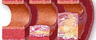
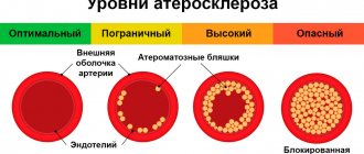
Areas of necrosis and scar tissue, implants, atherosclerotic plaques, blood clots, that is, any abnormal tissue, are subject to calcification. Impaired fat metabolism stimulates calcification, as cholesterol combines with calcium ions to form lime deposits. Therefore, atherosclerotic changes are considered as a stage preceding calcification.
These processes develop in places of greatest stress on the valves and vascular walls. The onset, as a rule, is damage to the aortic and then the mitral valve. Subsequently, the septum and left ventricle become calcified. The valve flaps lose their elasticity and mobility. Foraminal stenosis is formed. Calcinosis is the most common cause among acquired heart defects in adulthood.
Calcification has a developmental mechanism similar to bone formation. The primary process is described in the aorta, a tissue section of which contained bone marrow cells. The pathological process tends to constantly progress and worsen clinical manifestations.
Diagnostics
X-ray diagnostics are used to confirm the diagnosis. This method allows you to determine the nature and size of deposits, as well as the degree of damage to the organ in which the calcium conglomerate is localized. Additional research methods are prescribed:
- Ultrasound with Doppler sonography to study the state of the vascular system;
- ECG for studying the heart muscle;
- CT with the introduction of a contrast agent;
- MRI.
To identify the cause of tissue calcification, additional studies are prescribed, in the form of a general clinical and biochemical blood test. The latter method allows you to determine the level of calcium, phosphorus and magnesium in the blood. If renal function is impaired, a general clinical and bacteriological urine test is prescribed to assess the performance of the kidneys.
To exclude the malignant nature of neoplasms in the affected organ, a tissue biopsy is prescribed. This method involves collecting biological material and examining it under a microscope in a laboratory setting. A biopsy also helps to differentiate between benign and malignant neoplasms.
Diagnostic measures
It is extremely difficult to diagnose heart valve calcification, especially stage 1 of its development. Doctors are trying to visualize the cavities of the heart muscle. The most effective methods include:
- Ultrasound of the heart;
- cardiac catheterization;
- radiography;
- ventriculography;
- aortography;
- two-dimensional echocardiography;
- ELCG (electrocoagulography);
- ultrasonic densitometry.
More reliably and more often, calcification of the heart valves is detected using an electron-optical amplifier and X-ray television.
Treatment
To treat calcification, a therapeutic course is prescribed that will help cope with the underlying disease. So, if inflammation of an infectious nature has led to excessive calcium deposition, antibacterial drugs are prescribed.
If calcium absorption is impaired, medications containing magnesium, which is a calcium antagonist, are prescribed. Sufficient intake of magnesium from food and medications allows you to dissolve conglomerates and remove excess calcium from the body. During treatment, it is important to take diuretics, which will help speed up the process of excretion of the macroelement.
Vitamin D takes part in the process of calcium absorption, the excess supply of which also negatively affects the condition of the body. Therefore, during treatment, it is necessary to follow a special diet that excludes the consumption of large quantities of foods rich in calcium and vitamin D. These include fatty fish, leafy greens, dairy products, egg yolk, and nuts.
If conservative treatment methods are ineffective, as well as the formation of large conglomerates, their surgical removal is prescribed. The choice of surgical intervention method is carried out depending on the size of calcium accumulations, as well as their location.
Preventive measures
It is possible to prevent the occurrence of a disease such as microcalcification. Patience and adequate strength are necessary. Preventive measures include:
- Control your weight.
- Sustainable food diet.
- Monitor changes in parathyroid hormone levels.
- Run an active lifestyle.
There are also proven preventive measures. They are carried out in specialized medical institutions.
The onset of vascular calcification is difficult to diagnose. The disease remains asymptomatic for a long time. The first messengers appear after complications such as vascular stenosis occur, preventing proper blood flow. And with complete clogging or cracking in certain areas, it can be fatal.
Vascular calcification can cause the creation and progression of many serious diseases, such as: coronary disease, stroke, heart attack, pasmulation of the left atrium of the heart, atherosclerosis.
It is very important to prevent this disease and control the level of this mineral in the blood. If you follow your doctor's recommendations, you can put off this disease for a long time.
Calcinosis during pregnancy
Calcium deposition during pregnancy is most often diagnosed at the end of the third trimester of the gestational period. From a medical point of view, such a process is permissible and is associated with a modification of the placenta.
If calcification is diagnosed earlier, it can lead to premature maturation of the placenta. As a rule, calcinosis in pregnant women is associated with the consumption of large amounts of foods rich in calcium, infectious processes and metabolic disorders.
An excess of a macronutrient in a pregnant woman’s body is just as dangerous as its deficiency. May cause injury to the baby and mother during delivery.
Prevention
To prevent calcification of soft tissues and internal organs, special attention should be paid to proper nutrition. It is important to ensure a sufficient supply of all minerals and vitamins to the body in order to prevent the development of pathologies of various etiologies.
It is also important for people with congenital and acquired diseases of the cardiovascular system, kidneys, and endocrine pathologies to undergo regular scheduled examinations by specialists, which will help to promptly prevent the development of complications.
Treatment of various diseases should be carried out only under the supervision of a specialist and in accordance with his recommendations. Some groups of medications, including those for lowering blood cholesterol levels, blood pressure, as well as antibacterial and hormonal agents, can lead to an increase in calcium levels in the body and disruption of its metabolism.
To prevent calcinosis, you should lead an active lifestyle, which helps restore normal metabolism, and stop drinking alcoholic beverages and smoking.
Tissue calcification is a pathological process associated with a high concentration of calcium in the body. Affects the cardiovascular, nervous, musculoskeletal, digestive and urinary systems. To prevent illness, you need to eat right and lead a healthy lifestyle. As therapy, a course of medications is prescribed to eliminate the cause of the pathological process and normalize the level of calcium and magnesium in the blood.
What kind of disease is this
The human body contains 1 kg of calcium. Moreover, 99% of it is contained in bone tissue, and 1% is in the form of a solution. If a failure occurs and this ratio is disrupted, the person will develop calcification. Typically, this occurs when there is too much calcium in the body and it is not excreted naturally.
It is widely believed that calcium is a beneficial element. True, but everything is good in moderation. So, what is calcinosis and what are its symptoms?
Vascular damage by this disease occurs according to the following scheme:
- Calcium salts pass from a dissolved state to a crystalline state and are deposited on the walls of blood vessels.
- Over time, their entire interior, right down to the aorta and coronary arteries, becomes “porcelain.”
- As a result, the vessels lose their elasticity and become very fragile. In this condition, blood vessels can rupture as a result of a sharp increase in blood pressure.

