Causes
Symptoms
Prevention
Treatment
Causes of phlebitis
Slowing blood flow is the most common cause of phlebitis, especially when the venous wall is weakened and prone to varicose veins.
Phlebitis can also develop if a vein is compressed for a long time by tight clothing or, for example, squeezed by the edge of a chair. Another common cause is venous catheters that are left in a vein for several days to give infusions or medications. Phlebitis can also be caused by mechanical damage to the venous wall or the penetration of bacteria after operations and during prolonged bed rest. Another common cause of vein inflammation is damage to the vessel wall. This damage may be caused by:
- Infusions, e.g. during or after surgery,
- blood sampling,
- venous access (catheter) or
- a bite of an insect
The entry of bacteria can cause the development of inflammation of infectious phlebitis. Blood composition also plays an important role in the development of phlebitis. Blood clotting may increase due to illness or lack of fluid in the body. This can lead to the development of blood clots.
Diagnostics
When examining patients who have leg pain in the vein area, phlebologists pay attention to the location of the pain and the color of the skin over the vein.
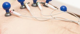
If a vein appears on the leg and it hurts, an ultrasound scan is performed. Doctors use 1, 2 or 3 research modes. The main one is B-mode. Additionally, color coding of blood flow or pulsed wave Doppler, as well as a combination of both, is used. The method allows you to simultaneously examine the vein in the leg that hurts, determine the direction of blood flow and its parameters. Patients who have pain and stretching of the veins in their legs must undergo an examination of the superficial and deep veins of both lower extremities. At the Yusupov Hospital, doctors for ultrasound scanning use devices equipped with linear sensors with a frequency of 5 MHz and more from the world's leading manufacturers. To scan deep veins, especially in obese patients, convex sensors with a lower radiation frequency (from 3.5 to 5.0 MHz) are used.
Doppler ultrasound allows you to obtain sound information, which doctors use to judge the presence or absence of blood flow through the main veins. By changing the sound signals during functional tests, blood reflux (reverse discharge) is detected. During X-ray contrast venography, the phlebologist examines the deep and superficial veins. He receives comprehensive information about the morphological changes of the venous system. This research method is used when planning surgery in patients with blocked or underdeveloped veins. Phlebologists at the Yusupov Hospital use the technique of transfemoral ascending phlebography.
Radionuclide phlebography allows you to obtain data on the nature and direction of blood flow through deep, superficial and perforating veins under conditions that are as close as possible to physiological ones. The study is carried out with the patient standing while simulating walking. Radiophlebography allows for an integrative assessment of blood flow throughout the entire system simultaneously.
Intravascular ultrasound examination is carried out to determine the shape and extent of the narrowed segment of the vein when its patency is impaired after thrombosis or compression of the venous vessel from the outside. Infrared thermography, computer thermal imaging, radiothermometry are used as an additional method of studying patients whose cause of pain in the veins of the legs is chronic venous insufficiency. Using this method, the dynamics of the inflammatory process in tissues is monitored, as well as the effectiveness of therapeutic measures is assessed.
Prevention of phlebitis
You can independently prevent the development of phlebitis and other venous diseases by minimizing the risk factors described below. Adviсe:
- Basic treatment: Use medical compression stockings daily if you are prone to varicose veins.
- Exercise: Avoid prolonged standing or sitting, and do venous support exercises to activate the musculovenous pump.
- Sports: Sports such as swimming, hiking and race walking support your veins.
- Drink enough water: Lack of fluid thickens the blood and increases the risk of blood clots.
- See your doctor: Varicose veins need to be treated.
- Risk factor: Take care of your health: stop smoking, because smoking changes the walls of blood vessels.
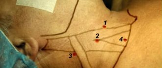
Additional cervical rib and other anomalies
If the hand becomes less sensitive, goes numb (and not the whole hand may go numb, but, as often happens, the fourth, fifth fingers and often the inner surface of the shoulder, forearm), certain movement disorders are observed, it is quite possible that the cause of the disease is related to the neurovascular thoracic outlet compression syndrome.
This disease occurs in women 6-7 times more often than in men, which is to a certain extent due to anatomical features. Thoracic outlet syndrome usually appears after the age of twenty; Compression of blood vessels and nerves can occur in different anatomical areas. Quite often this change is congenital (for example, a person has an extra rib, the so-called cervical rib). It happens that there is a narrow space between the first rib and the collarbone (due to an anomaly in the development of the upper ribs, collarbone, cervical vertebrae) or its narrowing with age, when the muscles weaken and the vessels are compressed...
In children, an additional cervical rib is usually detected at 12-13 years of age. Unfortunately, people are not interested in how many ribs a person has until complaints and complications arise. As a rule, an additional rib can only be seen on an image, but sometimes even radiologists do not “identify” it. It happens that the rib is not fully formed, and the dense fastial cord compresses the vessels and nerve trunks of the brachial plexus...
Elements of compression are often observed in people who consider themselves completely healthy. But compression syndrome can significantly reduce the quality of life and, most importantly, cause serious complications.
First of all, compression of the neurovascular bundle occurs in a certain position of the hand (“behind the head”, hand raised up and abducted, or turned “right-left”). As a result of microtrauma of the vascular wall repeated in this position and due to its compression, thrombosis of large main vessels or vessels of the hand can occur with the formation of small blood clots.
If, for example, thrombosis of the subclavian artery occurs as a result of the disease, this can lead to gangrene of the fingers and even the hand, and to the loss of a limb. In some cases, this disease leads to paralysis.
The clinical manifestations of hand diseases are so varied that a diagnosis can only be made by a specialist who is very well versed in this issue. Over the past thirty years, hand diseases have begun to be studied more closely in our country, and surgeons have developed appropriate operations. Moreover, it was Ukrainian specialists who took the leading positions in this matter.
Treatment of phlebitis
Treatment for venous inflammation depends on its type and severity. First, the doctor finds out whether the deep veins are affected.
Many patients with thrombophlebitis find that cooling the inflamed area relieves pain. Depending on the location and size of the clot, there are different treatment options. Treatment with anticoagulants to dissolve the blood clot is possible. The clot can also be removed surgically. In some cases, symptomatic treatment is sufficient.
Main treatment: Use of medical compression stockings for phlebitis
The main treatment involves the use of properly selected medical compression stockings. It is particularly effective in combination with physical exercise: the compression stockings and the activation of the muscular-venous pump ensure that the diameter of the veins is reduced. This means that blood moves faster towards the heart and does not accumulate in the vessels of the legs. Symptoms such as pain, swelling and pressure are significantly reduced, resulting in an improved quality of life.
What happens if phlebitis is not treated?
Easily diagnosed, superficial phlebitis is highly treatable and often resolves within a few days without complications. If phlebitis is not treated, it can progress and cause prolonged pain lasting up to several weeks. The situation becomes especially dangerous if the vein affected by phlebioma connects to the deep venous system. If in this case treatment is not started in a timely manner, deep vein thrombosis may develop with a high risk of pulmonary embolism.
Which doctor treats phlebitis?
If you develop phlebitis, you should immediately consult a doctor (phlebologist, vascular surgeon, dermatologist).
Veins hurt after training: how to relieve the condition
To relieve discomfort after exercise, doctors recommend resting in a lying position, raising your legs up (for example, placing your feet on the wall). Warm foot baths will also be helpful.
However, this is a symptomatic treatment.
“To avoid unpleasant sensations after training, you need to make sure that venous stagnation does not occur in the lower extremities,” comments Grigory Bashkirtsev.
For this it is recommended:
- Wear compression garments;
- Eliminate bad habits;
- Monitor your weight - reduce body weight if it is increased;
- Correct hormonal imbalances, etc.
“If you correctly follow all the recommendations, pain will not bother you,” adds Grigory Bashkirtsev.
Compression jersey medi
Thanks to the breathable and elastic material, compression jersey provides high wearing comfort. Modern medical compression hosiery is visually indistinguishable from model hosiery, but provides high medical effectiveness when used.
Here you can find more information about medi compression stockings.
The human body
How do veins work?
Vienna
Product Tips
Ideal compression product
Compression hosiery
Symptoms of thrombosis
The main symptom of venous thrombosis is swelling of the affected limb. The pathology begins acutely and progresses quickly. In most cases, the increase in symptoms occurs within 24 hours, much less often – within 2-3 days.
Also symptoms of thrombosis are:
- increasing pain in the affected area;
- thickening of the veins at the location of the blood clot;
- swelling, bulging of superficial veins;
- a sharp change in skin color at the location of the blood clot: first redness, and after a few hours - blue discoloration.