Types of myelocytes
Myelocyte in peripheral blood smear
Myelocytes are the precursor cells of mature granulocytes, a type of white blood cell.
The formation of myelocytes is an intermediate stage of granulocytopoiesis, which begins with the mitotic division of a stem cell.
In order to fully understand what myelocytes are and what place they occupy in hematopoiesis, it is necessary to indicate all the forms of cells of the granulocytic lineage of hematopoiesis:
- Myeloblasts are cells that arise from the division of a colony-forming stem cell. Myeloblasts lose pluripotency - the ability to differentiate into any other types of cells. Their main task is to ensure normal maturation of granulocytes.
- Promyelocytes are the largest cells in all stages of granulocyte formation. Already at this stage of maturation, primary granules appear in the cells, which divide promyelocytes into eosinophilic, basophilic and neutrophilic.
- Myelocytes - are formed after the third division of promyelocytes. At this stage of maturation, the granularity of the cells (inclusions) becomes strictly specific (secondary), which makes it possible to clearly distinguish future neutrophils, basophils and eosinophils. Myelocytes are actively dividing cells. The physiological activity and functionality of granulocytes depends on the completeness of myelocyte maturation.
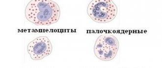
- Metamyelocytes (young leukocytes) - such cells have a low ability to divide. Thanks to them, the final stage of granulocyte maturation occurs. The nucleus of metamyelocytes undergoes changes, dividing the cells into two large groups - segmented and band.
- Granulocytes are the result of granulocytopoiesis. Such cells are called polymorphonuclear, since the cell type depends on their granularity.
Thus, myelocytes are the cells that determine the full growth of mature granulocytes. They belong to granular leukocytes and differentiate into three main types of white blood cells:
- neutrophils,
- eosinophils,
- basophils.
In peripheral blood, only mature forms are normal
The main hematopoietic organs - bone marrow, spleen and lymph nodes - finally acquire their specialization towards the end of intrauterine development and the birth of a person. The lymph nodes and spleen ensure the maintenance of the circulating pool of lymphocytes (lymphocytopoiesis), and the bone marrow is fully responsible for the formation of the formed elements of the myeloid and erythroid series - erythrocytes (erythropoiesis), monocytes (monocytopoiesis), platelets (thrombocytopoiesis), as well as the differentiation and maturation of granulocytes - granular white blood cells (granulocytopoiesis), the most numerous group in the leukocyte population.
general scheme of hematopoiesis
Leukocytes in the peripheral blood are represented only by mature cells: the already mentioned granular descendants of myelocytes - granulocytes, as well as non-granular ones - agranulocytes (monocytes, lymphocytes)
Granulocytes, in turn, are divided into:
- Neutrophils (segmented: 47-72% in the blood and band-nuclear: 1-6%) are mature, highly specialized cells that have a pronounced protective ability (phagocytosis) and high motor activity, which explains their significant number in the group of leukocytes of the granulocytic series. They are in the majority in the blood, and accordingly, their connection with myelocyte ancestors is maximum;
- Eosinophils (0.5-5% in the blood) – their phagocytic and motor activity is lower than that of neutrophils, their main task is to participate in allergic reactions;
- Basophils (0-1% in the blood) are a small group that is directly related to allergies and is involved in blood clotting processes.
leukocytes of the granulocyte series - descendants of myelocytes
Obviously, the blood of a healthy person does not report anything about what happens to the cells before they enter the bloodstream: everything is calm, “adult” granular leukocytes, being within their normal values, quietly perform the important functions assigned to them. Abnormalities can be suspected when conducting a qualitative hematological analysis of blood samples from a sick person.
The detection of proliferating representatives of the granulocytic series (myeloblasts, promyelocytes, myelocytes) and maturing cells (metamyelocytes or young ones, also not reaching the stage of full maturation) that are unusual in peripheral blood may indicate a serious hematological pathology.
Norms of myelocytes in the bone marrow
Blood cell maturation
Since immature forms of leukocytes must undergo all stages of maturation in the bone marrow, they should normally be present only in the bone marrow puncture.
If a person is healthy, then there is no reason for immature forms of blood cells to enter the systemic circulation.
To assess the consistency of granulocytopoiesis and all hematopoiesis in general, a research method such as bone marrow puncture (sternal puncture, trephine biopsy) is used.
Normally, the granulocytic (myelocytic) lineage of hematopoiesis will give the following indicators when assessing the myelogram:
| Cellular composition of the bone marrow (granulocytopoiesis) | Quantity, % |
| Undifferentiated blast cells | 0,1-1,1 |
| Myeloblasts | 0,2-1,7 |
| Promyelocytes | 1,0-4,1 |
| Myelocytes | 6,9-12,2 |
| Metamyelocytes | 8,0-14,9 |
| Rod | 12,8-23,7 |
| Segmented | 13,1-24,1 |
| Neutrophil Maturation Index | 0,5-0,9 |
| All eosinophils | 0,5-5,8 |
| Basophils | 0-0,5 |
Myelocytes: classification and norm
This is a type of bone marrow cell. Myeloblasts form promyelocytes. Promyelocytic division promotes the formation of myelocytes. The process precedes the stage of complete maturation of granulocytes. Myelocytes have a round shape. They contain a large cytoplasm and nucleus and have a granular structure. They have a size of about 12-20 microns. There are 3 types of cells:
- neutrophilic;
- eosinophilic;
- basophilic.
Neutrophil myelocytes contain pink protoplasm. Also, the first cells have coarse granularity. They are the most mature type relative to the other two. Basophilic myelocytes contain violet protoplasm, and eosinophilic myelocytes contain pink-red protoplasm with large inclusions. Their quantity in the bone marrow is determined by puncture. Cell content standards are presented in the table.
| Cell type | Quantity |
| Eosinophilic | 0,5-2% |
| Neutrophilic | 5-10% |
| Basophilic | 0,2-1% |
Causes of appearance in the blood
Severe infection may lead to the appearance of myelocytes
In a healthy person, there should be no myelocytes or other immature cells of the myelocytic lineage of hematopoiesis in the blood test. Even minor concentrations of dividing and maturing cells are considered a variant of the pathological condition.
The detection of juvenile forms of granulocytes indicates that the body is at risk and is struggling with one of the following diseases or pathological processes:
- acute bacterial and viral infections, most often complicated by purulent inflammation. These may be purulent tonsillitis, other severe infections of the ENT organs, acute pyelonephritis, pneumonia, cholera, sepsis, scarlet fever, tuberculosis, typhoid fever, brucellosis, paratyphoid fever, measles, mumps, rubella, etc.
- conditions after severe infectious and inflammatory processes;
- appendicitis and other acute surgical pathology;
- gangrene;
- severe burn disease;
- strokes, heart attacks;
- acute blood loss of any origin;
- metastasis to the bone marrow;
- tumor collapse syndrome;
- consequences of chemotherapy, radiation treatment;
- long-term use of cytostatic, immunosuppressive medications;
- lead poisoning;
- alcoholism;
- all types of coma;
- acidosis;
- shock;
- heavy and constant physical activity;
- many types of anemia;
- leukemia;
- myeloid leukemia;
- deficiency of cyanocobalamin and/or folic acid.
Blast crisis on blood smear
If the concentration of blast cells in the blood is increased slightly - up to 2% of the total leukocyte mass - then we are talking about chronic leukemia.
High values of blasts in the blood are a sign of severe disruptions in the activity of the bone marrow, which indicates acute leukemia.
An excess of the number of blast cells in the blood of more than 5% indicates the development of a blast crisis in patients with chronic myeloid leukemia, as well as the terminal stage in oncological pathology.
The detection of myelocytes, promyelocytes and metamyelocytes in the blood of no more than 5% does not indicate the presence of hematological diseases, but still indicates the presence of certain health difficulties.
But a significant increase from 10% and above is a very unfavorable indicator; it is a marker of myeloproliferative diseases - chronic leukemia, which originate from young cells of the myeloid lineage of hematopoiesis.
The most common reason for the detection of myelocytes and other maturing cells of leukocytopoiesis is chronic myeloid leukemia, the substrate of which is predominantly immature neutrophil myelocytes and other young forms.
At the initial stages of the disease, the growth of myelocytes is not pronounced. The progression of myeloid leukemia is accompanied by a significant increase in myelocytes in the blood, as well as mature eosinophils and basophils. A sharp increase in immature neutrophils, i.e., neutrophilic myelocytes, is an extremely unfavorable sign that worsens the course of leukemia and the prognosis.
Myeloid leukemia is a malignant disease of the blood and bone marrow in which an excessive number of granulocytes (neutrophils, eosinophils and basophils) and their precursors are formed. Granulocytes are a type of white blood cell and are responsible for protecting the body from infections. With myeloid leukemia, they cease to perform their functions and displace normal blood cells from the blood and bone marrow and penetrate other organs, disrupting their functioning.
There are many types of myeloid leukemia, distinguished depending on the speed of development of the pathological process, the maturity of leukemic cells, and changes in chromosomes. Most often, there are two main types of the disease: acute myeloid leukemia and chronic myeloid leukemia.
For any type of myeloid leukemia, complex and fairly long-term treatment is used. Every year more and more effective methods of treating this type of blood cancer appear. The prognosis of the disease depends on the type of myeloid leukemia, the stage of the disease at which treatment was started, and the age of the patient. In general, the prognosis for acute myeloid leuosis is favorable, especially in children. With chronic myeloid leukemia, the prognosis is worse, but with timely initiation of treatment, modern methods of therapy can permanently stop the progression of the pathological process.
Synonyms Russian
Myeloid leukemia, Ph-positive chronic myeloid leukemia, granulocytic leukemia, myeloid leukemia, myeloid leukemia, myelosis, multiple myeloma, acute myeloid leukemia, acute non-lymphoblastic leukemia, acute non-lymphoblastic leukemia in adults.
English synonyms
Childhoodacutemyeloidleukemia, adultacutemyeloidleukemia, acutemyeloidleukemia, acutemyeloblasticleukemia, acutegranulocyticleukemia, acutenonlymphocyticleukemia, chronicmyeloidleukemia, chronicgranulocyticleukemia.
Symptoms
Acute myeloid leukemia usually develops rapidly – within a few weeks. Its main symptoms are:
- weakness;
- irritability;
- dizziness;
- dyspnea;
- frequent infectious diseases;
- fever;
- frequent, prolonged bleeding, possible severe nosebleeds, bleeding gums;
- hemorrhages in the skin and mucous membranes;
- soreness, inflammation of the gums;
- heaviness in the stomach;
- swollen lymph nodes;
- headaches, nausea, vomiting, convulsions.
Chronic myeloid leukemia develops gradually and goes through 3 stages:
1) chronic – there are usually no symptoms;
2) progressive – weakness and abdominal pain appear;
3) blast crisis - at this stage the disease occurs with all the symptoms of acute myeloid leukemia.
General information about the disease
All blood cells develop from a single stem cell, which then gives rise to myeloid and lymphoid stem cells. Lymphocytes form from lymphoid cells, while myeloid cells give rise to the precursors of erythrocytes, platelets and myeloblasts. It is from myeloblasts that granulocytes and monocytes are formed as a result of a chain of successive divisions.
Granulocytes are a type of leukocyte and are called so because of their appearance - under a microscope they contain characteristic dark granules, as well as a nucleus consisting of several segments. There are several types of granulocytes - eosinophils, basophils and neutrophils. Monocytes also have a segmented nucleus, but their granules are light-colored. The main task of granulocytes and monocytes is to fight against foreign agents harmful to the body (viruses, bacteria).
In myeloid leukemia, the bone marrow produces an excessive number of abnormal granulocytes. Gradually, they displace normal blood cells from the blood and bone marrow, which leads to the appearance of characteristic symptoms. When the division and growth of red blood cells is suppressed, symptoms of anemia arise - pallor, dizziness, weakness; when platelet growth is suppressed - blood clotting disorders, frequent bleeding. Leukemia cells can penetrate other organs - the liver, spleen, lymph nodes, brain and spinal cord - causing disruption of their functions and characteristic manifestations. Pathological myeloid cells can also form clusters in the periosteum, mediastinum, and organs of the gastrointestinal tract (chloromas).
Malignant hematopoietic disorders occur due to damage to the DNA of myeloid cells. The DNA of a cell contains information about its growth, division and death and is presented in the cell in the form of chromosomes. Factors that damage the DNA of myeloid cells are not fully understood. The harmful effects of ionizing radiation, previous chemotherapy, and toxic substances such as benzene have been proven. Characteristic changes in the structure and number of chromosomes have also been identified in certain types of myeloid leukemia.
In acute myeloid leukemia, damage to the 8th, 15th, 16th, 17th and 21st chromosomes is often observed. A characteristic feature of chronic myeloid leukemia is the presence of the Philadelphia chromosome. It occurs in 95% of all cases of chronic myeloid leukemia and is formed as a result of the attachment of a section of the 9th chromosome to the 22nd chromosome. The Philadelphia chromosome activates the synthesis of special tyrosine kinase proteins that disrupt myelocyte division. As a result, both mature granulocytes and blast cells appear in the blood.
Acute myeloid leukemia occurs in both adults and children. With it, a large number of myeloblasts are found in the blood and bone marrow. In chronic myeloid leukemia, myeloid cells are more mature and “specialized.” The average age of patients with chronic myeloid leukemia is 55-60 years.
Who is at risk?
- Men.
- People over 60 years of age.
- Smokers.
- Exposed to radioactive radiation.
- Have undergone chemotherapy or radiation therapy for another form of cancer.
- People with Down syndrome and other genetic disorders.
- Suffering from myelodysplastic diseases (this is a group of chronic diseases in which the bone marrow does not produce a sufficient number of full-fledged blood cells).
Diagnostics
Laboratory examination methods
- Complete blood count (without leukocyte formula and ESR) with leukocyte formula. This study provides the doctor with information about the quantity, ratio and degree of maturity of blood elements. Leukocytes. In myeloid leukemia, leukocytes may be elevated, normal, or decreased. The leukocyte formula (the ratio of individual types of leukocytes) is determined from a blood smear. To do this, a thin smear of blood is applied to a glass slide, stained with special dyes, and then examined under a microscope. In this way, the doctor can not only determine the ratio of leukocytes, but also identify pathological, immature cells that differ in appearance from normal ones. Acute leukemia is characterized by the presence of specific inclusions in leukocytes - azurophilic granules and Auer rods. This is a characteristic feature of myeloblasts. In chronic myeloid leukemia, more mature leukocytes are found in the blood.
- Platelets, red blood cells and hemoglobin may be reduced.
- Determination of neutrophil alkaline phosphatase level. This is a special bactericidal substance that is detected only in mature leukocytes. In myeloid leukemia it is reduced, while in other diseases, such as infections, it can be significantly increased.
- Flow cytometry or immunophenotyping. In complex variants of myeloid leukemia, these techniques make it possible to accurately determine the type of malignant cells. Flow cytometry measures cell parameters using a laser beam. Immunophenotyping involves the detection of proteins specific to different cell types on the surface of the leukocyte membrane.
- Cytogenetic studies. Venous blood is usually taken for testing. Blood cells are fixed and stained, after which a specialist examines their karyotype under a microscope - a complete set of chromosomes that is identical in all cells of the human body. Used to identify chromosomal abnormalities characteristic of myeloid leukemia.
Other examination methods
- Spinal tap. It is carried out to determine leukemia cells in the cerebrospinal fluid that washes the spinal cord and brain. A sample of cerebrospinal fluid is taken using a thin needle, which is inserted between the 3rd and 4th lumbar vertebrae after local anesthesia.
- Chest X-ray - may show enlarged lymph nodes.
- Ultrasound examination of the abdominal organs. Helps identify enlarged liver and spleen.
Treatment
- Chemotherapy is the use of special drugs that destroy leukemia cells or prevent them from dividing.
- Targeted therapy is the use of drugs that have a targeted effect on certain types of malignant cells. They interact with certain proteins on the surface of leukemia cells and cause their destruction.
- Immunotherapy is the use of drugs that enhance the body's immune system's response to malignant cells. In the treatment of leukemia, interferon alpha, a specific protein with antiviral activity, is most often used.
- Bone marrow transplant - normal bone marrow cells are transplanted into the patient from a suitable donor. A course of chemotherapy or radiation therapy is first administered in high doses to destroy all pathological cells in the body.
- Radiation therapy is the destruction of leukemia cells using ionizing radiation. Can be used in acute myeloid leukemia to completely destroy leukemia cells before bone marrow transplantation.
Prevention
There is no specific prevention of myeloid leukemia. For timely diagnosis, it is necessary to undergo regular preventive examinations, and if alarming symptoms occur, consult a doctor immediately.
Recommended tests
- General blood analysis
- Leukocyte formula
- Cytological examination of punctates, scrapings of other organs and tissues
Myelocytes in children and pregnant women
Myelocytes can appear when the immune defense in children is reduced
The detection in the blood of any cell that has not completely completed its differentiation during granulocytopoiesis indicates that the bone marrow has become activated in response to some pathological transformation.
In a healthy child, myelocytes, like other young forms, should not be detected in the blood. The release of myelocytes into the blood, as well as other maturing granulocytes, is determined by the same factors as in adults. Also, the detection of immature cells in babies is often diagnosed with congenital heart defects, uncontrollable vomiting and dehydration. Very often, immature forms of granulocytes are found in children with weak immunity.
Severe physical stress will also lead to the appearance of a small number of myelocytes in the blood of healthy children.
As for pregnant women, fluctuations in the blood picture are allowed. The process of hematopoiesis during pregnancy is enhanced to maintain the vital functions of the entire body of the mother and baby. Additionally, the appearance of myelocytes and other young forms in the blood may be the result of an exacerbation of chronic diseases, for example, sinusitis, pyelonephritis, etc.
In expectant mothers, the concentration of myelocytes in the blood is allowed to be no more than 2-3%. But, in any case, this phenomenon requires further diagnosis in order not to miss the development of malignant pathology.
During pregnancy
Myelocytes in the blood are normal.
Granulocytes are increased, and a large number of their immature forms are released into the bloodstream. In peripheral blood their content reaches 3%.
However, the detection of myelocytes in pregnant women may also indicate pathological processes. Their appearance is associated with the body's response, for example, to inflammation.
In general, the appearance of myelocytes in the blood of expectant mothers does not negatively affect either their health or the health of the fetus.
How to determine myelocyte level
A myelogram will show the exact level of myelocytes
Determination of the level of myelocytes, as well as other components of the bone marrow, is carried out by taking a bone marrow puncture. The resulting myelogram will show the exact cellular composition of the bone marrow.
Bone marrow samples are taken from:
- sternum,
- iliac crest,
- from the calcaneus (this is how puncture is performed in small children).
After receiving the punctate, the myelogram data must be compared with the data of the general clinical blood test.
Treatment
Elimination of the cause leads to normalization of the blood picture
Since myelocythemia is caused by the development of a disease, it is the root cause that requires treatment.
Only after the cause of the pathological change in blood composition has been clarified, treatment is prescribed.
There are many reasons for the penetration of immature blood cells, in particular myelocytes. The vast majority of such triggers, unfortunately, are dangerous diseases that can quickly claim a human life. At the slightest transformation in the composition of the blood, it is necessary to urgently consult a doctor, diagnose and prescribe the necessary treatment methods.
The place of “birth”, division and differentiation is the bone marrow
The granular leukocyte pool originates in the bone marrow from pluripotent stem cells. Moving in their development from class to class through a relatively small number of unipotent precursor cells, future leukocytes reach morphologically distinguishable proliferating forms - blasts (myeloblasts) , which are subsequently destined to become full-fledged “adult” neutrophils, eosinophils, basophils (provided that hematopoiesis runs normally). As the myeloblast matures, it differentiates through the promyelocyte into the last cell of the granulocytic (granular) series, which retains the ability to divide and differentiate - the myelocyte .
Myelocytes in the bone marrow exist in the form of two generations: cells that are larger in size are maternal, and smaller ones are daughter cells. It is believed that mother cells lose the ability to proliferate and differentiate, but daughter cells have similar capabilities and, having passed the stage of metamyelocytes (young) and stab cells, are legally sent into the blood to circulate through the blood vessels and perform important tasks for the body - to provide primary anti-infective protection by phagocytizing (“eating”) microorganisms that have entered from the outside. That is, before the myelocyte turns into a full-fledged “adult” neutrophil, another stage of maturation must follow - metamyelocyte .
Metamyelocytes are called young; they sometimes normally enter the peripheral blood, but their number is tiny compared to cells that have reached maturity. In addition, in the peripheral blood there are (in small quantities, the norm is up to 6%) cells whose characteristics are as close as possible to the mature forms, these are band granulocytes . The rods are older than metamyelocytes (young), however, while still retaining signs of “youth”, they are still unable to take on such responsible tasks that fall within the competence of segmented neutrophils, therefore, in relation to the segments, they are young and their number in the analysis is completely normal not great. It turns out that everything is not so simple:
Under normal conditions, it is practically impossible for myelocytes to reach the blood in large quantities, except that a few may accidentally leak out. Therefore, an increase in any noticeable appearance of myelocytes occurs only in pathology.