© Author: Z. Nelly Vladimirovna, laboratory diagnostics doctor at the Research Institute of Transfusiology and Medical Biotechnology, especially for SosudInfo.ru (about the authors)
Seromucoids or seroglycoids are serum glycoproteins (complex proteins containing carbohydrates). Proteins and carbohydrates in the fraction of carbohydrate-protein complexes are in a ratio of 1:12, that is, for 1 part of all plasma proteins there are several groups of all carbohydrates present in the plasma, which consist of the same or different monosaccharide residues (heterooligosaccharides).
The main habitat of these glycoproteins is connective tissue, where they are concentrated in large quantities. However, if the connective tissue is subjected to mechanical stress or change and destruction for other reasons, then seroglycoids leave their usual conditions and are directed into the bloodstream (in such cases, seromucoids in the blood are increased). This circumstance long ago formed the basis for one of the biochemical tests (seromucoid in the blood), which, however, is gradually increasingly joining the category of outdated laboratory tests.
Meanwhile, diagnostic laboratories located in the “outback”, not fully equipped with advanced technologies, and therefore not having the opportunity to use the latest techniques, use the old-fashioned test for seromucoid to diagnose inflammatory processes of any localization. And, it should be noted, very successfully.
Seromucoids
Description of the analysis:
Seromucoids are one of the groups of proteins (glycoproteins) detected in blood serum and containing carbohydrate components. Seromucoids contain about 12% of the carbohydrates found in blood plasma, while seromucoids make up only 1% of the total plasma proteins.
Normally, seromucoids are part of the connective tissue of the human body, but when it is damaged, degraded or destroyed, they enter the bloodstream. An increase in the concentration of seromucoids, therefore, indicates activation of the inflammatory process, even in cases where the patient is not bothered by any symptoms.
An elevated level of seromucoids is an important diagnostic indicator for rheumatologists, as it indicates the likely development of rheumatoid arthritis.
In addition, it can indicate a wide range of diseases that are characterized by an inflammatory process:
- pleurisy;
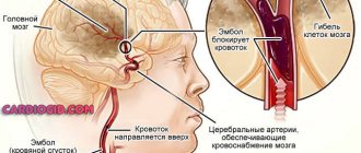
- stroke;
- myocardial infarction;
- tuberculosis;
- lymphogranulomatosis;
- diabetes;
- gout;
- glomerulonephritis;
- acute cholecystitis;
- pancreatitis;
- systemic lupus erythematosus;
- malignant neoplasms;
- damage to the respiratory tract (asthma, pneumonia, etc.);
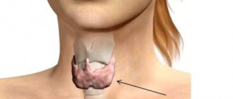
- severe pathologies of the thyroid gland, etc.
Indications for prescribing a blood test for seromucoids
Considering that an increase in the concentration of seromucoids is a universal marker of the inflammatory process affecting connective tissue, its purpose can be included in the diagnostic complexes of many diseases. Accordingly, the study can be prescribed by a wide range of specialists: from cardiologists and endocrinologists to gastroenterologists, oncologists and nephrologists.
But the study is of greatest diagnostic value for rheumatologists, as it allows one to identify the erosive and destructive effects on the joints, which are a constant companion to rheumatoid arthritis. Therefore, most often a test for the concentration of seromucoids in the blood is prescribed by a rheumatologist .
Meaning of the results
If the amount of seromucoids in a blood sample is in the range of 0.12-0.2 U, then the concentration of these proteins is within normal limits. The reason for the increase in their concentration may be an inflammatory process that develops during one of the above diseases.
A reduced concentration of seromucoids in the blood is also of diagnostic significance, which may indicate multiple sclerosis, liver disease (cirrhosis, hepatitis) or infertility.
By itself, an analysis for seromucoids cannot be a sufficient basis for making a particular diagnosis (including rheumatoid arthritis), but is used only in combination with other laboratory tests and diagnostic measures
Preparation for the study: the analysis is taken in the morning, on an empty stomach.
Material for research: venous blood.
Research method: biochemical.
Duration of the study: 2 working days.
Sign up for tests
Analysis of alpha-1-acid glycoprotein (seromucoid) in Moscow
Biological role of alpha-1 acid glycoprotein
The biological role of AGP is not fully understood, although numerous activities have been reported in vitro and in vivo, such as inhibition of platelet aggregation, modulation of lymphocyte proliferation, and drug transport.
AGP may be involved in various immunomodulatory or anti-inflammatory events for the following two reasons. First, AGP expression is regulated by both cytokines (interleukin-1, interleukin-6, and tumor necrosis factor-α) and glucocorticoids, in contrast to other acute phase proteins, including fibrinogen, ceruloplasmin, and α2-microglobulin. The regulation of AGP production in human hepatocytes by glucagon, interleukin-8, and interleukin-6 is thought to act through the mitogen-activated protein kinase pathway.
Second, it is well known that plasma AGP concentrations are influenced by several factors. For example, stress, inflammation, burns, infections, pregnancy, etc. can increase concentration. In addition, drugs such as phenobarbital and rifampicin can also increase plasma concentrations of AGP through mechanisms that are independent of the inflammatory pathway.
Action of AGP in response to ischemia and infection
Increasing the concentration of AGP severalfold in the circulation during the acute phase response may affect the biological functions of the molecule in humans. The researchers found that human AGP reduced ischemia/reperfusion injury to kidney tissue by suppressing apoptosis, TNF-α expression, and neutrophil influx. These results indicate that AGP could be used as a potential new therapeutic intervention in the treatment of acute liver and kidney failure, as seen after transplantation of chemically damaged liver and kidneys.
AGP has also been reported to have a protective effect against sepsis from Gram-negative infections. Scientists have shown that AGP interacts with bacterial lipopolysaccharide, which is the initiator of the acute inflammatory response associated with septic shock, leading to the formation of the AGP-LPS complex. These results suggest that increased AGP expression under infection conditions facilitates clearance of LPS, resulting in a protective effect against endotoxic shock resulting from infection.
Relationship between drugs and protein
Like plasma albumin, binding and transporting a number of endogenous and exogenous compounds is one of the main physiological functions of AGP. Therefore, drug binding to AGP is important in terms of proper understanding of drug pharmacokinetics, especially in the acute phase setting. Over the past twenty years, studies have been conducted on the drug binding specificity and pharmacokinetic properties of AGP using various biophysical and biochemical analytical techniques such as spectrophotometry and protein engineering.
AGP options
AGP exists as three major genetic variants with genes located in tandem on chromosome 9. AGP expression is under the control of three adjacent genes: AGP-A, which encodes the F1, F2 and S variants, while AGP-B and AGP-B code for variant A. All three genes are structurally similar to each other, the AGP B/B genes are identical while the AGP A gene contains 22 substitutions. The precursor product of the AGP-A gene is a 201 amino acid polypeptide with an 18-residue secretory N-terminal signal peptide.
A blood test for alpha-1 acid glycoprotein is prescribed when
:
- diagnosis of acute inflammatory conditions;
- assessing the effectiveness of treatment of inflammatory processes;
- diagnosis of collagenosis (connective tissue diseases), tuberculosis infection, malignant tumors; hemolysis (together with haptoglobin);
- diagnosis of disease relapses.
Research method
: immunoturbidimetry.
Research material
: deoxygenated blood.
What can tests say about the heart?
What are tests? Tests are a confirmation or exclusion of a particular disease about which an opinion was formed after a clinical examination of the patient. With their help, the doctor will find out what exactly is preventing your body from living and working normally, and what is the condition of its individual organs and systems.
So, what do these same tests say if there is pain in the heart area? The determination of enzymes contained inside cells is important in the diagnosis of diseases associated with myocardial damage. And depending on which and how many cells die, their values will change.
Biochemical blood test indicators:
ALT (alanine aminotransferase): up to 68 U/l, when assessing the level of this enzyme, it is worth considering that it is contained not only in the myocardium, but to a greater extent in the liver, therefore AST and ALT are always determined together, which helps in distinguishing between heart damage and liver. The timing of ALT increases is similar to AST.
AST (aspartate aminotransferase): up to 45 U/l, this enzyme is found in large quantities in the myocardium, and its increase, in most cases, indicates damage to cardiomyocytes - the muscle cells of the heart; An increase in AST in the blood serum is observed in myocardial infarction (95-98%) cases within 6-12 hours from the onset of the disease. The maximum increase is observed on days 2-4, and on days 5-7 the enzyme level returns to normal. There is a clear relationship between AST numbers and the size of the focus of cardiac muscle necrosis. Therefore, if the necrosis is less than 5 mm in diameter, it is possible to maintain the level of this enzyme within normal limits, which also must be taken into account.
LDH (lactate dehydrogenase) and the fractions that make up this indicator: up to 250 U/l, is considered a specific marker for AMI; an increase in the activity of the LDH1 and LDH2 isoenzymes, even with normal levels of general LDH activity, indicates the presence of minor necrosis in the heart muscle. With AMI, its level increases quickly on days 2-4, and normalizes only at 2-3 weeks. LDH levels provide valuable information about MI throughout the course of the disease. Other fractions LDH3 and LDH4 are enzymes of the lung tissue, LDH5 is of the liver.
CK (creatine phosphokinase) and the fractions that make up this enzyme: up to 190 U/l, creatine phosphokinase - is considered a specific marker (especially an increase of more than 10 times) in acute myocardial infarction. It increases in the acute period (in the first 4-8 hours from the onset of the disease), much faster than the activity of the above enzymes and is a marker for early diagnosis of AMI, especially the CPK-MB isoenzyme. After 8-14 hours, the CPK value can reach its maximum value, and normalization can occur after 3-4 days. Also, the CPK value may increase with myocarditis;
Troponin test: up to 0.4 mcg/l. Troponin is a specific contractile protein that is part of the structure of the heart muscle and skeletal muscles. This test is a diagnostic marker for suspected acute damage to myocardial cells and is one of the key results in diagnosing “acute myocardial infarction”;
myoglobin: 12-92 µg/l. A protein in muscle tissue involved in the process of cell respiration. If it appears in the blood, it is regarded as a product of the breakdown of the muscle tissue of the heart or skeleton, with the appropriate clinic, it may indicate necrosis (necrosis) of the heart muscle tissue, therefore it is also considered a specific marker of this pathology.
The indicators of ALT, AST, CPK, CPK-MB, LDH, myoglobin and troponin test closely correlate with the size of the necrosis focus in the heart muscle, and therefore have not only diagnostic, but also prognostic significance.
Acid phosphatase: 67-167 nmol/(s·l), increases in activity in patients with severe, complicated MI, mainly transmural;
C-reactive protein (CRP): up to 0.5 mg/l, its detection indicates the presence of a pathological process in the body, in particular inflammatory or necrotic. It belongs to the so-called “acute phase” proteins. A sharply positive reaction to CRP indicates the severity of the inflammatory process.
sialic acids: 2.0-2.36 mmol/l, the content of sialic acids may increase with endocarditis, MI;
electrolytes are mainly represented by K+ ions (normal 3.6 – 5.2 mmol/l), Na+ (normal 135 – 145 mmol/l), Cl- (normal 100 – 106 mmol/l), Ca2+ (normal 2, 15-2.5 mmol/l). An increased amount of potassium in the serum may be accompanied clinically by cardiac arrhythmia, which is confirmed by an ECG. Atrioventricular blockade of the conduction system of the heart may develop, the syndrome of premature excitation of the ventricles, ventricular fibrillation, and such a serious disorder as cardiac arrest may develop. Therefore, patients with heart rhythm disturbances need to monitor the content of K+ ions in the body. On the other hand, a decrease in potassium in the blood can also lead to adverse consequences in these patients - myocardial hyporeflexia. A decrease in the level of sodium ions may be accompanied by the development of cardiovascular system failure, since the ratio of K+ and Na+ ions, as regulators of processes in the cell, is in constant interaction and a decrease in one leads to an increase in the other ion. Hyperchloremia occurs in patients with kidney disease and may also lead to the development of cardiovascular disease;
lipid spectrum, is associated by the common person with the word “cholesterol”. In this case, substances (lipoproteins of various densities, triglycerides) that are involved in the metabolism of cholesterol (CH) are determined (normal blood levels are 3.1 – 5.2 mmol/l). In addition to the value of total cholesterol, an important indicator is the atherogenicity coefficient (norm up to 4), which shows the ratio of “good” and bad” lipids involved in the metabolism of fats and cholesterol, and the threat of development or progression of atherosclerosis and all the ensuing consequences. An increase in the fractions of lipoproteins and triglycerides can be either a physiological condition (of a nutritional nature) or a pathological condition. Increased lipids are characteristic of widespread atherosclerosis, obesity that accompanies and causes arterial hypertension. But it would be more accurate to say that this disruption of the functioning of internal organs and intermediate links in the metabolism of lipids and triglycerides, expressed in an increase in the atherogenicity index, causes the deposition of cholesterol in vessels of various diameters, the deposition of “spare fat,” which leads to the above diseases. Therefore, with widespread atherosclerosis, in this blood test, you can see increased values of ß-lipoproteins and total cholesterol. At the same time, a decrease in phospholipid concentration can be seen. But it is also necessary to take into account that there are age-related fluctuations in blood fats.
coagulogram is an analysis by which you can look at the “viscosity” of the blood, or in other words, whether there is a threat of blood clots, which can lead to the formation of blood clots with different localizations, which in turn can be complicated by pulmonary embolism, which causes instant death. Or, on the contrary, see how high the probability of bleeding is and whether it can stop on its own after surgery, for example, heart valve replacement.
Any analysis or research provides the doctor with additional information that helps to more accurately make a diagnosis, determine the stage of the disease, and prescribe treatment. Tests also help to monitor the course of the disease, the effectiveness of the prescribed treatment, and ensure the safety of therapy. But sometimes additional research is required to confirm or complement the results of past analyzes.
Fedorova Lyubov Alekseevna – doctor of the first category, therapist, medical cardiologist.
Treatment and prevention
An increase in seromucoids in the blood is not an independent disease. This is only a symptom of pathological processes occurring in the body, the etiology of which can be completely different. Therefore, there is no single medicine to normalize the level of seromucoids - it is necessary to establish the cause of the disease and purposefully treat it.
For example, for rheumatism, the patient is prescribed complex therapy consisting of antibiotics, immunomodulators, hormonal and anti-inflammatory drugs. Usually this treatment is effective and after completing the full course, the level of seromucoids decreases.
If the increase in seromucoids is caused by a malignant tumor, then the entire arsenal of anti-oncology drugs is used - chemotherapy, radiotherapy, and other methods of fighting cancer.
There is no prophylaxis against increased seromucoid levels as such. Often this condition is caused by diseases whose occurrence is genetically determined (asthma, rheumatism, cancer). Therefore, the only way to prevent it is to maintain a healthy lifestyle, which reduces the likelihood of such diseases. It should be reflected in the absence of bad habits, moderate regular physical activity, a balanced diet and compliance with personal and public hygiene standards.
Prevention of tuberculosis (which also results in an increase in the concentration of seromucoids) consists of timely vaccination, avoiding contact with infected people, and observing safety and hygiene rules when working in damp, dark, dirty rooms.
How does the analysis procedure work?
Venous blood is used to study the concentration of seromucoids. The material is collected in the morning, on an empty stomach. One day before the test, it is necessary to exclude the use of alcohol and medications (or inform the attending physician about their use), and 12 hours before - smoking. It is necessary to avoid physical and emotional stress the day before donating blood. Women should consult with their doctor in advance about choosing to donate blood, as their test results may be affected by the timing of their menstrual cycle.
It is contraindicated to be nervous on the day of the test. In addition, after donating blood, it is recommended to avoid physical activity and follow a moderate diet to quickly restore lost blood volume. If blood is to be donated to a child, parents need to ensure that the child is calm and not subjected to excessive physical activity.
The test is carried out by separating the blood serum and then analyzing the serum particles using a spectrophotometer or photometer. The principle of the analysis is that the serum particles of the substance absorb light in different ways. Both an increase and a decrease in the concentration of seromucoids may indicate unfavorable processes. An increase in concentration is caused by:
- various inflammatory conditions;
- Jaundice;
- rheumatism;
- pulmonary tuberculosis;
- exacerbation of chronic diseases, for example, cholecystitis;
- myocardial infarction and stroke.
A decrease in seromucoid concentration indicates that the liver does not synthesize protein sufficiently; this is an indicator of liver dystrophy, cirrhosis, hepatocellular and liver failure, alcoholic and viral hepatitis, or hepatocellular carcinoma. Low levels of albumin, seromucoid, prothrombin and protein in general are a reason to seriously check the liver.
Alpha-1-acid glycoprotein (AGP, orosomucoid) is one of the main “acute phase” proteins. The concentration in blood serum can increase 3-4 times during inflammatory processes, infections, and malignant neoplasms. A dynamic study of AGP allows one to assess the course of the inflammatory process and diagnose relapse after surgical treatment of tumors. The analysis is used as a prognostic sign of the development of heart failure after myocardial infarction, as well as to detect hemolysis (together with haptoglobin).
English synonyms
AGP.
Research method
Immunoturbidimetry.
Units
G/L (grams per liter).
What biomaterial can be used for research?
Venous blood.
How to properly prepare for research?
- Do not eat for 2-3 hours before the test; you can drink clean still water.
- Do not smoke for 30 minutes before the test.
General information about the study
The synthesis of all protein substances occurs in the liver. Most of them are produced constantly, as they ensure the vitality of the body. But there are also those whose production starts under certain stressful circumstances. Alpha-1-acid glycoprotein (orosomucoid) is an acute phase protein of inflammation that suppresses immune reactivity, changes the functionality of platelets, and binds hormones. To date, scientists have not been able to determine the exact purpose of this protein, but it has been reliably recorded that it begins to appear in the plasma in the presence of various kinds of inflammatory and degenerative (destructive) pathological processes in the body. Its determination in the blood is used for diagnosis, monitoring of acute inflammatory processes, and for assessing intravascular destruction of red blood cells (in combination with the haptoglobin test). It suppresses immunoreactivity, changes platelet adhesiveness, binds hormones and drugs with essential properties.
Alpha-1-acid glycoprotein is a glycoprotein with a MW of 41-43 KDa with a pI of 2.8-3.8. Its peptide part is a simple chain of 183 amino acids. It is synthesized mainly by parenchymal cells of the liver. It is one of the main acute-phase human proteins. Its plasma levels increase 3-4 times in most cases of inflammatory processes, infection and tissue necrosis. Alpha-1-acid glycoprotein is perhaps one of the most accurate markers of ulcerative colitis. Like haptoglobin and prealbumin, its concentration increases in the presence of endogenous (eg, Cushing's syndrome) or exogenous (with treatment with prednisone or dexamethasone) glucocorticoids. Estrogens, nephrotic syndrome, and protein-losing enteropathy cause a decrease in plasma levels of alpha-1-acid glycoprotein.
The principle of the method used in the analysis is that the alpha-1-acid glycoprotein present in the sample precipitates in the presence of human anti-alpha-1-acid glycoprotein. Light dispersion caused by antigen-antibody complexes is proportional to the concentration of alpha-1-acid glycoprotein and can be measured by turbidimetry.
What is the research used for?
- To identify acute inflammatory conditions.
When is the study scheduled?
- Monitoring the treatment of inflammatory processes;
- diagnosis of collagenosis (connective tissue diseases), tuberculosis infection;
- diagnosis of malignant tumors;
- diagnosis of relapses;
- diagnosis of hemolysis (together with haptoglobin).
What do the results mean?
Reference values: 0.5 - 1.2 g/l.
Indicator increase:
- inflammatory processes (bacterial and viral infections, especially in newborns);
- Crohn's disease, myocardial infarction;
- rheumatic diseases;
- systemic lupus erythematosus and other autoimmune diseases;
- Itsenko-Cushing's disease;
- injuries, surgical interventions, tumor processes;
- inflammation of the vascular wall;
- Hodgkin's disease.
Decrease in indicator:
- early stages of pregnancy;
- severe liver damage (end stage of hepatocellular disease);
- nephrotic syndrome;
- gastroenteropathy with protein loss, fasting;
- taking medications - antimicrobial agents, cyclosporins, penicillamine, tamoxifen, oral contraceptives, estrogens.
With severe hemolysis, the levels of alpha-1-glycoprotein and haptoglobin increase simultaneously. If an elevated level of alpha-1-glycoprotein is detected with normal haptoglobin levels, this indicates moderate hemolysis.
The level of orosomucoid during the inflammatory process, as a rule, correlates with the concentration of other positive acute-phase proteins (CRP, haptoglobin, alpha-1-antitrypsin, etc.).
What can influence the result?
Content enhancement:
- age, male gender, physical activity;
- menopause;
- obesity, smoking;
- taking medications - carbamazepine, anticonvulsants, androgens, glucocorticoids.
Content reduction:
- starvation;
- early pregnancy;
- early childhood;
- taking medications - antimicrobials, cyclosporins, penicillamine, tamoxifen, oral contraceptives.