The human heart is an amazing organ, thanks to which the normal functioning of all organs and systems is possible. Contraction of the heart muscle is ensured by electrical impulses that pass through special pathways. If there are additional pathways for these impulses, certain changes occur that can be monitored using an electrocardiogram (ECG). With changes in the ventricular complex we are talking about the phenomenon of WPW (premature excitation of the ventricles). For minor disorders, characterized only by a change in the speed of impulses between the ventricles and the atrium, CLC syndrome is diagnosed.
What is the disease
Clerk-Levy-Christesco syndrome or CLC is a congenital pathology of the heart structure, one of the types of premature ventricular excitation syndrome. The anomaly is due to the presence of a James beam. Thanks to this bundle, the atrioventricular node is connected to one of the atria. This feature is accompanied by the transmission of a premature impulse to the ventricles.
Pathology can be diagnosed using an ECG. Often, CLC syndrome occurs accidentally in completely healthy people, and the patient does not have any symptoms. There are known cases of this disease being detected in children.
Even in the absence of symptoms, one should not assume that the pathology is harmless. Ventricular preexcitation syndrome can cause arrhythmia. The condition is accompanied by increased heart rate (sometimes over 200 beats per minute). This is especially felt in older people; young patients tolerate arrhythmia more easily.
Syndrome and phenomenon of shortened PQ interval
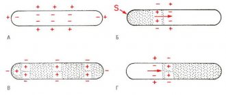
The PQ interval is a purely electrocardiographic criterion that allows one to evaluate the transmission time of an electrical impulse from the sinus node in the atrium to the contractile fibers located in the ventricles. In other words, it reflects the operation of the atrioventricular junction, a kind of “switch” that redirects electrical excitation from the atria to the ventricles. Normally, it is no less than 0.11 seconds and no more than 0.2 seconds:
- An increase in the interval beyond the specified time indicates a slowdown in conduction through the atrioventricular node,
- Shortening means that the excitation is carried out too quickly. In fact, there is more frequent impulse of the ventricles, with the so-called “reset” of excitation.
The shortening of this interval is due to the presence of additional conduction bundles in the conduction system of the heart. It is through them that additional pulses are reset. Therefore, at certain moments the ventricles receive double impulses - physiological in a normal rhythm (60-80 per minute), and pathological, through bundles.
There may be several pathological bundles, and all of them are named after the names of the authors who first discovered them. Thus, the Kent and Maheim bundles are characteristic of the SVC syndrome, and the James bundles are characteristic of the CLC syndrome. In the first case, the pathological discharge of impulses goes from the atria directly to the ventricles, in the second, the James bundle passes through the atrioventricular node, that is, the node is stimulated first, and then the ventricles. Due to the “throughput” capacity of the AV node, part of the impulses conducted to the ventricles returns through the same bundle to the atria, therefore such patients have a high risk of developing paroxysmal supraventricular tachycardia.
What is the difference between a syndrome and a phenomenon?
Many patients, having seen the concepts of the phenomenon or CLC syndrome in the ECG conclusion, may be puzzled which of these diagnoses is worse. The CLC phenomenon, subject to a correct lifestyle and regular monitoring by a cardiologist, does not pose a great danger to health, since the phenomenon is the presence of signs of PQ shortening on the cardiogram, but without clinical manifestations of paroxysmal tachycardia.
CLC syndrome, in turn, is an ECG criterion accompanied by paroxysmal tachycardias, most often supraventricular, and can cause sudden cardiac death (in relatively rare cases). Typically, patients with short PQ syndrome develop supraventricular tachycardia, which can be quite successfully stopped at the stage of emergency medical care.
Why does short PQ syndrome occur?
As already indicated, the anatomical substrate of this syndrome in adults is a congenital feature, since additional conduction bundles are formed in the prenatal period . People with such bundles differ from ordinary people only in that they have an additional tiny “thread” in the heart, which takes an active part in conducting the impulse. But how the heart behaves with this beam will be discovered as the person grows and matures. For example, in children, CLC syndrome may begin to manifest itself both in infancy and adolescence, that is, during the rapid growth of the body. Or it may not manifest itself at all, remaining only an electrocardiographic phenomenon throughout adult life until old age.
No one can name the reason why the syndrome begins to manifest itself as paroxysmal tachycardia. However, it is known that in patients with organic pathology of the myocardium (myocarditis, heart attack, hypertrophic cardiomyopathy, heart disease, etc.) attacks of tachycardia occur much more often and clinically occur with a more pronounced clinical picture and with a severe general condition of the patient.
But the provoking factors that can cause paroxysm can be listed:
- Physical activity that is significantly or not significantly greater than the patient’s usual physical activity,
- Psycho-emotional stress, stressful situation,
- Hypertensive crisis,
- Eating large amounts of food at one time, drinking very hot or very cold liquids,
- Visiting a bathhouse, sauna,
- Changes in external temperatures, for example, going out into severe frost from a very hot room,
- Increased intra-abdominal pressure, for example, during severe coughing, sneezing, defecation, pushing during childbirth, lifting heavy objects, etc.
How does short PQ syndrome manifest?
The clinical picture of shortened PQ syndrome is caused by the occurrence of paroxysmal tachycardia, since during the interictal period the patient usually does not present any complaints from the cardiovascular system. Symptoms of tachycardia are the following:
- The sudden, sharp onset of an attack, caused by provoking factors or occurring without them, in itself,
- Feeling of a strong heartbeat, sometimes with a feeling of interruptions in the heart,
- Autonomic manifestations - severe weakness, hyperemia or paleness of the face, sweating, coldness of the extremities, fear of death,
- Feeling of suffocation or lack of oxygen, feeling of insufficient inspiration,
- Unpleasant discomfort in the heart area of a pressing or burning nature.
If the symptoms described above appear, you should definitely seek medical help by calling an ambulance or going to a clinic.
Diagnosis of shortened PQ
The diagnosis is established after recording an ECG and interpreting its data by a doctor. The main ECG signs of CLC syndrome:
- Increased heart rate - 100-120 per minute or more, sometimes reaching 200 beats per minute,
- Shortening of the PQ interval between the P wave and the ventricular QRST complex is less than 0.11-0.12 seconds,
- Unchanged ventricular complexes in case of supraventricular tachycardia, and dilated, deformed - in case of ventricular tachycardia, which is a life-threatening condition,
- Correct sinus rhythm in supraventricular tachycardia.
- After the diagnosis has been established and the paroxysm has been relieved, the patient is prescribed an additional examination to exclude gross cardiac pathology (heart defects, myocarditis, heart attack, etc.). Of these, the use of the following is justified:
- Ultrasound of the heart,
- Installation of an ECG monitor during the day,
- Examination of the electrocardiogram after physical activity (stress tests using bicycle ergometry, treadmill, tests with a load of pharmacological drugs),
- TEE, or transesophageal electrophysiological study and electrical stimulation of the heart muscle by inserting a probe into the esophagus,
- In especially unclear clinical cases, endovascular or intravascular EPI (endoEPI). The plan for further examination and treatment of the patient is determined only by the attending physician. Treatment of shortened PQ syndrome
- The shortened PQ phenomenon, also called the CLC phenomenon, does not require treatment. It is quite enough to correct the lifestyle and undergo regular examinations with a doctor - a cardiologist or arrhythmologist, for a child - once every six months, for adults - once a year.
- Treatment of shortened PQ syndrome (CLC - Clerk-Levi-Christesco syndrome) consists of providing first aid at the time of tachycardia paroxysm and further taking prescribed medications. First aid can be provided by the patient independently - this is the use of vagal tests. These manipulations are based on a reflex effect on the vagus nerve, which slows down the heart rate. Vagal tests can be used at the time of paroxysm only if the patient has had an attack of tachycardia not for the first time, he has been diagnosed and has not previously had ventricular tachycardia. In addition, vagal tests should be explained to the patient in detail by the doctor. The most effective techniques include the following:
- Straining test (Valsalva maneuver),
- Simulating a cough or sneeze
- Lowering your face into a basin of cold water, holding your breath,
- Finger pressure with moderate force is applied to the closed eyeballs for three to five minutes. Restoration of the correct heart rhythm is carried out by a doctor or paramedic and is carried out by administering medications intravenously. As a rule, this is asparkam, verapamil or betaloc. After hospitalization of the patient in a cardiology hospital, treatment of the underlying heart disease, if any, is carried out. In the case of frequent attacks of tachyarrhythmia (several per month, per week), as well as a history of ventricular arrhythmias, a hereditary history of sudden cardiac death or death from cardiac causes in young people, the patient is indicated for surgical treatment. The operation involves applying radio frequencies, a laser or a cold factor to an additional beam. Accordingly, radiofrequency ablation (RFA), laser destruction or cryo-destruction are performed. All indications and contraindications are determined by an arrhythmologist, cardiologist and cardiac surgeon. Many patients are interested in the possibility of permanent cardiac pacing. An pacemaker can be installed if the patient has a tendency to paroxysmal ventricular tachycardia, ventricular fibrillation and there is a high risk of clinical death with cardiac arrest (asystole). Then you can consider installing a cardioverter-defibrillator, which, unlike an artificial pacemaker, does not impose the correct rhythm, but “restarts” the heart when such fatal arrhythmias occur. Is it possible to develop complications with PQ shortening? The phenomenon of shortened PQ does not lead to any complications can not. Due to the fact that the manifestation of PQ syndrome is an attack of tachyarrhythmia, there will be corresponding complications. These include the occurrence of sudden cardiac death, fatal arrhythmias (ventricular fibrillation), thromboembolism of the cerebral and pulmonary arteries, the development of myocardial infarction, arrhythmogenic shock and acute heart failure. Of course, not every patient develops such complications, but everyone needs to remember them. Prevention of complications is timely seeking medical help, as well as timely surgery if indications for it are discovered by a doctor. Prognosis Determining the prognosis for patients with CLC syndrome is always difficult, since it is not possible to predict in advance the occurrence of certain rhythm disturbances, the frequency and conditions of their occurrence, as well as the appearance of their complications. According to statistical data, the life expectancy of patients with shortened PQ syndrome is quite is high, and paroxysmal arrhythmias most often occur in the form of supraventricular rather than ventricular tachycardias. However, in patients with underlying heart pathology, the risk of sudden cardiac death remains quite high. The prognosis regarding the phenomenon of shortened PQ remains favorable, and the quality and life expectancy of such patients do not suffer. Prepared by cardiorheumatologist RNPCSM Taraleva T.A.
Features of the pathology
Pre-excitation of the cardiac ventricles can be asymptomatic, in which case we are talking about the “pre-excitation phenomenon”. When signs of pathology appear in a patient, the disease is classified as “pre-excitation syndrome.”
There are several types of disease:
- Breschenmash (atriofascicular) - here the right atrium is connected to the trunk of the His bundle;
- Maheima (nodoventricular) - in this case, the right side of the interventricular septum is connected to the atrioventricular node;
- Kent (atrioventricular) - here the atria and ventricles are connected bypassing the atrioventricular node;
- James (atrionodal) - impulses pass between the lower part of the atrioventricular and sinoatrial node.
Important! Sometimes there are several paths of abnormal conduction of impulses. The number of such patients is no more than 10% of all cases of the disease.
CLC syndrome is clearly monitored by performing an electrocardiogram
Republican Scientific and Practical Center for Sports Medicine
President of Uzbekistan Islam Karimov signed the Resolution “On the creation of the Republican Scientific and Practical Center for Sports Medicine under the National Olympic Committee of the Republic of Uzbekistan”
- 100027, Tashkent, Shaykhantakhur district, Olmazor street 6
- +998 ( 71 ) 241-52-45 +998 ( 71 ) 241-52-46
- [email protected]
Links
News
Insurance
Articles
Prices
What Causes CLC?
In order to understand how CLC syndrome occurs, you should remember the anatomy. For the normal filling of the ventricles of the heart with blood and their contraction after contraction of the atria, there is a special filter located between them in the structure of the organ. This filter is called the atrioventricular node. When an impulse is transmitted to this node, the excitation passes slowly, after which it is transmitted to the ventricles. Thanks to this process, blood enters the aorta and pulmonary trunk.
With some congenital pathologies, a person may have bypass pathways for conducting cardiac impulses. Often the James beam and some other channels act in this way. In this case, the ECG shows minor deviations from the norm or severe disturbances, for example, with serious deformation of the ventricles. The syndrome of premature excitation of the ventricles in medical practice is called WPW.
How does the disease progress?
Pathology can manifest itself at any age of the patient. For a long time, the disease can occur without obvious signs and symptoms. The CLC phenomenon is accompanied by a shortening of the PQ interval during an ECG. At the same time, the patient’s well-being is not impaired, the person leads a full-fledged lifestyle.
Signs of pathology often include attacks of tachycardia and some other manifestations
With CLC syndrome, many patients experience sudden attacks of strong heartbeat - paroxysmal tachycardia. The following symptoms are observed:
- the appearance of an attack of rapid heartbeat. Heart rate is up to 220 beats per minute. The onset of an attack is accompanied by a strong shock in the sternum or neck;
- dizziness;
- noise in ears;
- increased sweating;
- bloating;
- sometimes nausea occurs, accompanied by vomiting;
- copious amounts of urine at the beginning or end of an attack, sometimes involuntary urination.
Less commonly, the pathology is accompanied by atrial fibrillation, a rapid heartbeat of an irregular nature.
Causes and risk factors
The area where this compression occurs is called the superior thoracic outlet or thoracic outlet. The arm and neck are in constant motion, therefore, with the congenital narrowness of the gap between the collarbone and the first rib, constant trauma to the nerves of the cervical brachial plexus, subclavian arteries and veins occurs. Additional cervical ribs and shortening of the scalene muscles can lead to the development of compression of the vessels and nerves of the shoulder girdle. Common causes of superior outlet syndrome can be work-related or sports injuries.
The cause of compression of the neurovascular bundle of the arm is most often a congenital narrowing of the space between the first rib and the collarbone. However, thoracic outlet syndrome develops only under certain conditions:
- Anatomical defects
Congenital accessory cervical ribs located above the first rib or a shortened anterior scalene muscle can compress the neurovascular bundle. Sometimes the subclavian bundle can be compressed by the tendon of the pectoralis major muscle due to the anatomical features of the shoulder girdle.
- Poor posture
The habit of sitting and standing with drooping shoulders can cause compression of the vascular bundle at the exit from the chest.
- Injury
A traumatic event, such as a car accident with a fracture of the clavicle or sternum, can cause changes in anatomical relationships and lead to compression of blood vessels and nerves.
- Same type of movements in the shoulder girdle
Sports activities, work on a conveyor belt and other activities associated with constant raising of arms above the shoulders can contribute to the development of clinical signs of compression of the neurovascular bundle.
- Pressure on the shoulder joints
Carrying heavy bags on the shoulder or backpacks can be a provoking factor for the onset of the syndrome.
First aid during an attack
Patients with CLC syndrome who frequently experience an attack of increased heart rate are advised to adhere to the following recommendations during an attack:
- Perform carotid sinus massage. Impact on this area between the division of the carotid artery into the external and external helps to normalize the heart rate.
- Gently press on the eyeballs.
- To alleviate the condition, you can lower your face into a container of cool water, holding your breath for 5 to 10 minutes.
- Take a deep breath, strain, hold your breath for a few seconds, exhale slowly.
- Sit down several times, tensing your whole body.
Important! If attacks occur frequently, do not delay visiting a doctor. Early diagnosis of pathology will help prevent complications and bring the disease under medical control.
Forecast
In most cases, it is possible to treat radicular syndrome in the lumbosacral spine conservatively (without surgical intervention) and restore ability to work. The duration of treatment may vary from 4 to 12 weeks depending on the severity of symptoms. Patients should continue to perform exercises at home to improve their posture, stretching, strengthening, and stabilization. These exercises are necessary to treat the condition causing radicular syndrome.
Diagnostics
The main method for diagnosing CLC syndrome is an electrocardiogram. An ECG can detect the following disorders:
- reduction of PQ intervals;
- D-wave in the presence of an ascending part of the R-wave with a simultaneous decrease in the Q-wave;
- the appearance of an extended QRS set;
- deviation of the CT segment and T-wave in the opposite direction from the D-wave.
Much attention is paid to the spatial vector electrocardiogram. Using the method, it is possible to accurately determine the location of additional conductive channels of the heart.
Timely diagnosis of CLC syndrome allows you to take the pathology under medical control and prevent its complications
For a more detailed study, magnetocardiography is used. The method allows you to give a complete assessment of the activity of the heart muscle. This happens thanks to equipment that records the magnetic components in the electromotive force of the heart.
Instrumental research methods such as electrophysiological examination of the heart (EPI), endocardial and epicardial mapping can provide an accurate assessment of the activity of the heart muscle.
Prevention
The development of radicular syndrome in the lumbosacral spine can be prevented. To reduce the likelihood of developing this condition you should:
- Practice good posture while sitting and standing, including while driving.
- Use proper body mechanics when lifting, pushing, pulling, or performing any activity that places additional stress on the spine.
- Maintain a healthy weight. This will reduce the load on the spine.
- No smoking.
- Discuss your profession with a physical therapy doctor, who can analyze work movements and suggest measures to reduce the risk of injury.
- Muscles should be strong and elastic. It is necessary to consistently maintain a sufficient level of physical activity.
Features of pathology treatment
Patients diagnosed with asymptomatic pathology do not require special treatment. Attention is paid to patients who have a history of sudden death in their family. People involved in heavy physical work, athletes, divers, and pilots are also at risk.
If a patient experiences paroxysms of supraventricular tachycardia, therapy consists of prescribing medications to relieve the attack. In this case, the nature of the heart rhythm disturbance and the presence of concomitant diseases of the cardiovascular system must be taken into account.
To get rid of orthodromic reciprocal supraventricular tachycardia, blockades are used aimed at inhibiting the conduction of impulses in the atrioventricular node.
Drug therapy includes the following drugs:
- Adenosine;
- Verapamil.
Often, to reduce the manifestation of pathology, beta-blockers are prescribed, which have the ability to dilate the bronchi, arteries and veins. Drugs in this group activate the production of glycogenolysis by the liver and skeletal muscles and stimulate insulin secretion.
Amiodarone is a medication often prescribed for CLC syndrome.
In the absence of the desired therapeutic result, doctors use Novocainamide, which helps block impulses passing through the accessory atrioventricular canal. The medicine is relatively safe and has a good effect in the treatment of many cardiac pathologies.
In addition to those described above, the following medications are used:
- Amiodarone;
- Sotalol;
- Propafenone;
- Flecainide.
Important! The use of medications is permitted strictly as prescribed by the attending physician. Many of them have serious contraindications.
Non-drug treatment methods include transthoracic depolarization and atrial pacing. Surgical treatment of accessory pathways is used in special cases when conservative therapy does not produce a positive effect. The operation is performed using the open method in emergency cases when there is a serious threat to the health and life of the patient. The method involves destroying additional pathways of the heart.
A positive prognosis for patients with impaired conduction of cardiac impulses is possible with strict adherence to the doctor’s recommendations
Treatment of CLC syndrome
Patients with this syndrome are treated symptomatically. During attacks of arrhythmia, it is recommended to immerse your face in cold water and hold your breath or massage a special point on the neck. If these actions do not help, then you need to call an ambulance . The emergency doctor will be able to remove the attack with the help of special drugs, such as sodium adesine phosphate, amiodarone and others, which are injected intramuscularly.
Only a doctor can stop attacks of tachycardia, because some forms of tachycardia cannot be stopped with certain drugs. Do not self-medicate, but call a doctor immediately . After stopping an attack of arrhythmia, the patient is recommended to take a drug that helps restore heart rhythm. In the hospital, the heart rhythm can be restored using a special machine. This apparatus delivers electrical impulses that stop the circular excitation described above.
If the attacks recur very often, which interferes with the patient's daily work and normal life, then the patient is offered surgical removal of the arrhythmia. The surgical intervention takes place through a special probe, which is inserted through the vessel. This operation is called radiofrequency catheter ablation.
Before such an intervention, the patient first undergoes an electrophysiological study in order to find out where the accessory pathway is located, which conducts impulses bypassing the filter. We must not forget that each patient may have several workarounds for the impulse, then the operation becomes more complicated. If the accessory pathway is localized on the right side, then the catheter is inserted through the right femoral vein, and if on the left, then through the femoral artery. The operation is considered non-traumatic and the patient recovers quickly, returning to normal life.
One hundred percent recovery is observed in 96 percent of those operated on. However, there is 1 percent for whom this operation does not bring results or ends in death. Only surgical treatment can eliminate the problem of the syndrome forever and forget about tachycardia and rapid pulse.