Cardiac muscle tissue, or myocardium, is a specialized type of muscle tissue that forms the heart. This muscle tissue, which contracts and releases involuntarily, is responsible for maintaining the heart's pumping of blood throughout the body.
The human body contains three different types of muscle tissue: skeletal, smooth and cardiac. The only tissue present in the heart is cardiac muscle tissue containing cells called myocytes.
In this article, we will discuss the structure and function of heart muscle tissue. We also cover medical conditions that can affect heart muscle tissue.
Definition of cardiac muscle
The cardiac muscle, also known as the heart muscle, is a layer of muscle tissue that lies between the endocardium and epicardium. These inner and outer layers of the heart, respectively, surround the heart muscle tissue and separate it from the blood and other organs. Cardiac muscle is made up of sheets of cardiac muscle cells. These cells, unlike skeletal muscle cells, are usually single-celled and are connected to each other through specially inserted discs. These specialized cell connections and arrangement of muscle cells allow the heart muscle to contract quickly and repeatedly, pumping blood throughout the body.
Age and gender factor
Statistical studies prove the connection between the spread of the disease and the gender and age of the patient.
In the age group up to 65 years, women suffer from congestive heart failure to a lesser extent; above this age, the rates become equal. Death as a result of acute heart failure is recorded in men after 45 years.
In old age, symptoms of congestive heart failure are more common, this is explained by chronic diseases, poor lifestyle, and reduced immunity.
Structure of the heart muscle
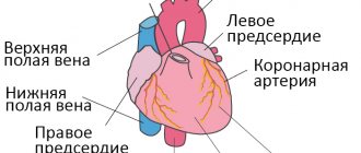
Cardiac muscle exists only in the heart of animals. It is a specialized form of muscle that contracts repeatedly and continuously to circulate blood throughout the body. The heart is a relatively simple organ. Through all the twists and turns and various chambers, there are only three layers. The outer layer, known as the epicardium or visceral pericardium, surrounds the outside of the heart muscle. This helps protect it from contact with other organs. The parietal pericardium attaches to this outer layer, creating a fluid-filled layer that helps lubricate the heart. The inner layer, or endocardium, separates the muscle from the blood it pumps into the chambers of the heart. Between these two sheets lies the heart muscle. The heart muscle is sometimes referred to as the myocardium. This can be seen in the image below.
When we look a little at the heart muscle, we see that it is arranged in layers of cells that are interconnected in a lattice fashion. Where two cells meet, a specialized junction called an intercalated disc locks the two cells in place. While this area appears as a dark disk under a microscope, it is actually a blocking of hundreds of finger-like projections from each cell. These projections have small holes, gap junctions, that can transmit impulse to the connected cells. Between and around these cells are nerves and blood vessels that carry signals and oxygen to the heart muscle.
At the microscopic level, cardiac muscle is very similar to skeletal muscle. Both muscle tissues are cross-striated, which means they appear as dark and light stripes when viewed under a microscope. These groups are created by highly organized sarcomeres. A sarcomere is a bundle of protein fibers that respond to a signal and a contract. In both skeletal and cardiac muscle, these sarcomeres are composed of actin and myosin and are supported by the same proteins. Tropomyosin is a protein that wraps around actin and prevents myosin from binding to it. Troponin is a protein that holds tropomyosin until a signal to contract is received. These proteins are the same in both skeletal and cardiac muscles.
Degrees of chronic pathology
There are several degrees of chronic heart failure. The earlier a problem is identified, the easier it is to treat. In the later stages of the disease, mortality is 50%.
The first stage of chronic insufficiency is characterized by shortness of breath and rapid fatigue after active physical work or sports. Treatment at this time is most effective. Early detection of symptoms of chronic deficiency will not only help to cope with it, but also to determine the true cause.
In the second stage of heart failure, symptoms become more pronounced:
- cyanosis, or blue discoloration of the fingertips, nose and nasolabial folds, lips. The reason is a change in blood composition, insufficient oxygen content.
- With low-intensity loads, typical symptoms appear: shortness of breath, rapid heartbeat, weakness.
- Dry cough not associated with a cold.
The second stage is characterized by damage to the pulmonary circulation, the lungs suffer, so a cough occurs, and the blood is poorly saturated with oxygen.
Another variant of the manifestation of the second stage is damage to the systemic circulation. First of all, swelling appears. They can be hidden: fluid accumulates in body cavities, for example, filling the abdominal cavity. Swelling of the legs is more noticeable. This condition is called dropsy.
In addition, stagnation begins in all organs: liver, kidneys, digestive system, brain. Associated pathologies develop and the functioning of these organs is disrupted.
At the third stage, irreversible changes occur. Usually the functioning of the entire circulatory system is disrupted. Lesion of the small circle is characterized by a strong cough with sputum production, the appearance of pink foam not only when coughing, but also at rest. This is explained by a violation of the permeability of numerous vessels, lymphatic fluid fills the lungs, and edema occurs. The condition is fatal and requires urgent hospitalization. Shortness of breath accompanies a person at rest. Worsens when lying down. Periodically there are attacks of suffocation, accompanied by fear of death. They force the patient into a sitting position with his legs down.
At the same time, all vital organs suffer. Signs of congestive liver damage:
- increase in organ volume;
- formation of sclerotic and connective tissue;
- cirrhosis of the liver.
Congestive kidney damage leads to chronic renal failure. At the same time, the amount of urine excreted decreases, swelling increases and no longer goes away under the influence of medications. Intestinal dysfunction occurs, constipation or diarrhea is possible.
The third stage is irreversible. A person completely loses the ability to be active. Even household chores become beyond his power. Usually the patient is assigned a certain form of disability. The survival rate in this case is 50%. Medicine in this case can only offer supportive therapy.
Important! The third stage of the formation of congestive heart failure occurs as a result of the lack of treatment for the disease. If you consult a doctor in a timely manner in the early stages, you can not only stop the progression of the disease, but also be completely cured.
If you notice the following signs of heart failure, you should immediately contact a cardiologist:
- Shortness of breath with minor physical exertion;
- Blueness of fingers, lips, nose;
- Unreasonable dry cough, sometimes accompanied by sputum production;
- Pain in the chest, under the shoulder blade;
- Swelling of the legs, enlargement of the abdomen, which may be hidden edema.
Mild heart failure responds well to treatment, so a preventive examination by a therapist and cardiologist and the necessary diagnostic methods will help avoid serious consequences.
Cardiac muscle function
As in skeletal muscle, the signal to contract is an action potential. However, for skeletal muscles, this signal usually comes from the somatic, or voluntary, nervous system. The cardiac muscle is controlled by the autonomic nervous system. Cells in your brain and cells embedded in your heart act to release well-timed nerve impulses that signal your heart cells to contract in the correct pattern. Although the source of the signals is different, the signal reception and the rest of the reduction are very similar.
An action potential, or nerve impulse, at the cell surface stimulates a specialized organelle to release calcium ions (Ca2+). This organelle is called the sarcoplasmic reticulum and is derived from the endoplasmic reticulum found in the common cell. Ca2+ ions released into the cytoplasm influence the protein troponin, causing it to release tropomyosin. Tropomyosin changes position and myosin is allowed to attach to actin. Myosin then used the energy stored in ATP molecules to move along the actin filaments and shorten the length of each sarcomere. When the impulse disappears, Ca2+ is rapidly reabsorbed into the sarcoplasmic reticulum. Troponin reattaches to tropomyosin and the heart muscle cells are released. This general process happens every time your heart beats.
Because all the muscle cells work in unison, a force can be exerted in the chambers of the heart. The sheets of cardiac muscle are laid so that they run perpendicular to each other. This creates the effect that when the heart contracts, it does so in multiple directions. The ventricles and atria of the heart are compressed from top to bottom and side to side as these multiple layers of muscle fibers contract. This creates a strong pumping and twisting force in the ventricles, forcing blood throughout the body.
Acute form
Along with chronic, there is an acute form of heart failure. The reasons for its occurrence are:
- myocardial infarction;
- major stroke;
- structural defects of the heart, congenital and acquired;
- hypertension.
A distinctive feature of acute cardiovascular failure is the suddenness of the attack and the absence of the listed stages of development. The person's condition quickly deteriorates and death is possible within a few minutes. The occurrence of pathology is indicated by a sharp deterioration in condition, suffocation, severe cough with foam or red sputum, blue discoloration of the skin, and cold sweat.
In such cases, first aid is to call an ambulance. Then the victim should be taken out into the open air, the collar and other tight clothing should be unbuttoned. The best pose is considered to be sitting, with legs down. A nitroglycerin tablet should be given under the tongue. Repeat the dose every 10 minutes until the ambulance arrives.
Important! In acute heart failure, you should not take a supine position.
quiz
1. Which of the following answers lists components unique to skeletal and cardiac muscle? A. Striated, intercalated discs, branched B. Striated, sarcomeres C. Actin and myosin
Answer to question #1
B is correct. Skeletal muscle and cardiac muscle are similar in cross section. These dark bands are produced by highly organized sarcomeres within. All muscles, even smooth muscle, rely on actin and myosin, as well as a number of other cellular processes that depend on motor proteins. Intercalated discs and arborizations are unique to cardiac muscle tissue.
2. AED (automated external defibrillator) is a device used to restart a stopped heart. The AED pulses with a strong shock of electricity to the heart. Why does it restart the heart? A. Electricity is similar to an action potential B. The jolt causes the brain to work C. Electricity compresses the heart and allows blood to flow
Answer to question No. 2
right. Nerve impulses are essentially electricity. As they travel to your heart muscle from the brain and from the pacemakers in the heart, they can become restless. A stopped heart occurs when these signals are interrupted. An AED essentially starts the heart pumping and resets the pacemaker cells, allowing a steady heartbeat to resume.
3. The cardiac muscle present in the atria of the heart is thinner than that in the ventricles. Why is this so? A. The atria do not contract B. The atria are part of the blood vessels C. The atria function more like pressure tanks, holding applied pressure
Answer to question number 3
C is correct. The atria contract and pump blood, but in a different way. Both atria take in blood and must keep it under pressure to quickly move it to the next chamber or vessel. To do this, the force must be given by the muscles. However, the force is less than that produced by the ventricular muscles, which are used to increase pressure and push blood.
Classification
There is a classification of CHF according to the stages of development of the disease (N.D. Strazhesko, V.Kh. Vasilenko):
Stage I (initial) - signs of deficiency appear only during physical activity, there are no symptoms at rest.
Stage II is divided into two periods:
- period A – clinically pronounced disorder of the right or left side of the heart, congestion in the pulmonary or systemic circulation, shortness of breath and symptoms occur with little physical effort;
- period B – stagnation in both circles of blood circulation (manifested by shortness of breath, swelling), performance is sharply reduced.
Stage III (final) - changes in the structure of organs and tissues, due to impaired blood supply and trophism, shortness of breath at rest.
The severity of the disease is determined by the functional class (FC), which shows how limited the patient’s physical activity is.
- I FC - no restrictions on physical activity. Normal physical activity does not cause excessive shortness of breath, fatigue, or palpitations;
- FC II - minor limitation in physical activity. Feel comfortable at rest, but normal physical activity causes shortness of breath, fatigue, or palpitations;
- III FC is a clear limitation of physical activity. Feeling comfortable at rest but doing less physical activity than usual causes excessive shortness of breath, fatigue, or palpitations;
- IV FC - inability to perform any physical activity without discomfort. Symptoms may be present at rest. With any physical activity, discomfort increases.
The disease should not be allowed to develop; timely diagnosis will help prevent the occurrence of pathology. Our cardiology center of the Federal Scientific and Clinical Center of the Federal Medical and Biological Agency invites you to undergo a comprehensive heart examination. Timely identification of the cause of heart failure and its treatment will help maintain your quality of life.