Sometimes we experience a special kind of discomfort - it seems that either our head or our eyes hurt. This pain occurs in the sinus area or at the back of the eye. Sometimes it is pulsating, sometimes it is constant. This condition frightens us very much and we want to know what causes this pain? What can be done to alleviate it? Could there be something wrong with my vision?
Let's first answer the last question.
Scientists from the American Academy of Ophthalmology defined "eye pain" as "physical discomfort caused by an eye or other disease." But scientists emphasize that “the location of the pain does not necessarily indicate the cause of the pain.”
In most cases, the cause of a headache felt in the eyes may be hidden in a completely different place. We feel pain in this particular location because of a network of interconnected sensory nerves that penetrate all tissues of the body.
“Almost every pain-sensitive structure in the head transmits the sensation of pain to the eye area,” says Dr. Mark W. Green, MD, professor of neurology at the Icahn School of Medicine at Mount Sinai Hospital in New York City. “Just because the eye hurts does not mean that the problem is in the eye itself. In fact, this happens quite rarely."
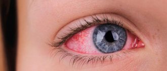
Green advises to remember one useful rule: if the white part of the eye (sclera) is not red, and there are no complaints about vision - a blurred or distorted picture, it is very unlikely that the headache is associated with the eyes themselves.
Causes
The most common reasons:
- Glaucoma. This disease is associated with increased intraocular pressure. The development of pathology is accompanied by pressing pain in the eye. The list of typical symptoms also includes fog before the eyes, a sharp decrease in visual acuity, and sometimes nausea. Glaucoma progresses quickly, so you should consult an ophthalmologist as early as possible.
- Neuritis is inflammation of a nerve. In the case of pain in the eye, the pathological process affects the optic nerve. Discomfort increases with eye movement. More often, neuritis develops against the background of infection.
- Inflammation of the iris. Painful sensations are usually accompanied by photophobia.
- Migraine. The pain is accompanied by loss of part of the visual field. After the attack ends, vision is restored.
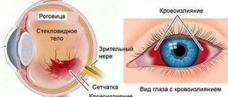
- Eye injury.
There are also reasons not related to the function of the eye: for example, sinusitis and increased intracranial pressure.
Sinusitis is an inflammation of the paranasal sinuses. If it affects the upper sinuses, the headache may spread to the eyes. As a rule, the development of the disease is accompanied by an increase in temperature and a deterioration in general well-being.
Increased intracranial pressure can be chronic (due to vascular diseases), as well as acute (occurs due to head injuries or the development of infectious diseases).
In some cases, pressing pain in the eyes may appear due to visual fatigue. This often happens after working at the computer for a long time or with small print, or when working in insufficient lighting. In such cases, it is necessary to ensure working conditions that comply with visual hygiene recommendations. Set up a comfortable computer brightness, color scheme, and ensure adequate lighting of the workplace.
When to see a doctor if you have eye pain
If your eyes hurt, you cannot do without medical advice. This is necessary to make a diagnosis and begin proper treatment.
Sometimes the cause of pain in the eye is immediately clear (for example, when pain occurs as a result of a foreign body entering the eye), but in most cases it is necessary for the diagnosis to be made by a specialist - an ophthalmologist.
It should be remembered that pain in the eye can mean the threat of a sharp deterioration or even loss of vision.
You should definitely consult a doctor if:
- foreign body getting into the eye;
- bruise in the eye area;
- dull pain in the eye lasting more than two days;
- sharp, piercing pain in the eye;
- pain in the eye accompanied by blurred vision;
- pain in the eye, accompanied by symptoms such as nausea, vomiting, increased sensitivity to light.
If a child’s eyes hurt, he should definitely be shown to a pediatric ophthalmologist.
Intraocular hypertension and glaucoma
Intraocular hypertension is a pathological increase in intraocular pressure. The pressure that the intraocular fluid and vitreous body exert on the membranes of the eyeball. It can only be measured at an appointment with an ophthalmologist.
At CELT you can get advice from an ophthalmologist.
- Initial consultation – 3,900
- Repeated consultation – 2,000
Make an appointment
Temporary increases in blood pressure can be caused by drinking alcohol, smoking, or eye strain. If the sensation of pain does not go away for a long time, then this is a symptom of glaucoma - one of the most common and dangerous diseases. It manifests itself as a decrease in visual acuity and over time can lead to complete blindness.
The insidiousness of glaucoma is that it develops gradually: in the first stages, a slight pressing pain may be the only symptom. The disease develops at any age, but most often in older people.
Iphthalmotonus standard
The constant accepted norm of pressure inside the eyes in an adult lies in the range from 10 to 22 mm Hg. Art. (on average, for most people these indicators are 15-17) and is characterized by constancy. During one day, the pressure fluctuates only within 3-4 mmHg. Art. — in the morning it is usually higher, decreasing slightly in the evening. Consistency is necessary for the stable functioning of the inner membranes of the eye, especially the retina, and depends on a number of physiological mechanisms responsible for regulating the filling of the vessels inside the eye with blood, the influx and outflow of aqueous humor. In addition, normal ophthalmotonus is important for maintaining the optical properties of the retina.
Treatment
Before prescribing treatment, the ophthalmologist will conduct an examination and make a diagnosis. As a rule, the main stages are measuring intraocular pressure, examining the fundus, and examining with a slit lamp. This set of examinations allows you to identify the main and most common eye diseases.
In the case of glaucoma, special medications will need to be dripped into the eyes to reduce intraocular pressure. In most cases, you will need to prepare for surgery - glaucoma does not respond to conservative treatment.
If the pain is caused by an inflammatory disease, then you will need to identify the pathogen and start taking appropriate medications: antibacterial or antiviral.
Treatment for pain caused by fatigue involves following basic visual hygiene recommendations. The patient will have to be more attentive to his health. This pathology is most often found among office workers, and it has even been called visual fatigue syndrome. An ophthalmologist may prescribe eye exercises and recommend coming for preventive examinations.
Prevention of increased eye pressure
The pathology is usually caused by age-related changes, so all persons over 40-45 years of age must follow certain rules and precautions, namely:
- reduce the load on the visual organs, reduce the time spent in front of the TV screen and computer monitor;
- periodically perform eye exercises;
- enrich the menu with fresh vegetables and fruits, regularly include blueberries, carrots, fish and seafood in the diet;
- spend enough time outdoors, take walks, play sports;
- monitor blood pressure levels;
- in sunny times, protect your eyes with sunglasses;
- give up bad habits, follow the rules of proper sleep.
The main rule of prevention is to visit an ophthalmologist at least once a year upon reaching the age of 45 for a general examination, determining visual acuity and measuring eye pressure. If the indicators are elevated, visits to the ophthalmologist should be more regular – once every three months. Following the rules will help maintain sharp vision for many years.
Our services in ophthalmology
The administration of CELT JSC regularly updates the price list posted on the clinic’s website. However, in order to avoid possible misunderstandings, we ask you to clarify the cost of services by phone: +7
| Service name | Price in rubles |
| Pneumotonometry | 500 |
| Tonometry (according to Maklakov) | 1 000 |
| Goldmann tonometry (Icare tonometer) | 1 000 |
| MSCT of orbits | 7 000 |
All services
Make an appointment through the application or by calling +7 +7 We work every day:
- Monday—Friday: 8.00—20.00
- Saturday: 8.00–18.00
- Sunday is a day off
The nearest metro and MCC stations to the clinic:
- Highway of Enthusiasts or Perovo
- Partisan
- Enthusiast Highway
Driving directions
What pressure values indicate the presence of pathology?
In a situation where IOP has elevated values, this is a sign to sound the alarm, since its increase indicates the presence of glaucoma at different stages. Here is a table from which you can find out how dangerous you are from this disease 1. A value from 10 to 22 mm Hg is considered normal. Art. (usually 15-17). 2. A pressure of 22 to 25 mmHg may indicate primary signs of glaucoma, in which case a thorough examination should be performed. 3. The figure 25-27 mmHg most likely confirms the presence of the initial stage of glaucoma. 4. When the ophthalmotonus level is 27-30 units, we can say that glaucoma is actively developing. 5. IOP value is above 30 mm Hg. Art. means a severe degree of development of the disease.
We remind you that the change in pressure inside the eyes during the day should not exceed the norm of 3-4 mm Hg. Art. - higher in the morning, lower in the evening. What factors may prompt you to contact a specialist to check your eye pressure?
Diagnostics
An acute attack of increased IOP is diagnosed based on patient complaints, concomitant symptoms, changes in IOP, and biomicroscopy results. To determine intraocular pressure, you can use the palpation method or non-contact tonometry.
In diagnostic measures, it is important to differentiate an acute attack of increased IOP from other conditions:
- Pupillary block against the background of secondary glaucoma (phacomorphic with the presence of a diffusely cloudy, slightly pearlescent lens in the lumen of the pupil and a gradual decrease in vision, phacotopic with entrapment of the lens); bombardment of the iris (when it bulges like a sail with circular posterior synechia).
- Blocking the angle of the anterior chamber against the background of secondary glaucoma (neoplastic glaucoma with an uneven anterior chamber, swelling of the iris or ciliary body, phacotopic glaucoma with dislocation of the lens in the anterior chamber of the eye).
- Glaucomocyclic crisis syndrome, accompanied by mild pain and mild corneal edema.
- Eye diseases manifested by “red eye” syndrome (acute conjunctivitis without decreased vision, increased IOP and iris edema; acute iridocyclitis with increased IOP, precipitates on the corneal endothelium, deep injection, etc.).
- Eye injury.
- Hypertensive crisis.
Risk factors that may cause increased IOP
Increased pressure inside the eyeball can also be caused by some common human diseases. That is why the doctor collects in detail all the data about your lifestyle, hereditary diseases, and even what activities you like to do in order to accurately establish the clinical picture. For example, an increase in ophthalmotonus can provoke disturbances in the functioning of internal systems or organs.
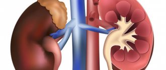
1. Diabetes mellitus. This is a group of endocrine diseases characterized by increased blood sugar, as well as the inability of the pancreas to produce the hormone insulin. The body experiences constant jumps in sugar from normal levels to high or, conversely, low. In this regard, problems begin with the condition of the blood vessels, causing an increase in arterial and intraocular pressure. 2. Vegetovascular dystonia. Persons suffering from VSD may complain of interruptions in heart function, decreases or surges in blood pressure, as well as headaches and dizziness. At the same time, the functioning of many body systems is disrupted. VSD can also cause an increase in ophthalmotonus. 3. Diseases of the cardiovascular system. Their list is quite extensive. These may be atherosclerosis, congenital heart disease, impaired elasticity of blood vessels, varicose veins and many other ailments affecting the normal functioning of the heart and blood vessels. 4. Kidney diseases. Severe and long-term glomerulonephritis, as well as a wrinkled kidney, can lead to retinal lesions and increased eye pressure. 5. The presence of uveitis, astigmatism, farsightedness and some other disorders can also cause an increase in IOP. 6. Post-traumatic glaucoma. Occurs after mechanical or chemical damage to the organs of vision. 7. Spending a long time in front of a computer monitor. Research by scientists has long confirmed the fact that prolonged exposure to a screen and intense visual work can provoke an increase in ophthalmotonus. The eyes are under constant tension, the person begins to blink several times less often, a headache may occur and the IOP may increase.
We have listed several factors that can cause increased intraocular pressure. In addition, people over 40 years of age are at risk, since the older the age, the more common the disease. However, increased eye pressure can also occur in children, so parents should be vigilant. If he has any visual impairments (myopia, astigmatism, lazy eye syndrome, etc.), then this is a reason to keep eye pressure readings in children under control.
Treatment of vision problems caused by hypertension
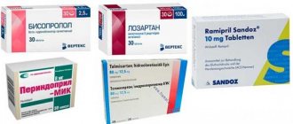
If a person manages to start treatment in a timely manner and normalize blood pressure, then most retinal problems can be avoided. This requires regular ophthalmoscopic examinations at your eye doctor's office to evaluate the effects of high blood pressure on your vision. The condition of the arteries and arterioles is assessed only by examining the fundus. As a rule, normalizing pressure to normal levels is sufficient to eliminate the signs and consequences of hypertensive retinopathy. To do this, the patient undergoes a course of drug treatment aimed at taking vasodilators. Sometimes systematic medication is required to maintain normal blood pressure (often after 40 years).
If pathological processes are detected in the organs of vision (with advanced hypertension), an additional course of ophthalmological treatment is necessary.
The sooner eye pathology is detected and the sooner a course of rehabilitation is selected, the more favorable the outcome. Ophthalmologists say that people with hypertension have every chance of maintaining 100% vision, subject to systematic examination and compliance with recommendations.
The Ochkov.Net website presents a large assortment of contact lenses from world brands. Correctly selected optical products will allow you to enjoy a clear perception of the world and comfortable wearing.