In this article we will tell you:
- Causes of eye hemorrhage
- Types and differences of eye hemorrhage
- Symptoms of eye hemorrhage
- Treatment of eye hemorrhage
- Bottom line
Bleeding in the eye may indicate serious problems with the organ of vision. Many people ignore the disease, hoping that the hematoma will resolve on its own. The danger lies in the fact that hemorrhage can be a consequence of diseases of both the organ of vision and the body as a whole. Therefore, if the hematoma occupies a significant space, spreads throughout the eye, causing pain, other unpleasant phenomena, increased tearing or a feeling of pressure, blurred vision, you should consult an ophthalmologist as soon as possible.
Causes of eye hemorrhage
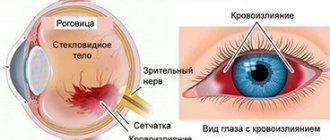
Hematomas appear as a result of a violation of the integrity of the vessels of the eyeball and arise for various reasons. Moreover, bleeding can affect various formations: sclera, conjunctiva, retina, vitreous body.
Among the most common causes of hemorrhage in the eye are:
- A sleepless night. Sleep is essential for the entire body, including the eyes. During sleep, the eyelids close, the membrane is protected by tear fluid. With a lack of sleep, blood vessels in the sclera often burst, causing itching or pain. Such hemorrhages are safe for vision.
- Dry eye syndrome. Accompanied by dryness, pain, and minor hemorrhages. Manifests itself with vitamin A deficiency, taking oral contraceptives or diseases of the lacrimal glands (blockage, autoimmune lesions of Sjögren's disease).
- Increased intracranial pressure. An increase in blood pressure during hypertensive crises can cause one of the most dangerous hemorrhages - in the retina. In this case, patients experience blurred vision.
- Infectious diseases (leptospirosis, hepatitis). Leptospirosis can cause hematomas in the conjunctiva and sclera. With hepatitis, liver function is impaired - regulation of blood clotting, synthesis of coagulation factors.
- Diseases of the liver and pancreas.
- Inflammatory processes: conjunctivitis, iridocyclitis, episcleritis, allergies.
- Taking medications that reduce blood clotting (anticoagulants, antiplatelet agents).
- Injuries or surgeries, childbirth. During a difficult birth, there is a risk of retinal detachment and bleeding.
- Vascular damage in rheumatoid diseases (vasculitis).
- Diabetes.
- Vitamin K deficiency, calcium deficiency, hemophilia.
If the cause is vascular disease, then hemorrhages in the eye appear as the disease progresses. At the first stages they are small, but over time more and more vessels burst and the hematomas can be quite large. This can often be observed in patients suffering from atherosclerosis and hypertension. But it is not always the disease that causes hemorrhage in the eye. It can be triggered by heavy physical activity, sports, childbirth in women, and even a severe cough. Whatever the cause of this phenomenon, during hemorrhage you should contact an ophthalmologist as quickly as possible.
Where can you buy Emoxipin in the regions of the Russian Federation
Emoxipine is available in the regions of the Russian Federation:
- FarmLand pharmacy (Tyumen) - 213.00 Russian rubles;
- online pharmacy "April" Krasnodar - regular price 316 rubles, but if you have a club card 217.00;
- online platform "Zdravcity" - 244.00 (Arkhangelsk region);
- “Planet of Health” - 226 Russian rubles 00 kopecks. (in the Tomsk region);
- MAGNIT-pharmacy - 231 Russian rubles.
Also, the manufacturer points out that in some pharmacies and online stores there is an o in the description - in this case, you will not be able to buy the product without a doctor’s permission. The drug is produced by a domestic pharmaceutical company and is therefore available even in isolated regions. Using an online application, you can order Emoxipin to the nearest post office or have it delivered by courier to your address.
When ordering drops, do not forget to look at the description, expiration date and manufacturer. After receiving your order, immediately check the integrity of the packaging.
Types and differences of eye hemorrhage
Depending on the location of the bleeding, there are several types of disease:
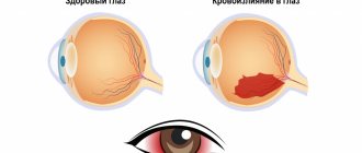
- Retinal hemorrhage is the most dangerous type of disease. After all, the retina is the shell of the eye that perceives light and transmits impulses further to the brain, thanks to which we see images. Blood not only prevents light from reaching the retina, but also has a toxic effect on its structure and function. All this leads to significant, sometimes irreversible loss of vision. Such hemorrhages are the most difficult to treat.
- Hemorrhage into the sclera is the most common type of disease. Its symptoms are pronounced. The cause may be injuries, pressure surges, fragility of blood vessels. Such hematomas are superficial, usually do not pose a threat and go away without intervention within a few days.
- Hemorrhage into the eye cavity is a case that requires immediate examination by a doctor. May manifest as slight blurred vision. The patient often sees a “cobweb” before his eyes. If nothing is done, this can lead to significant vision loss. A small hemorrhage in the eye cavity can resolve on its own, but a serious hematoma requires medical or surgical treatment to restore normal functioning of the organ of vision.
Interaction with other drugs
The drug Emoxipin is compatible with all medications, except eye drops. It is prohibited to use several types of funds at the same time. Otherwise, the manufacturer cannot be held responsible for the absence of side effects and therapeutic effect stated in the instructions for use.
If the ophthalmologist prescribed additional eye drops, then it is imperative to fulfill the condition - Emokispin is instilled last, maintaining an interval of at least 15 minutes.
Symptoms of eye hemorrhage
The disease manifests itself differently, depending on which part of the eye the hemorrhage occurs. Regardless of how much blood leaks into the retina, the symptoms of this disease are not always pronounced.
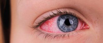
When hemorrhage occurs, the eye becomes red and other symptoms appear:
- The patient's vision deteriorates and the sharpness of perception decreases. The picture becomes less clear, dark spots or “spots” appear before the eyes. If the bleeding is significant, the consequences can be very serious, including loss of vision.
- With hyphema (hemorrhage in the anterior chamber of the eye), in most cases there is no loss of quality of vision. But with heavy bleeding, the pupil may close. This leads to limited vision. After complete removal of the hematoma, it is restored.
- If there is a significant leakage of blood into the vitreous body, the consequences can be serious, including loss of vision. Patients diagnosed with hemophthalmos complain of the appearance of light flashes or dark spots in front of their eyes that constantly move.
- In some cases, if the disease is caused by the development of vasculitis, symptoms such as bulging eyes (exophthalmos) occur. The motor functions of the eye are impaired due to the fact that the eyeball moves forward.
To diagnose the disease, an ophthalmologist examines the eye and assesses the nature and extent of hemorrhage. An ultrasound examination is also performed to determine the degree of hemorrhage and prescribe effective treatment.
Release form
Emoquispin is available in the form of drops in a concentration of 1%. A 5 ml glass bottle is tightly closed with an aluminum dropper cap and sealed with a rubber stopper sealed around the edges. Packaged in a cardboard box with an image of an eye on a white background, which indicates the expiration date and composition of the medicinal product. Comes with instructions in Russian. There is a label attached to the bottle. The solution is transparent or with a slight yellowish tint, without a pronounced odor.
You can order eye drops on the website https://emoxypin.ru/
Treatment of eye hemorrhage
Since the causes of hemorrhage can be different, treatment is also prescribed individually, depending on the factors that caused it. Of course, the first symptoms require immediate consultation with an ophthalmologist. Depending on the clinical case and complexity of the disease, the patient may be prescribed hospital treatment. Bed rest is often recommended. The main goal of treatment is to stop the bleeding, and after that, therapy is carried out to resolve blood clots.
- If the patient has a slight hemorrhage caused by rupture of blood vessels, after some time the hematoma decreases (several hours), this indicates that the damage is not severe and the hematoma will quickly go away. This usually happens if a blood vessel bursts due to overexertion (for example, playing sports). Such hemorrhage does not require special treatment.
- If the hemorrhage is small and resolves on its own after a few hours, but appears regularly, it is recommended to consult a doctor, since there is a problem with the organs of vision or blood vessels. If left untreated, it will only get worse.
- Eye bleeding should not be taken lightly as it can cause complications. Blood cells that break down produce toxic substances. In the early stages of hemorrhage they can damage the cornea, and in the later stages they can lead to the development of retinal detachment.
It is not recommended to self-medicate and use traditional methods. Various rinses, lotions and other traditional medicine tips can aggravate the problem. Before starting treatment, you need to find out the cause of the hemorrhage and only after that the appropriate therapy is selected.
Treatment is often limited to ordinary drops. But they must be selected correctly for a specific clinical case to ensure maximum effect.
Where can you buy Emoxipin in Moscow
Here are the best deals for buying Emoxipin eye drops.
Moscow
- online store apteka.ru - 263.00 with free delivery to 892 pharmacies in Moscow;
- online pharmacy stolichki.ru - promotional price 221.00, subject to ordering on the website. Reservations for a day are possible;
- network "Main Pharmacy" - 255.00 rubles;
- "ABC-pharmacy" - 278.00 rubles;
- 24-hour pharmacy "NEOPHARM" - 358.00.
Bottom line
Bleeding in the eye can indicate quite serious illnesses, so it should not be ignored. It is recommended to immediately contact an ophthalmologist who will give advice and provide professional medical assistance. This will minimize the risk of consequences that may arise due to untimely treatment or attempts to recover using traditional methods. Therapy is selected depending on the nature and severity of the hemorrhage. The doctor selects medications individually, depending on the clinical case.
Precautionary measures
When using Emoxipin drops, no allergic reactions or side effects were identified. In reviews of eye drops, users note the affordable price, but some complain about the lack of effect. The manufacturer warns against purchasing the medicine from hand, not from pharmacy chains or spontaneous retail outlets.
Improper storage, transportation and counterfeit goods will have a detrimental effect on the organs of vision. Also pay attention to the prices - too high or low should raise doubts about the originality of the drug.
You can buy the medicine throughout the Russian Federation and the Republic of Belarus in the public domain without prior ordering from official pharmacy chains.
Another important indicator that must be checked is the release date, expiration date, series and lot number. They must match on the cardboard packaging and glass bottle.
Do not use a drug you are not sure about.
Cost of some services for hemophthalmia:
| № | Service name | Price in rubles | Make an appointment |
| 2003004 | Repeated appointment with the leading surgeon | 1400 | Sign up |
| 2003006 | Repeated appointment with an ophthalmologist | 1500 | Sign up |
| 2003005 | Initial appointment with an ophthalmologist | 2500 | Sign up |
| 2011039 | Vitrectomy for uncomplicated hemophthalmos or vitreous opacities of the second category | 64500 | Sign up |
| 2011038 | Vitrectomy for uncomplicated hemophthalmia or vitreous opacities of the first category | 54000 | Sign up |
| 2011040 | Vitrectomy for uncomplicated hemophthalmia or vitreous opacities of the third category | 78600 | Sign up |
Making an appointment Today: 13 registered
Indications for use
It is worth considering that eye drops can be used for the following reasons:
- mechanical impact that led to injury;
- injuries of any etiology;
- rupture of the ocular vessels;
- diseases of hematological etiology;
- hypertension;
- atherosclerosis;
- diabetes mellitus and other diseases of the endocrine system;
- blood diseases and blood clotting disorders;
- ophthalmological diseases – iritis, uveitis;
- pathological changes in the retina.
Hemorrhage that occurs as a result of injury requires immediate medical examination. Such manifestations should not be ignored. With the help of special diagnostics, the doctor will be able to determine the etiology of such lesions.
Vitamin preparations for the prevention of vascular disorders
The human visual organs in the modern world experience enormous loads every day. Most people, spending time at the computer during the day, switch to watching TV or picking up a mobile gadget in the evening. Discomfort in the eyes, dryness, burning, blurry images have long become common symptoms for many, but few pay attention to them, attributing them to banal fatigue. Meanwhile, such signs indicate chronic fatigue, which leads to the development of serious diseases of the retina or optic nerve, and, as a consequence, to complete loss of vision. That is why, for prevention purposes, you need to use strengthening eye drops.
Eyelid injury
Superficial wounds of the eyelids damage only the skin and muscle layer, while penetrating wounds damage all layers of the eyelid. Since the skin of the eyelids is characterized by great extensibility and looseness of the subcutaneous tissue, swelling and hemorrhages appear here very early. The skin of the eyelids becomes tense, and the color ranges from dark blue to purple. The swelling may spread to the eyelid of the other eye.
A small external eyelid wound may conceal massive internal damage that requires immediate attention from an ophthalmologist.
Based on the appearance of the wound, one cannot draw a conclusion about the degree of damage to deeper tissues. A small external eyelid wound may conceal massive internal damage that requires immediate attention from an ophthalmologist.
If the wound is located vertically, then its edges gape due to rupture of the transverse muscle fibers. When the eyelid is injured, subcutaneous emphysema can form. This indicates a violation of the integrity of the bones of the paranasal sinuses.
Minor eyelid wounds end favorably, but if the wound becomes infected, scarring and deformation of the eyelid are possible.
If eyelid wounds become infected, scars can form, which in turn leads to cicatricial eversion of the eyelid. If the muscle that lifts the upper eyelid is damaged, ptosis of traumatic origin may appear. If you suspect the introduction of a foreign body into the tissue of the eyelids, orbit or lacrimal organs, you need to take an x-ray of the orbit.
Treatment
First aid for a wound of the eyelid - the skin around the wound should be treated with an antiseptic (miramistin, ethacridine, picloxidine, boric acid), and if the wound is contaminated, it should be cleaned and rinsed with a solution of hydrogen peroxide, and then apply an aseptic bandage.
If the eyelid wound is small and horizontal, then it does not require surgical intervention. If the wound gapes, surgical help is necessary. If it is impossible to carry out primary surgical treatment in a timely manner, it must be carried out later - even after a few days and in the absence of signs of suppuration. When treating eyelid wounds, it is necessary to take care of the damaged tissues and prevent their excision. If the wounds of the eyelids are through, then suturing is used “in two levels”: “first” - sutures on the conjunctiva and cartilage, and “second” - sutures on the skin of the eyelid.
Foreign body of the conjunctiva
A foreign body of the conjunctiva (usually small particles of earth, coal, stone or metal, grains of sand, hairs of cereal plants) can remain on the surface or penetrate into it, violating the integrity of the epithelial cover, in the latter case an inflammatory infiltrate is formed.
In the clinic, symptoms of eye irritation predominate: photophobia, pain, blepharospasm and, naturally, the sensation of the presence of a foreign body. During examination or biomicroscopy, you can see a foreign body on or inside the conjunctiva.
A foreign body can move due to blinking reflex movements of the eye. Often it lingers in the groove of the eyelid, on its inner surface. Then, when the eyelid is everted, an inflammatory infiltrate with papillae in the center of which is a foreign body (hair of cereal plants, etc.) is discovered.
Often, damage to the conjunctiva leads to subconjunctival hemorrhage. This can happen due to trivial rubbing of the eye. A blood-red spot appears on the sclera. Despite the “terrible appearance,” such hemorrhage is not dangerous: it resolves on its own in two to three weeks and does not lead to visual impairment. But sometimes a perforation of the eyeball can be missed behind a bloody spot. Therefore, at the slightest suspicion, the victim should be sent for examination to an ophthalmologist.
Treatment
If it is located superficially, the foreign particle can be easily removed with a cotton swab soaked in a disinfectant solution. If penetration occurs deep into the tissues of the eye, you need to drip a solution of tetracaine (0.5%) into the eye and then remove it with a special needle or tweezers. If it is impossible to remove in this way, a section of the conjunctiva is excised along with the foreign particle.
Large fragments of coal, stone, glass, metal that cause severe irritation should be removed. And small particles of sand and stone that do not cause irritation do not need to be removed; they will “come out” on their own thanks to the blinking movements of the eye.
General information about the retina and the current state of retinology
The retina is a layer of special light-sensitive receptor cells in the fundus of the eye that perceives the image focused by the eye. Next, it is transmitted to a special nerve, which, with its terminal disc, enters the most developed macular region of the retina () and serves as a conductor of the visual signal from the neuroepithelium to the visual zones of the cerebral cortex, where, through the processing and fusion of two separate signals, a single binocular image is built. Normal vision implies complete safety, consistency and correct “tuning” of all elements of the optical conduction tract.
At the same time, the human visual system is evolutionarily protected not as reliably and rigidly as some other systems of the body, and has significantly fewer resources for regeneration and damage compensation. Therefore, when the functional failure of ocular structures and tissues is caused by injury or disease, a person most often has to replace them - either with donor material (for example, corneal transplantation) or with synthetic material (for example, the well-known implantation of an artificial lens). As for the retina, firstly, it plays a completely exceptional role in the visual system, and secondly, it is too complex for the current stage of technological development, i.e. cannot yet be replaced by, say, an electronic transceiver device at the neural level.
Donor retinal transplantation today is also inaccessible, has not been sufficiently studied statistically and, in fact, remains only an experimental procedure, very expensive and complex. The first relatively successful attempts at such operations were made quite recently - in 2008 in Europe and then in 2014 in Japan, where the world's first retinal transplant grown from stem cells was performed. Regarding donor retinal transplants, the relativity of success is emphasized - vision is only partially restored, the stability of the effect is unknown due to the extremely short period and volume of observations. Reports on the functioning of the brainstem retina are generally distinguished by restraint and stinginess (“the patient’s field of vision has become brighter”).
Conservative, drug treatment of the retina by itself is ineffective and is prescribed mainly as additional or maintenance therapy. In fact, the only and most proven method of treating an affected, damaged, degenerating and/or detached retina is ophthalmic surgery (and timeliness is the decisive factor), especially in combination with excimer laser coagulation methods - in this case, the probability of success is immeasurably higher and is, according to various sources, from 70% to 90%.
From this brief review, a logical conclusion follows that folk wisdom came to a long time ago: the retina of the eye should be protected “like the apple of the eye,” and in case of the slightest problem, seek qualified help immediately. Otherwise, the risk of a pronounced decrease or complete loss of vision is very high.
How do strengthening eye drops work?
When blood vessels are weakened, blood circulation is disrupted and, accordingly, the flow of oxygen to the tissues of the eyes is disrupted. Metabolic processes deteriorate, the quality of vision decreases. As a result, the capillaries weaken even more and, as soon as they become overfilled with blood, they begin to burst, and visible hemorrhages appear on the whites of the eyes. The active components of medicinal liquids help nourish all parts of the visual organs: lens, optic nerve, retina. They help strengthen damaged blood vessels and restore normal blood circulation. This result is achieved with the help of vitamins and minerals that are included in ophthalmic preparations in this category.