Intraocular pressure (IOP) is the pressure of the fluid that fills the eye. Obviously, ideally it should be normal. Most people are afraid of high blood pressure, but low blood pressure is also bad: the eye cannot maintain its correct shape, and the lack of fluid leads to deterioration in the cleansing of neighboring tissues.
Excessive pressure causes increased inflow or impaired outflow of fluid.
Among the reasons for this deviation from the norm are:
- anatomical features
- heredity
- age-related changes
- problems with the cardiovascular system
If elevated intraocular pressure is not normalized, it can lead to a serious disease - glaucoma. The disease is insidious, in advanced cases it leads to complete and irreversible blindness.
Normal blood pressure is considered to be between 10 and 20 millimeters of mercury (mmHg).
The norm is relative, as, for example, with blood pressure. It is impossible to start from dry population statistics and use them in a stereotyped manner in the treatment of a specific person. There are people whose blood pressure is 20 mm Hg. Art. no glaucoma, but 50% of people with diagnosed glaucoma have pressure below 21 mmHg. Art. The disease depends not only on pressure, but IOP is the only factor that we can influence, lowering it with drops or laser.
How to measure eye pressure
Methods for measuring IOP are divided into two groups: contact (when there is contact with the cornea of the eye) and non-contact.
- Palpation is the oldest method, requiring highly qualified doctors, experience and special sensitivity of the fingertips. IOP was considered significantly increased if the eyeball felt hard to the touch, and if it was soft, the pressure was normal. Of course, this type of diagnosis (even if you have the necessary skills) does not provide accuracy in determining the degree of deviation from the norm, so it is not used now.
- The tonometric method involves contacting the device with the cornea. For this reason, the measurement is performed under local anesthesia. To visualize the result, dyes are used. The line of contact is clearly visible. The IOP value is calculated using special tables.
- Non-contact is a modern and painless method that makes it possible to accurately measure eye pressure (used in our clinic). Non-contact tonometers have left contact methods behind, because:
- anesthesia and dyes are not needed;
- the possibility of corneal damage, infection or discomfort is excluded;
- the procedure does not take time - IOP is measured in a few seconds;
- all necessary measurements are provided automatically.
Non-contact tonometry is suitable for children and patients with increased sensitivity.
- Icare rebound tonometry is our main method. Indispensable for round-the-clock monitoring of intraocular pressure. Previously, for this control, people were forced to go to the hospital, but now, thanks to the latest technology - the Icare Home tonometer, patients with glaucoma can measure their pressure at home, even every 10 minutes. The rhythm of life remains unchanged, which enhances the veracity of the data obtained. In the hospital, the patient simply lies and does nothing, and the pressure decreases accordingly.
By the way, the demand for this device is so great that we almost never see it in the clinic.
The rebound principle of measurement involves touching the cornea with the lightest (disposable!) sensor literally for an instant. It is a safe, painless and hygienic procedure.
Anesthesia, drops or special skills are not needed for this tonometry.
Measurement according to Maklakov
- The patient lies down on the couch, and the doctor administers anesthesia by instilling several drops of dicaine into each eye in turn.
- Then the head is fixed and asked to look at one point.
- A small weight treated with special marking paint is carefully lowered onto the open eye, under the pressure of which the eyeball should be slightly deformed.
- Now the weight is lowered onto a sheet of paper to see how much paint is left on it. Intraocular pressure is determined by the intensity of the imprint.
- The procedure is repeated again in both eyes to avoid the possibility of misinterpretation.
Naturally, some amount of paint from the load will remain on the surface of the eyeball, but it will quickly be washed away with tears. Instead of weights, ophthalmologists sometimes use a portable device that looks like a ballpoint pen. They also apply pressure on the eye, having previously treated the eyeball with an anesthetic.
This method also has an alternative – non-contact tonometry. No weights are placed on the eye, but instead a controlled air flow is used. Many patients find this method more acceptable, but in reality it is rarely used - it is not as accurate.
Important! In patients with glaucoma, eye pressure fluctuates much more noticeably throughout the day than in healthy people. Having such suspicions, the doctor may ask the patient to come to the clinic several times throughout the day. To ensure the accuracy of the diagnosis, you need to measure your blood pressure at least three times before lunch, and the same number in the evening.
How to measure the pressure inside the eye yourself?
Without the help of a doctor and special equipment, it is impossible to determine with what force the fluid secreted by the ciliary body presses on the eye. Nevertheless, it is possible to understand that blood pressure is greatly increased, and ophthalmologists recommend that everyone master this simple technique.
Close your eyes and relax. Now gently press your index finger on the eyeball. You should feel an elastic ball that gives in to your pressure - this is normal. If the eye is very hard and practically does not deform, the IOP level is most likely elevated and it is better to consult a specialist to make sure that its value is not critical
Increased eye pressure: symptoms
Direct symptoms are not observed until the pressure exceeds a very high level - 40 or 50 mm. rt. Art. It is then that the patient already feels acute discomfort. This could be a headache, nausea or some other ailment. With average elevation, the eye does not turn red, there is no pain, and it does not affect vision in any way. You can't determine anything at home.
Without regular preventative examinations, it is easy to miss the onset of glaucoma, which is why it has been dubbed the “silent thief of vision.”
Statistics by age
Anyone can get sick, regardless of age. Let us only note the fact that children are less susceptible to this disease than adolescents and adults.
- If you are over 65 years old, you have a 20% chance of having glaucoma.
- From 70 years old – 30%
- From 85 years old – 40%
As we age, the natural drainage of fluid from the eyes becomes worse. The process can be aggravated by cataracts, in which the thickened lens begins to take up more space. As sad as it is, aging is a factor that increases the likelihood of developing glaucoma.
Prevention of irreversible consequences of elevated IOP is an examination by an ophthalmologist, especially after reaching 40 years of age. Ideally, once every 6 months, in extreme cases, at least once a year.
A timely and correct diagnosis is the most important thing. There is no other prevention, and the degree of damage directly depends on the lost time. The earlier the disease is detected, the faster measures are taken to preserve vision. Nowadays, no one should go blind from glaucoma! Blindness occurs when glaucoma is discovered late and the optic nerve is too damaged.
A patient at the reception complains: when I park, I always hit the wall, and even without a car, sometimes I hit my shoulder on the door frame. Lateral vision deteriorates. Unfortunately, this is already an advanced case. Treatment should begin when there are no signs. And in order to “catch” glaucoma at an early stage, you need special equipment, which today can detect the disease 7-8 years earlier than was possible until recently. The patient is armed with knowledge and begins to follow the recommendations, monitor IOP, measure regularly, and, as soon as the pressure rises, treat. Thus, literally changing your destiny, because the importance of vision in everyday life cannot be overestimated.
Why does intraocular pressure increase?
Two parts are responsible for the regulation of intraocular pressure - the nervous system and some hormones. That is why most often a temporary increase in IOP is associated with increased mental work, stress and the experience of violent emotions. In women, the risk of glaucoma can develop during menopause, when large-scale hormonal changes occur in the body.
So, the true cause of IOP most often needs to be looked for in one of the following directions.
- Chronic stressful situations, prolonged mental or physical stress.
- Problems with the cardiovascular system that provoke surges in blood pressure.
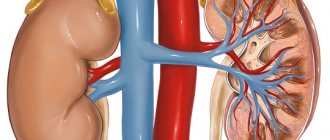
- Some kidney diseases, due to which a lot of fluid is retained in the body.
- Endocrine pathologies, in particular increased levels of adrenal hormones in the blood, hypothyroidism.
- Anatomical pathology in the structure of the eyeball. People suffering from atherosclerosis should be especially careful in this regard.
In any case, it is important to understand that intraocular pressure does not suddenly increase on its own, it is always a consequence. Sometimes - hidden pathological processes occurring in the body, and sometimes - diseases associated directly with the eyes. So, a jump in IOP can be provoked by:
- an eye tumor that puts pressure on the inner membranes and chambers of the eye, thereby interfering with the normal outflow of fluid;
- inflammatory disease of the iris (iritis), ciliary body (cyclitis), choroid (uveitis);
- severe eye injury, after which inflammation, swelling and stagnation of blood in the vessels inevitably appears.
Important! Under any of the above circumstances, IOP cannot be critically high all the time. It increases periodically, in jumps, which depend on the characteristics of the course of the disease that provokes it. But if the pathology is not found and treated, then with age, increased IOP can transform into glaucoma. Gradually, due to strong pressure, the retinal cells will be destroyed, the optic nerve will atrophy, and eventually the person will completely lose vision.
And yet, what factors can affect IOP?
Eye fatigue. Vision is a passive process. It doesn't matter if you spend most of your day staring at a computer screen, constantly knitting, or working as a seamstress. Fatigue is manifested by dry eyes - they may begin to water, itch, turn red, “burn”, “prick”, etc. This is due to the fact that those looking at the seam or monitor blink less often. Instead of the norm 10-15 times per minute - only 5-6 times.
It turns out that to avoid the harmful effects of fatigue, all you need to do is blink more often?
Of course, but it is important not to “skip” visiting the doctor. We never tire of reminding you: the patient’s chances in diagnosis, treatment and results are limited by the doctor’s capabilities – his level of education, experience and class of equipment. And there is only one place where we have collected all the newest and best... just kidding, of course, but one of these places is the Vasily Shevchik Clinic, where your vision will be taken seriously.
What is ophthalmotonus?
Intraocular pressure is normal when approximately 2 mm³ of fluid enters and flows out of the eye every minute. It is with this ratio that it has an indicator of 12 to 22 mmHg. The value may depend on age, physical activity, even the pressure is different in the morning and evening. However, so far it does not exceed 22 mm Hg. and does not fall below 12 mmHg, then it is considered normal. If the process of inflow and outflow is unstable, then the pressure is not normal. There are three types of such violations.
- Transient - there is a short-term deviation from the usual indicator and a return to the limits.
- Labile - pressure increases occur on a regular basis.
- Stably elevated blood pressure is constantly observed, and this poses a great danger to the health of the visual organs.
However, IOP varies depending on age. Until the age of 40, it is 12-22 mm Hg; at 50-60 years, a value of 23-25 mm Hg is considered normal, and after 70 years, this figure increases to 23-26 mm Hg. These values do not indicate a pathological condition.
For information or treatments for glaucoma
Many ophthalmologists begin treatment for glaucoma with eye drops. Drop until the medications begin to lose their ability to reduce the patient's eye pressure. The Sevcik Clinic recommends considering another option: selective laser trabeculoplasty (SLT). This is a type of laser surgery designed to treat glaucoma by reducing intraocular pressure by activating the eye's natural drainage pathways. SLT is more often used as the next step in treatment. However, modern studies have shown that additional laser treatment resulted in better and longer-lasting control of IOP on the one hand, and the use of fewer medications by patients, on the other. Depending on the individual characteristics of the disease, SLT may be the best first step of treatment.
Treatment with medications
The doctor prescribes drops that lower IOP and also improve blood circulation and metabolism. Sometimes this is quite enough to stop the progression of the disease.
Surgical treatment
For more serious forms of glaucoma, surgery is prescribed. Depending on the type of glaucoma and the lesion, the operation is performed with a laser or the eyeball is opened. Unfortunately, even surgery does not provide a complete cure. The main goal is to reduce IOP to stop the progression of the disease. To avoid recurrence subsequently, regular ophthalmological examinations are required.
When drawing up a treatment plan, the doctor starts from the stage to which the disease has developed. To correctly assess it, a comprehensive examination is necessary under the supervision of a highly qualified specialist. At the Vasily Shevchik Eye Microsurgery clinic, excellent ophthalmologists work according to the most modern American and European protocols, and they have the latest equipment at their disposal for diagnosing and treating disorders caused by abnormalities in intraocular pressure.
How to maintain normal ophthalmotonus
To avoid long-term treatment with medications and not lead to surgical intervention, you need to follow simple rules for the prevention of increased ophthalmotonus:
- sleep at least 8 hours a day and avoid stressful situations;
- if there is no need, do not spend a long time in front of a computer monitor;
- work in good lighting to reduce eye strain;
- do not wear clothes with tight collars and buttons to ensure normal blood flow in the cervical-collar area;
- eat right, limiting the amount of salt in dishes, avoiding fried, spicy and smoked foods;
- drink 1.5 liters of clean water per day;
- Raise the pillow during night sleep to ensure proper drainage of aqueous fluid from the eye.
It has been established that bad habits (drinking alcohol and smoking) contribute to pathological narrowing of blood vessels.
As a result, the blood supply to internal organs, the brain and the organs of vision deteriorates. Otherwise, it is fraught with the development of hypertension, other systemic diseases and increased ophthalmotonus. Therefore, if you are prone to eye pressure, it is better to make a choice in favor of a healthy lifestyle.
It must be remembered that normal eye pressure is different for everyone, but it is necessary to adhere to generally accepted standards. If you notice symptoms such as eye fatigue, redness and dryness of the apples, migraine-type headaches, deterioration of visual function, you should immediately visit an ophthalmologist. This will allow you to start treatment on time, if necessary, and reduce the risk of developing glaucoma.
Interesting Facts
- Eye pressure behaves differently in different nationalities and nations.
- African Americans are 10 times more likely to have glaucoma even when the IOP is normal, but people from Eastern Europe often have a special type of glaucoma that behaves very strangely compared to “normal” glaucoma. With the so-called pseudoexfoliation glaucoma, flakes are formed that settle in the drainages of the eye, and their presence provokes the occurrence of cataracts and glaucoma.
- Regular sunglasses cannot be worn if you have glaucoma, because they cause an increase in IOP. In this case, special ones with green lenses are suitable.
- Glaucoma affects almost 9% of the world's population.
- Cases have been described that suggest the likelihood of a viral nature of glaucoma. When the disease simultaneously manifested itself in the same way in people who lived together for a long time in close contact, but were not blood relatives.
What is low ophthalmotonus
In addition to increased IOP, low blood pressure also occurs, although much less frequently. This condition may be caused by the following reasons:
- retinal detachment;
- inflammatory processes in the eyes;
- eye injuries;
- low blood pressure;
- dehydration and some others.
With reduced ophthalmotonus, the following symptoms are observed: dry eyes, painful sensations when blinking, pop-up dots, burning, blurred vision. Low IOP is no less dangerous than high IOP. Clinical examination of this condition reveals papilledema, venous congestion, maculopathy, papillar atrophy, vitreous opacities, and keratopathy. Visual functions deteriorate significantly.
Cost of services for glaucoma
| № | Service name | Price in rubles | Make an appointment |
| 2010025 | Set of disposable consumables for antiglaucomatous surgery | 36000 | Sign up |
| 2010024 | Molteno valve implantation | 54000 | Sign up |
| 2010023 | Implantation of the EX-Pres shunt valve | 54000 | Sign up |
| 2010022 | Ahmed valve implantation | 54000 | Sign up |
| 2010021 | Summing collagen or silicone drainage | 9000 | Sign up |
| 2010020 | Pupilloplasty | 30000 | Sign up |
| 2010019 | Iridoplasty | 22200 | Sign up |
| 2010018 | Basal iridotomy | 10800 | Sign up |
| 2010001 | Sinutrabeculectomy (STE) | 42000 | Sign up |
| 2010002 | Non-penetrating deep sclerectomy (NPDS) | 46200 | Sign up |
| 2010004 | Antiglaucomatous surgery for primary glaucoma of the first category of complexity | 23400 | Sign up |
| 2010005 | Antiglaucomatous surgery for primary glaucoma of the second category of complexity | 30600 | Sign up |
| 2010006 | Antiglaucomatous surgery for primary glaucoma of the third category of complexity | 37200 | Sign up |
| 2010007 | Antiglaucomatous surgery for secondary or refractory glaucoma of the first category of complexity | 29500 | Sign up |
| 2010008 | Antiglaucomatous surgery for secondary or refractory glaucoma of the second category of complexity | 42000 | Sign up |
| 2010009 | Antiglaucomatous surgery for secondary or refractory glaucoma of the third category of complexity | 48000 | Sign up |
| 2010010 | Antiglaucomatous surgery with drainage of the anterior chamber angle for primary glaucoma of the first category of complexity | 28200 | Sign up |
| 2010011 | Antiglaucomatous surgery with drainage of the anterior chamber angle for primary glaucoma of the second category of complexity | 39300 | Sign up |
| 2010012 | Antiglaucomatous surgery with drainage of the anterior chamber angle for primary glaucoma of the third category of complexity | 45600 | Sign up |
| 2010013 | Antiglaucomatous surgery with drainage of the anterior chamber angle for secondary or refractory glaucoma of the first category of complexity | 33360 | Sign up |
| 2010014 | Antiglaucomatous surgery with drainage of the anterior chamber angle for secondary or refractory glaucoma of the second category of complexity | 43800 | Sign up |
| 2010015 | Antiglaucomatous surgery with drainage of the anterior chamber angle for secondary or refractory glaucoma of the third category of complexity | 54000 | Sign up |
| 2010017 | Reconstruction of the anterior chamber angle in secondary glaucoma | 27000 | Sign up |
Making an appointment Today: 8 registered
Fundus examination
Fundus examination (ophthalmoscopy) is an examination of the retina, its vessels, optic nerve, and choroid. The method is based on the reflection of light rays from the fundus of the eye. Diagnosis is carried out using an ophthalmoscope. The doctor directs a light beam from the device lamp into the eye. In certain positions, the macula, the retina and its vessels, and the periphery of the eye are examined.
Ophthalmoscopy is one of the most commonly used eye examinations. There is an indirect ophthalmoscopy, in which the doctor receives an image with approximately 15x magnification.
Direct ophthalmoscopy involves magnifying the image approximately 5 times.
Diagnostics of fundus pressure is carried out after standard procedures - checking the visual field, visual acuity, measuring the size of the organ of vision. To be able to examine the fundus of the eye in more detail, the doctor instills drops that dilate the pupil for a short time.
It is recommended to do ophthalmoscopy once every 2 years. But if you are unfavorably prone to glaucoma, this procedure is done more often (ideally, once every six months, so that you can notice the signs of an incipient eye disease as early as possible.
More about eye drops
What drops do ophthalmologists most often prescribe to their patients diagnosed with ophthalmotonus?
- First of all, these are analogs of prostaglandins F2α, for example: Latanoprost or Xalatan.
- Secondly, beta-blockers, including drugs such as Timolol and Betaxolol.
- The third category is M-cholinomimetics, which include: Pilocarpine and Aceclidine.
- It is very important not to use more than two types of medications at the same time.
How to treat illness and its prevention?
The most effective treatment for elevated intraocular pressure is surgical intervention. This is the most radical way. They resort to it if it was not possible to achieve the desired result with the help of physiotherapeutic procedures such as vacuum massage or color pulse therapy.
prophylaxis will help prevent both the operation and, in fact, the onset of illness . If you are at risk, it is incredibly important to adjust your own diet. You should limit the consumption of salt and fast carbohydrates, and enrich the menu with vegetables, red fruits, and dark chocolate.
You should also avoid clothes that have tight collars. You should not tighten your tie and unbutton the top button. In another case, the outflow of blood that comes from the veins of the head is disrupted.
It is recommended to minimize visual and physical stress. Try to avoid emotional stress and overload. As a preventive measure, it is better not to drink excessive amounts of liquid and forget about tea and coffee altogether.
Prevention of relapse
Before re-treating ophthalmotonus disorders, you need to try simple preventive measures to ensure the maintenance of lasting results of the therapy received:
- do not take breaks in the use of medications if the doctor has prescribed their instillation on an ongoing basis;
- avoid stress, get proper rest, do not overload the nervous system;
- be in more light so as not to provoke dilation of the pupils and an increase in intraocular pressure;
- follow a diet with limited salt, fried, smoked and spicy foods;
- stop smoking, alcohol and caffeinated drinks;
- drink at least 1.5-2 liters of clean water per day.
Patients prone to changes in ophthalmotonus are recommended to take blueberry-based vitamin preparations for the eyes and include more carrots and fish in their diet. When working at a computer, every hour you need to pause and perform a series of gymnastic exercises for the eyes. Once a year it is necessary to visit an ophthalmologist to monitor the condition of the retina, vision and ophthalmotonus.
Dangerous consequences of increased pressure in the eyes include the development of glaucoma and complete blindness, so you should not consider ocular hypertension a non-serious disease. If regular signs of problems with IOP appear (redness and fatigue of the eyes, dry mucous membranes, loss of luster and blurred vision), you should consult a specialist.
At the initial stages of the pathology, medications, exercises and measures to prevent relapse will help relieve symptoms. And with advanced forms of the disease, surgical intervention is no longer possible. Therefore, if you have eye diseases, it is better to visit a doctor again than to develop glaucoma and lose your vision.
Normal IOP in children
The normal intraocular pressure in children is the same in both sexes. For young patients, it is allowed to use tonometry to determine the indicator of eye functioning in question.
Parents need to pay attention to their child's behavior. When intraocular pressure changes, the child may experience heaviness in the eyes, pain in the head, and feel tired. Fatigue is especially noticeable in the evening.
At the first symptoms of increased ophthalmic tone in a child, he should be urgently shown to a pediatrician or ophthalmologist. After measuring the IOP value, he will develop further measures. It is necessary to pay attention to the fact that glaucoma develops extremely rarely in children. An increase in IOP in young children often indicates a malfunction of the thyroid gland.
We recommend reading: Eye pressure in children
Additional Treatments
Even the most innovative drops for reducing eye pressure work best only in combination with a healthy lifestyle and eye exercises. Also, do not forget about some simple, but at the same time effective rules that will help stabilize vision and prevent the progression of the disease. Ophthalmologists strongly recommend watching TV or working at a computer for no more than two hours at a time, and then doing gymnastic exercises and giving your eyes a rest. Limit consumption of salt, coffee, energy drinks and strong teas, as well as alcohol. Additionally, if you have been diagnosed with chronic eye pressure, it is not recommended to lift heavy objects, as this can cause damage to the optic nerve. Experts, in addition, advise sleeping on a volumetric pillow - this way the head will be higher than the level of the body, which, in turn, will help to avoid increased pressure. You should use the drops strictly according to the instructions and constantly visit a specialist to monitor the disease.