Blood flow speed is the speed of movement of blood elements along the bloodstream in a certain unit of time. In practice, experts distinguish between linear velocity and volumetric velocity of blood flow.
One of the main parameters characterizing the functionality of the body’s circulatory system. This indicator depends on the frequency of contractions of the heart muscle, the quantity and quality of blood, the size of blood vessels, blood pressure, age and genetic characteristics of the body.
Types of blood flow speed
Linear speed is the distance traveled by a blood particle through a vessel over a certain period of time. It directly depends on the sum of the cross-sectional areas of the vessels that make up a given section of the vascular bed.
Consequently, the aorta is the narrowest section of the circulatory system and has the highest blood flow speed, reaching 0.6 m/s. The “widest” place is the capillaries, since their total area is 500 times larger than the area of the aorta, the blood flow speed in them is 0.5 mm/s. , which ensures excellent exchange of substances between the capillary wall and tissues.
Volumetric blood flow velocity is the total amount of blood flowing through the cross-section of a vessel over a certain period of time.
This type of speed is determined:
- the difference in pressure at opposite ends of the vessel, which is formed by arterial and venous pressure;
- vascular resistance to blood flow, depending on the diameter of the vessel, its length, and blood viscosity.
Movement of blood through vessels
Blood in the human body moves through a closed system of blood vessels
. The continuous movement of blood occurs due to the contractions of the heart, which works like a pump.
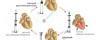
With each contraction of the heart, the left and right ventricles push 60–80 milliliters of blood into the aorta and pulmonary artery
.
The amount of blood ejected by the ventricle in one contraction
, is called
stroke
or
systolic volume
.
If we multiply the heart rate by the systolic volume, we calculate the minute volume of blood
.
At rest it is about 5 liters.
When blood is pushed into the aorta and pulmonary artery, they are stretched and
pressure
.
Blood, like any other fluid, flows from an area of higher pressure to an area where it is lower
.
To better understand how blood moves, let’s compare a blood vessel to a river, the bed of which sometimes narrows and sometimes widens. From a physics course or life experience, you probably know that in narrow places the flow is faster, and in wide places it is slower
.
However, the same amount of water flows past each point on the shore at the same time. The speed of blood flow in different sections is determined by the total area of the bloodstream
. Blood velocity is inversely proportional to the total cross-sectional area of the blood vessels.
The capillaries have the largest cross-sectional area (in total), the aorta has the smallest
.
Therefore, the highest speed of blood flow in the aorta
is approximately
0.5 meters per second
,
in the veins
-
0.25 meters per second
.
In capillaries,
the speed of blood movement is the lowest and ranges
from 0.5 to 1.2 millimeters per second
.
Despite the fact that blood enters the aorta and pulmonary trunk in portions, it flows through the vessels in a continuous stream. The walls of the aorta and arteries are elastic
, they stretch during contraction of the ventricles and accommodate the blood ejected by the heart. Due to elasticity, the vessels reduce their capacity and push blood forward, resulting in an expansion of the wall and an increase in pressure in the adjacent area.
In places where large arteries pass close to the skin, for example at the temples, on the sides of the neck, at the base of the hand, tremors
, which are called
pulse
.
Pulse
is
a periodic oscillation of the walls of blood vessels associated with changes in their blood supply during one cardiac cycle
.
When the left ventricle contracts, blood is thrown into the aorta with great force and stretches its walls. This creates a wave of vibrations that quickly spreads along the walls of the arteries. Each pulse beat corresponds to one heartbeat
.
By measuring the pulse, you can find out the speed, strength and rhythm of heart contractions, and judge the work of the heart.
The pressure that blood exerts on the walls of blood vessels is called blood pressure
. In a healthy person, blood pressure levels are constant, as is body temperature.
Blood pressure is regulated by the nervous system
and is ensured by changes in the lumen of blood vessels and the strength of heart contractions.
During the cardiac cycle, aortic pressure ranges from 115 – 140 millimeters of mercury to 60 – 85 millimeters of mercury. These fluctuations arise as a result of the rhythmic activity of the heart. The pressure
at the moment of contraction of the ventricles is called systolic
, or
maximum, blood pressure
.
The pressure at the moment the ventricles relax
is called
diastolic
or
minimum blood pressure
. As blood moves through the vessels, its pressure gradually decreases.
In small arteries the pressure drops to 70 millimeters of mercury, in capillaries it is approximately 40 millimeters of mercury, in small veins it is 20 millimeters of mercury, and in the vena cava near their entry into the right atrium it approaches zero.
Due to the difference in blood pressure in the arterial and venous sections of both circulatory systems, blood moves through the vessels and returns to the heart
.
For the first time, blood pressure was measured by Gaels. He inserted a glass tube into the horse's artery and observed how, with each contraction of the heart, blood under pressure rose up the tube to a height of up to two meters. This method of measuring pressure has been technically improved and is still used in animal experiments. It is called straight or bloody.
You can measure blood pressure using a tonometer
.
The patient is placed on the shoulder with a hollow cuff chamber, which is connected to a pressure gauge and a rubber balloon to inflate air. The phonendoscope
is applied to the elbow bend where the brachial artery passes.
Next, air is pumped into the cuff until the pulse disappears below the cuff. No sounds can be heard in the phonendoscope at this time. While releasing air, the doctor records the moment the pulsating noise appears and notes the pressure gauge readings. This is how systolic blood pressure is established. The moment the noise disappears corresponds to the level of diastolic blood pressure.
Currently, automatic blood pressure monitors are widely used. They do not require the use of a phonendoscope. Their electronic circuit registers fluctuations in air pressure in the cuff and converts them into digital values.
In a healthy person
the blood pressure is maintained at a constant level and is
120 millimeters of mercury in the brachial artery
, and 70 millimeters of mercury when they relax. Typically, blood pressure is depicted as a fraction, the numerator of which indicates the upper pressure, the denominator the lower, for example, 120 over 70.
Blood pressure depends
from many factors: the time of day, the psychological state of a person (under stress, blood pressure increases), taking various stimulants (coffee, tea) or medications that increase or decrease blood pressure. Blood pressure, which increases as a result of physical activity or nervous tension, soon returns to normal.
In the human body, blood is constantly redistributed: more of it goes to some organs, less to others. If the organ is working, then blood flows to it intensively, bringing additional oxygen and nutrients. At the same time, blood flow to non-functioning organs decreases.
French researcher Angelo Mosso performed the following experiment. He placed a man on a scale so that one half of the body balanced the other. After this, he asked the subject to solve an arithmetic problem. The subject began to think, and his head began to drop down. When the problem was solved, the scales balanced. Next, Mosso asked the person lying on the scale to move his toes. The scales dropped towards the legs. This experience proves to us that blood is constantly redistributed in the human body.
Lesson summary. The highest speed of blood movement is in the arteries, the lowest in the capillaries, and in the veins it increases again. The reason for the movement of blood is the difference in pressure in the vessels at the beginning and end of the journey. The highest pressure is in the aorta, the lowest in the vena cava. The pressure at the moment of blood ejection into the aorta is called upper or systolic. The lowest pressure at the moment of relaxation of the heart is called lower or diastolic. The pulse is the rhythmic vibration of the artery walls. It can be used to determine the frequency and strength of heart contractions.
Importance and severity of the problem
Determining such an important parameter as blood flow velocity is extremely important for studying the hemodynamics of a specific section of the vascular bed or a specific organ. If it changes, we can talk about the presence of pathological narrowing along the vessel, obstacles to blood flow (parietal blood clots, atherosclerotic plaques), and increased blood viscosity.
Currently, non-invasive, objective assessment of blood flow through vessels of different sizes is the most pressing task of modern angiology. The success of solving it depends on the success of early diagnosis of such vascular diseases as diabetic microangiopathy, Raynaud's syndrome, various occlusions and vascular stenoses.
Why is blood velocity measured?
Measuring blood flow velocity is important for diagnostic medicine. Thanks to the analysis of data obtained as a result of measurements, it is possible to determine:
- vascular condition, blood viscosity indicator;
- level of blood supply to the brain and other organs;
- resistance to movement in both circles of blood circulation;
- microcirculation level;
- condition of the coronary vessels;
- degree of heart failure.
The speed of blood flow in vessels, arteries and capillaries is not constant and the same value: the highest speed is in the aorta, the smallest is inside the microcapillaries.
Why is blood flow velocity measured in the vessels of the nail bed?
The speed of blood flow in the vessels of the nail bed is one of the clear indicators of the quality of blood microcirculation in the human body. The vessels of the nail bed have a small cross-section and consist not only of capillaries, but also of microscopic arterioles.
When there are problems related to the circulatory system, these capillaries and arterioles are the first to suffer. Of course, it is impossible to judge the state of the entire system solely on the basis of a study of blood circulation in the nail bed area, but it is worth paying attention if the blood flow in this area is too low or high.
In medicine, to obtain the most reliable information, measurements of blood circulation parameters are carried out over large areas of the blood circulation.
Blood flow speed
There are linear and volumetric blood flow speeds. Linear velocity of blood flow (V-lin) is the distance that a blood particle travels per unit time. It depends on the total cross-sectional area of all vessels forming a section of the vascular bed. Therefore, the narrowest section of the circulatory system is the aorta. Here the highest linear speed of blood flow is 0.5-0.6 m/sec. In arteries of medium and small caliber it decreases to 0.2-0.4 m/sec. The total lumen of the capillary bed is 500-600 times greater than that of the aorta, so the speed of blood flow in the capillaries decreases to 0.5 mm/sec. Slowing down the blood flow in the capillaries is of great physiological importance, since transcapillary exchange occurs in them. In large veins, the linear speed of blood flow increases again to 0.1-0.2 m/sec. The linear velocity of blood flow in the arteries is measured by ultrasound. It is based on the Doppler effect. A sensor with an ultrasound source and receiver will be placed on the vessel. In a moving medium—blood—the frequency of ultrasonic vibrations changes. The higher the speed of blood flow through the vessel, the lower the frequency of reflected ultrasonic waves. The speed of blood flow in the capillaries is measured under a microscope with divisions in the eyepiece, by observing the movement of a specific red blood cell. Volumetric blood flow velocity (vol.) is the amount of blood passing through the cross section of a vessel per unit time. It depends on the pressure difference at the beginning and end of the vessel and the resistance to blood flow:
1 — ——
where is the pressure at the beginning and end of the vessel,
Previously, in the experiment, the volumetric velocity of blood flow was measured using Ludwig's blood clock. In the clinic, volumetric blood flow is assessed using rheovasography. This method is based on recording fluctuations in the electrical resistance of organs to high-frequency current when their blood supply changes during systole and diastole. With an increase in blood supply, the resistance decreases, and with a decrease it increases. To diagnose vascular diseases, rheovasography is performed on the extremities, liver, kidneys, and chest. Plethysmography is sometimes used. This is a registration of fluctuations in organ volume that occur when their blood supply changes. Volume fluctuations are recorded using water, air and electrical plethysmographs. The speed of blood circulation is the time during which a blood particle passes through both circles of blood circulation. It is measured by injecting fluorescein dye into a vein in one arm and timing its appearance in the vein of the other. On average, the speed of blood circulation is 20-25 seconds.
Blood pressure.
As a result of contractions of the ventricles of the heart and the ejection of blood from them, as well as resistance to blood flow in the vascular bed, blood pressure is created. This is the force with which blood presses on the wall of blood vessels. The amount of pressure in the arteries depends on the phase of the cardiac cycle. During systole it is maximum and is called systolic, during diastole it is minimal and is called diastolic. Systolic pressure in a healthy young and middle-aged person in large arteries is 100 - 130 mmHg. Diastolic 60-80 mmHg. The difference between systolic and diastolic pressure is called pulse pressure. Normally its value is 30-40 mmHg. In addition, the average pressure is determined. This is so permanent, i.e. non-pulsatile pressure, the hemodynamic effect of which corresponds to a certain pulsating one. The average pressure value is closer to diastolic pressure, since the duration of diastole is longer than systole. Blood pressure (BP) can be measured by direct and indirect methods; to measure with the direct method, a needle or cannula connected by a tube to a pressure gauge is inserted into the artery. Now a catheter with a pressure sensor is inserted. The signal from the sensor is sent to an electric pressure gauge. In the clinic, direct measurements are made only during surgical operations. The most widely used are the indirect Riva-Rocci and Korotkoff methods. In 1896, Riva-Rocci proposed measuring systolic pressure by the amount of pressure that must be created in a rubber cuff to completely compress the artery. The pressure in it is measured by a pressure gauge. The cessation of blood flow is determined by the disappearance of the pulse in the radial artery. In 1905, Korotkoe proposed a method for measuring both systolic and diastolic pressure. It is as follows. The cuff creates pressure at which blood flow in the brachial artery completely stops. Then it gradually decreases and at the same time the sounds that arise are heard using a phonendoscope in the ulnar fossa. At the moment when the pressure in the cuff becomes slightly lower than systolic, short rhythmic sounds appear. They are called Korotkoff sounds. They are caused by the passage of portions of blood under the cuff during pregnancy. As the pressure in the cuff decreases, the intensity of the tones decreases and at a certain value they disappear. At this moment, the pressure in it approximately corresponds to diastolic. Currently, to measure blood pressure, devices are used that record vibrations of the vessel under the cuff as the pressure in it changes. The microprocessor calculates systolic and diastolic pressure. For this purpose, arterial oscillography is used. This is a graphical recording of the pulsations of large arteries when they are compressed by a cuff. This method allows you to determine systolic, diastolic, mean pressure and elasticity of the vessel wall. Blood pressure increases during physical and mental work, and emotional reactions. During physical work, systolic pressure mainly increases. This is due to the fact that systolic volume increases. If vasoconstriction occurs, both systolic and diastolic pressure increases. This phenomenon occurs with strong emotions. Long-term graphical recording of blood pressure reveals three types of fluctuations. They are called waves of 1st, 2nd and 3rd orders (Fig.). First-order waves are pressure fluctuations during systole and diastole. Second order waves are called respiratory waves. As you inhale, blood pressure increases and as you exhale, it decreases. With brain hypoxia, even slower third-order waves occur. They are caused by fluctuations in the tone of the vasomotor center of the medulla oblongata.
In arternoles, capillaries, small and medium-sized veins, the pressure is constant. In the arternoles its value is 40-60 mmHg, at the arterial end of the capillaries 20-30 mmHg, at the venous end 8-12 mmHg. Blood pressure in the arternoles and capillaries is measured by inserting a micropipette connected to a manometer. Blood pressure in the veins is 5 mmHg. In the vena cava it is equal to 0, and on inspiration it becomes 3-5 mmHg, below atmospheric. The pressure in the veins is measured by a direct method called phlebometry. An increase in blood pressure is called hypertension, a decrease is called hypotension. Arterial hypertension occurs with aging, hypertension, kidney disease, etc. Hypotension is observed with shock, exhaustion, and dysfunction of the vasomotor center.