Cerebral infarction or ischemic stroke are both names for a stroke caused by a blockage of a blood vessel in a person's brain.
This is the most common type of stroke among Russians, Ukrainians and Belarusians. A blood clot or fatty plaque forms in the patient's brain and blocks a blood vessel - an embolus.
This is focal necrosis of the brain caused by complete and prolonged ischemia, which affects all tissue elements, neurons, glia and blood vessels.
Ischemic infarctions cause focal neurological deficits in the patient. With embolic infarctions they appear abruptly. In atherothrombotic infarctions, they develop over a period of time, usually several hours. Atherothrombotic infarctions are often preceded by transient ischemic attacks (TIAs).
A TIA is a focal neurological deficit that lasts less than 24 hours and resolves.
Causes of cerebral infarction:
A cerebral infarction (stroke) is mainly caused by a blocked artery (ischemic stroke) or a ruptured blood vessel (hemorrhagic stroke). Our specialists see categories of patients who may experience only a temporary disruption of blood flow to the brain (transient ischemic attack or TIA), which does not cause permanent damage to the brain.
Types of ischemic stroke include:
Approximately 70% of strokes are ischemic strokes. Ischemic strokes occur when arteries to a patient's brain become narrowed or blocked, causing severely reduced blood flow (ischemia). The most well-known ischemic strokes include:
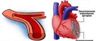
- Thrombotic stroke. A thrombotic stroke occurs when a blood clot (thrombus) forms in one of the arteries that supplies blood to the brain. The clot may be caused by fatty deposits (plaques) that accumulate in the arteries and cause decreased blood flow (atherosclerosis) or other artery diseases.
- Embolic stroke. An embolic stroke occurs when a blood clot forms from your brain - usually in the heart - and travels through the bloodstream to form in narrower arteries in the brain. This type of blood clot is called an embolus.
- Hemorrhagic stroke. A hemorrhagic stroke occurs when a blood vessel in the brain leaks or ruptures. Brain hemorrhage can be the result of many conditions that affect the blood vessels.
These include:
- Uncontrolled high blood pressure – arterial hypertension;
- Weak blood vessel walls (aneurysms);
- A less common cause of hemorrhage is the rupture of an abnormal tangle of thin-walled blood vessels (arteriovenous malformation).
Types of hemorrhagic stroke include:
- Intracerebral hemorrhage. In intracerebral hemorrhage, a blood vessel in the brain bursts and spills into the surrounding brain tissue, damaging brain cells. The brain cells outside the leak are deprived of blood and are also damaged.
- High blood pressure, trauma, vascular malformations, high dose use of blood thinning medications, and other patient factors can cause intracerebral hemorrhage in the brain.
- Subarachnoid hemorrhage. In a subarachnoid hemorrhage, an artery on or near the surface of the brain bursts and enters the space between the surface of the brain and the skull. This bleeding is often signaled by a sudden, severe headache. Subarachnoid hemorrhage is usually caused by the rupture of a small sac- or berry-shaped aneurysm. After a hemorrhage, the blood vessels in your brain may widen and narrow randomly (vasospasm), causing damage to brain cells by further restricting blood flow.
- Transient ischemic attack (TIA). A transient ischemic attack (TIA)—sometimes known as a mini-stroke—is a temporary period of symptoms. A temporary reduction in blood supply to part of the brain causes Thias, which can last only three to five minutes.
Risk factor for cerebral infarction:
- The patient has an unhealthy lifestyle and constant emotional stress (work, relationships with family, financial losses, etc.);
- Overweight or obesity (often our patients are obese due to constant stress);
- Insufficient physical activity;
- Smoking, exposure to secondhand smoke and alcohol;
- Drug use;
- Blood pressure readings above 120/80 millimeters of mercury (mmHg);
- High cholesterol level in the blood (regular laboratory diagnostics of CBC, BAC, OAM);
- Diabetes;
- Cardiovascular diseases;
- Genetic predisposition;
- Age—people aged 55 years and older have a higher risk of stroke than younger people;
- Floor. Men have a higher risk of stroke than women;
- Hormones—use of birth control pills or hormone therapy that includes estrogen.
Pain syndrome during an angina attack
M.V. KUZNETSOVA
, Candidate of Medical Sciences, Head of the Day Hospital Department, Doctor of the Highest Category,
State Research Center for Preventive Medicine
Coronary heart disease (CHD) has been the main cause of mortality in many economically developed countries for many years. Currently, cardiovascular diseases (CVD) play a decisive role in the evolution of overall mortality in Russia. Mortality from CVD in Russia in 2013 was 857 cases per 100 thousand inhabitants. Of this number, almost 50% are deaths from ischemic heart disease. The problem of treating and alleviating the clinical manifestations of these diseases is one of the priority issues of modern cardiology.
IHD occurs in both acute and chronic forms. This is sudden coronary death, angina pectoris (tension, unstable, new onset, progressive. Early post-infarction or post-operative, spontaneous - vasospastic, variant, Prinzmetal), silent myocardial ischemia, cardiac X-syndrome - microvascular angina, acute myocardial infarction, post-infarction cardiosclerosis, cardiac failure, rhythm and conduction disturbances.
Statistics show that the most common type of coronary artery disease is angina pectoris (the name of the disease comes from the Greek words “stenos” and “kardia”, which literally translates as narrowing or compression of the heart). It can be stable and unstable. A distinction is made between angina pectoris, which occurs when psychophysical stress increases significantly, and angina pectoris at rest. A dangerous variant of the course is “silent” angina, when the typical pain syndrome is absent in the clinical picture, the patient complains of a feeling of numbness in the arm, shortness of breath during physical activity.
According to the State Research Center for Medical Sciences, in the Russian Federation, almost 10 million of the working-age population suffer from coronary artery disease, more than a third of them have angina pectoris. The incidence of angina increases with age: in women from 0.1–1% at the age of 45–54 years to 10–15% at 65–74 years, in men - from 2–5% at 45–54 years to 10–20% at 65–74 years old. In most European countries, the prevalence of angina pectoris is 20,000–40,000 per 1 million population. According to the Framingham study, angina pectoris is the first symptom of coronary artery disease in men in 40.7% of cases and in 56.5% in women.
As shown by the international study ATP-Survey (Angina Treatment Patterns), conducted in 2001 in 9 European countries (including 18 centers in Russia), among Russian patients, patients with class II–III angina pectoris predominated. It is important that in the population only about 40–50% of all patients know that they have the disease and receive appropriate treatment, while in 50–60% the disease remains unrecognized. Patients diagnosed with stable angina die from coronary artery disease 2 times more often than those without this disease. Data from the State Research Center for Medical Sciences state that men suffering from coronary artery disease live on average 8 years less compared to those who do not have this pathology.
According to the results of the Framingham study, in patients with stable angina, the risk of developing non-fatal myocardial infarction and death from coronary artery disease within 2 years is respectively: 14.3 and 5.5% in men and 6.1 and 3.8% in women.
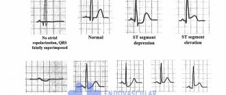
Angina occurs when the work of the heart and the myocardial oxygen demand exceed the ability of the coronary arteries to supply the corresponding areas of the myocardium with a sufficient amount of oxygenated blood. It is believed that pain during angina pectoris is a direct manifestation of myocardial ischemia, leading to the accumulation of under-oxidized metabolites in the heart muscle. As myocardial ischemia develops, the pH of the blood in the coronary sinus decreases, intracellular potassium is lost, and instead of lactate being utilized, its increased production begins. Pathological changes in the ECG appear, the mechanical performance of the ventricles is disrupted. During an attack of angina, diastolic pressure in the left ventricular cavity often increases, sometimes so much that pulmonary congestion occurs or shortness of breath develops.
The main factors determining myocardial oxygen demand are heart rate (HR), systolic tension or systolic blood pressure and myocardial contractility. An increase in any of these indicators against the background of reduced coronary blood flow can cause an attack of angina. Thus, physical stress in a patient with a critical degree of coronary artery stenosis causes an attack of angina pectoris, which is relieved with rest. During an attack that occurs spontaneously at rest, the subjective sensation of pain is usually accompanied by some increase in heart rate and an increase in blood pressure, sometimes significant. If the attack is not eliminated, these shifts provide the basis for the inclusion of potentially dangerous biofeedback: the higher the blood pressure and heart rate, the greater the discrepancy between the myocardial oxygen demand and the degree of its satisfaction.
Angina pectoris in most patients is caused by a critical decrease in the lumen of the coronary arteries due to atherosclerosis.
The course of angina pectoris largely depends on the severity and rate of progression of coronary artery disease underlying coronary insufficiency. In some cases, the functional class of angina pectoris may remain unchanged for many years, in others there is a slow but constant increase in symptoms. Angina pectoris of any functional class, as well as one that arises for the first time, can, for reasons that are not always known, suddenly acquire a rapidly progressive course and end in myocardial infarction.
The most typical manifestation of angina is discomfort or pain, which forces the patient to see a doctor. The characteristics of discomfort associated with myocardial ischemia (angina) can be divided into four categories: location, nature, duration, relationship with stress and other aggravating and eliminating factors.
With angina pectoris, a painful attack occurs during physical (sometimes emotional) stress and usually goes away within 1–2 minutes after its cessation. The duration of a severe attack can reach 20–30 minutes; with a longer duration, focal dystrophic or necrotic lesions of the myocardium usually develop. In the vast majority of cases, the pain is localized deep behind the sternum, most often at the level of the upper third of the sternum. Sometimes the area of most severe pain is shifted slightly to the left or right of the midline. Patients define the pain as squeezing, pressing, sometimes as aching, bursting or burning. The intensity of pain varies: from difficult to bear to barely pronounced, comparable to a feeling of discomfort. Irradiation of pain is not always observed; in typical cases, pain radiates to the left shoulder, left arm, shoulder girdle (right-sided irradiation is less typical) or to the neck, lower jaw, interscapular space. Rarely, atypical irradiation of pain is observed - in the epigastrium, the left half of the celiac region. When an attack occurs, the patient usually freezes; if the pain occurs while walking, he stops. More often this is walking uphill, on an incline, or against the wind, or in cold weather. Objectively, during a severe attack, hyperemia or pallor of the face, the appearance of droplets of sweat on it, sometimes increased sweating on the torso, slight tachycardia, a slight increase or decrease in blood pressure, and dilated pupils may be observed. Shortness of breath may accompany angina. Chest discomfort may also be accompanied by symptoms such as fatigue or dizziness, nausea, burning, restlessness, or a feeling of doom. Dyspnea may be the only symptom of stable angina, which can cause difficulty in verifying the diagnosis and require differential diagnosis with bronchopulmonary disease.
Worsening symptoms after eating a large meal or waking up in the morning are classic signs of angina. Angina can be reduced by further exercise (walk-through angina) or by a second exercise (warm-up angina). The threshold for angina and therefore symptoms can vary significantly from day to day and even within a single day.
It should be noted that there are some difficulties in verifying the diagnosis of angina pectoris. Thus, in older people, only 50% of cases are diagnosed with the classic form of stable angina. Most often, the equivalent of pain is shortness of breath. Decreased physical activity is the reason why patients do not reach the threshold when pain occurs. An important role in diagnosis is played by memory loss and the presence of concomitant diseases. Women often experience cardialgia, which, coming to the fore, makes it difficult to diagnose angina and determine its functional class. 50% of patients with diabetes do not have the classic form of stable angina, and the disease is often asymptomatic. Chronic refractory angina is severe angina, refractory to full drug therapy, occurring in patients for whom intravascular interventions or coronary bypass surgery are contraindicated or difficult. Refractory angina corresponds to class III–IV angina according to the Canadian Society of Cardiology classification.
There are several clinical forms of stable angina: early morning angina, occurring between 6 and 11 a.m., caused by activation of the sympathetic nervous system with increased heart rate and increased blood pressure; starting angina due to increased myocardial oxygen demand without prior inclusion of collaterals; angina pectoris after digestion, associated with redistribution of blood and “robbing” the heart; “cold” angina, due to spasm of peripheral arteries; angina pectoris in the supine position (decubitus) as a result of increased venous return and increased preload on the heart with increased myocardial oxygen demand; angina pectoris that occurs after stressful situations when the sympathetic nervous system is activated; “tobacco” angina (tobacoangina), caused by vasospasm, activation of the sympathetic nervous system with an increase in heart rate and blood pressure; angina pectoris of the “second” breath - overcoming the pain syndrome and continuing the load due to the production of substances in the ischemic myocardium that can increase coronary blood flow; “static” angina, caused by a sharp static load, the mechanism of which is the rise of the diaphragm, the rotation of the heart, an increase in intrathoracic pressure, an increase in venous return of blood and preload on the heart.
There are also typical and atypical angina. With atypical angina, most often the chest pain is similar to that with typical angina: the same localization and reaction to nitrates, but without a clear connection with provoking factors. The pain is often described as resting, starting at a low level of intensity that gradually increases in intensity, persists for 15 minutes, and then slowly decreases in intensity. This characteristic should alert the clinician to the possibility that coronary vasospasm is present. Another atypical presentation is pain, location and characteristics of angina, which occurs during exercise, but stops some time after exercise or can be stopped by taking nitrates. Most often it occurs in patients with microvascular angina. Below is a classification of chest pain.
Traditional clinical classification of chest pain
• Typical angina (proven) - all three complete characteristics are met: - chest discomfort of a typical nature and duration - provoked by physical or psycho-emotional stress - relieved by rest and/or taking nitrates for minutes. • Atypical angina (probable) - the presence of two of these characteristics. • Non-anginal chest pain - the presence of only one or no of the above characteristics
The pain syndrome stops or decreases when the load is stopped. Taking nitrates in the vast majority of cases completely stops the attack within 1-2 minutes.
Thus, the main task of the doctor is to relieve pain.
Currently, a range of nitrates is widely represented on the pharmacological market. Nitroglycerin (glycerol trinitrate) has been used in clinical practice since the second half of the 19th century. But only almost 100 years later, new drugs were created (isosorbide dinitrate, isosorbide 5-mononitrate) and new dosage forms (for intravenous administration, for use in the form of an aerosol, for transdermal use in the form of an ointment, a patch), including forms with a slow release of the drug in the gastrointestinal tract. Active study of the mechanism of action of nitrates led to an expansion of the range of indications for their use and made it possible to establish at the end of the twentieth century that the nitric oxide (NO) molecule plays an important role in ensuring the normal function of the vascular endothelium. It takes part in the regulation of vascular tone, has an antiaggregation effect, inhibits the adhesion and infiltration of monocytes, and inhibits the migration and proliferation of smooth muscle cells in the arterial wall. Impaired endothelial function, including a decrease in NO formation, has also been found in coronary artery disease.
Nitrates are widely used as antianginal agents for various forms of coronary artery disease (angina pectoris, myocardial infarction, etc.). The therapeutic effect of nitrates (nitrovasodilators) is based on the ability to substrate increase the content of nitric oxide, which is an endothelial relaxing factor. This factor stimulates guanylate cyclase, increases the content of cyclic guanosine monophosphate inside the cell and ultimately causes vasodilation. Nitrovasodilators include organic nitrates, as well as sydnonimines (molsidomine) and other nitric oxide donors (amyl nitrite). The most common in clinical practice are organic nitrates (nitroglycerin, isosorbide dinitrate, isosorbide mononitrate). Recently, not only new compounds and dosage forms of organic nitrates, including prolonged action, have been developed, but also the indications for their use have been clarified and expanded. Dosage forms of nitrates ensure penetration of the active substance into the bloodstream through the oral mucosa (tablets, wafers, aerosols), after oral administration (tablets, capsules for oral administration), transdermally, i.e., when applied to the skin (ointments, patches, discs ) or used for injection.
The antianginal effect of nitrates is due to the restructuring of hemodynamics associated with the general vascular effect of the drug, relaxation of the smooth muscles of peripheral vessels under its influence, especially its venodilating effect. Under the influence of nitrates, venous blood flow to the heart decreases, pressure in the right atrium and in the pulmonary artery system decreases, and peripheral vascular resistance decreases. As a result, the myocardium is unloaded, its tension in overcoming the resistance of blood ejection is reduced, energy costs and the need for oxygen are reduced. The resulting decrease in cardiac tension and coronary lytic effect help restore blood flow through the small vessels of the myocardium and improve blood flow in the epicardial coronary arteries. Along with a decrease in ischemia, pain impulses from the ischemic focus gradually disappear and the pain syndrome stops. The effect of nitrates is not limited to the effect on the coronary vessels, they cause dilation of other blood vessels (cerebral vessels, peripheral vessels, abdominal vessels). Nitrate intake can modulate fibrinolysis by changing the ratio in plasma between the two main components of this system - tissue plasminogen activator and its inhibitor, which may be both a consequence of changes in hemodynamics and the result of the inhibitory effect of NO on platelet aggregation and adhesion.
In addition to the use of nitrates as the drugs of choice for the initial treatment of most patients with angina (relief and prevention of attacks), their prescription to patients with a variable threshold for ischemic pain is justified, since they prevent intermittent coronary vasoconstriction in vasospastic angina. For the purpose of relief and prevention of a predicted attack of angina, dosage forms of nitrates that are absorbed in the oral cavity are more often used (Table 1).
| Table 1. Comparison of nitroglycerin (tablets) and Isoket spray | ||
| Properties | Nitroglycerin (sublingual tablets) | Isoket spray |
| Fast absorption | + | +++ |
| Instant effect | + | +++ |
| Duration of action | up to 30 min | up to 120 min |
| Hygiene | — | +++ |
| No special storage conditions | — | +++ |
Isoket spray is an antianginal drug from the group of organic nitrates. Isoket contains an active substance - isosorbide dinitrate - which has vasodilating properties. Isosorbide dinitrate reduces the need of the heart muscle for energy and oxygen, reduces pre- and afterload on the myocardium. In addition, the drug reduces diastolic pressure in the ventricles of the heart, eliminates spasm of the coronary vessels, as a result of which coronary blood flow in ischemic areas of the myocardium is normalized. The drug has a vasodilating effect mainly on the veins, however, when using isosorbide dinitrate, some dilatation of the arteries is also observed. Under the influence of isosorbide dinitrate, the resistance of the pulmonary vessels decreases and the pressure in the pulmonary circulation decreases.
The drug has virtually no effect on heart rate. Promotes the expansion of cerebral vessels. By stimulating the sympathetic nervous system and reducing afterload, it improves systolic function.
The main advantages of Isoket spray, in contrast to an aerosol, are that the drug is supplied from a cylinder (aerosol) by creating excess pressure in it, and extraction occurs by opening the valve. When using a spray, the drug is supplied by mechanically squeezing it out with a micropump piston, while the pressure in the bottle is equal to atmospheric pressure. An important point in the design is that when the piston moves to its original position, the cavity from which the drug is squeezed out is filled with a new portion. This serves as a liquid seal, preventing air from entering the bottle, i.e. the drug in the bottle does not come into contact with the external environment and is not exposed to aggressive factors, the bottle is hermetically sealed, which prevents spraying of the drug into the external environment.
The mechanism of action of the drug is based on its ability to increase the amount of nitric oxide in the vascular endothelium (Fig. 1). NO is necessary for the activation of guanylate cyclase, which is involved in the synthesis of cyclic guanosine monophosphate (cGMP). An increase in cGMP concentration leads to vascular dilatation (due to a decrease in the tone of the smooth muscle layer). A decrease in preload when using isosorbide dinitrate occurs due to a decrease in vascular tone, venous deposition of blood and a decrease in its venous return to the heart. Due to the decrease in venous return of blood to the heart, the pressure on the walls of the left ventricle is significantly reduced and the need for oxygen in the heart muscle is reduced. Direct dilatation of coronary vessels and eccentric coronary stenoses, as well as a slight decrease in their resistance, cause an improvement in myocardial perfusion and oxygen supply without the development of “steal syndrome”.
An increase in tolerance to physical activity was noted in patients suffering from coronary heart disease and angina pectoris taking the drug Izoket spray. Isosorbide dinitrate helps reduce the load on the myocardium, improving the quality of life in patients suffering from heart failure. In addition, by reducing blood flow to the right atrium, the drug reduces pressure in the pulmonary circulation and helps reduce the symptoms of pulmonary edema.
When using the drug in the form of a spray, isosorbide dinitrate is quickly absorbed into the systemic circulation. The active substance is absorbed over the entire area of the oral mucosa, so the therapeutic effect occurs faster than when using the drug in tablet form. The effect of the drug develops within 1–3 minutes after application. The peak concentration of the active substance in the blood is observed 3–6 minutes after application. When using the drug in the form of a spray or aerosol, there is no effect of the first passage of isosorbide dinitrate through the liver. The drug is metabolized in the liver to form pharmacologically active metabolites: isosorbide-2-mononitrate and isosorbide-5-mononitrate. The bioavailability of the drug is about 60–100%, which is significantly higher than the bioavailability of oral forms of isosorbide dinitrate. Excreted by the kidneys in the form of pharmacologically active metabolites, the half-life of isosorbide-2-mononitrate is from 1.5 to 2 hours, isosorbide-5-mononitrate - from 4 to 6 hours.
Thus, Isoket spray has a number of significant advantages for pain relief in comparison with sublingual nitrates (Table 2). It should also be noted that experimental studies on animals did not reveal the teratogenic effect of isosorbide dinitrate.
| Table 2. Comparison of Isoket spray with sublingual nitrates | |
| Isoket spray | Sublingual tablets |
| No passage through the liver | Part of the drug (swallowed) undergoes the first stage of metabolism through the liver |
| Large suction area | Smaller suction area |
| Fast absorption | Longer absorption |
| To achieve the effect, a smaller amount of active substance is required | More drug is required to achieve effect |
| Faster onset of clinical effect | The clinical effect occurs more slowly |
Prompt relief of pain to alleviate the subjective condition of the patient during an attack of angina is the primary task of the doctor. Isoket spray for sublingual use has a rapid onset of action due to the dosage form in the form of an aerosol, has a long-lasting protective effect due to long-acting metabolites, allows for precise dosing of the drug, and has a convenient release and packaging form. Each time you press the sprayer, an equal amount of solution is sprayed in the form of tiny droplets. It easily penetrates the mucous membrane of the mouth and enters the bloodstream in a matter of seconds. When using isosorbide dinitrate in the form of a sublingual spray, the effect of the drug begins after 30 seconds and continues for 120 minutes (2 hours).
Literature
1. Recommendations of the European Society of Cardiology - 2013. 2. Belenkov Yu.N., Oganov R.G. National Guidelines for Cardiology. GEOTAR-Media, 2012. 3. Mazur N. The role of nitrates in the treatment of cardiac patients in accordance with the principles of evidence-based medicine and recommendations for their practical use. Mediasfera, 2007. 4. Zmushko E.I., Belozerov E.S. Medications. St. Petersburg: Peter, 2000. 5. Kosarev V.V. Pharmacotherapy. Samara: Medicine, 1994. 6. Mashkovsky M.D. Medicines: In 2 volumes. T. 1. M.: Novaya Volna, 2002. 7. Sinev D.N., Gurevich I.Ya. A manual for pharmacists. M.: Medicine, 1982. 8. Pharmacological dictionary. Comp. P.M. Fadeeva. M.: Medicine, 2003. 9. Moncada S, Higgs A. The L-arginine - nitric oxide pathway. N Engl J Med 1993, 329: 2002-2012.4. 10. Cohn JN, Archibald DG, Zische S et al. Effect of vasodilator therapy on mortality in chronic congestive heart failure: results of veterans administration Cooperative Study. A comparison of enalapril with hydralazine-isosorbide dinitrate in the treatment of chronic congestive heart failure. N Engl J Med 1991, 325: 303-310. 11. Horowitz JD. Tolerance induction during therapy with long — acting nitrates: how extensive is the “Collateral demage”? Cardiovasc Drugs Ther 2004, 18:1:11-12.16. 12. Kosmicki MA, Szwed H, Sadowski Z. Antiischemic response to sublingual nitroglycerin during oral administration of isosorbide dinitrate in patients with stable angina pectoris: when does cross tolerance occur? Cardiovasc Drugs Ther 2004, 18:1:47-55.17. 13. Webb DJ, Muirhead GJ, Wulff M et al. Sildenafil citrate potentiates the hypotensive effects of nitric oxide donor drugs in male patients with stable angina. J Am Coll Cardiol 2000, 36:25-31. 14. Doucet S, Malekianpour M, Theroux P et al. Randomized trial comparing intravenous nitroglycerin and heparin for treatment of unstable angina secondary to restenosis after coronary artery angioplasty. Circulation, 2000, 101: 955-61. 15. Bussmann WD, Passek D, Seidel W, Kaltenbach M. Reduction of CK and CK-MB indexes of infarct size by intravenous nitroglycerin. Circulation, 1981, 63: 615-22.
Prevention
Knowing your risk factors for stroke, following the advice of highly trained professionals, an annual comprehensive body check-up and a healthy lifestyle are the best steps you can take to prevent stroke. If you have had a stroke or transient ischemic attack (TIA), these steps may help prevent another stroke.
The help you receive in the clinic plays a significant role in stopping the acute period of a cerebral infarction.
The risk factors for possible complications after an attack will depend on how qualified and prompt assistance a team of neurologists, cardiologists, neurosurgeons, rehabilitation therapists and physiotherapists will provide you.
Many stroke prevention protocols are the same as cardiovascular disease prevention protocols. Basic recommendations for a healthy lifestyle include:
- Constant blood pressure monitoring. This is one of the most important things you can do to reduce your risk of stroke. If a patient has already had a stroke, lowering blood pressure may help prevent a subsequent TIA or stroke.
- Exercise, manage stress, maintain a healthy weight, and limit sodium, alcohol, and tobacco. In addition to recommending lifestyle changes, our doctor may prescribe medications to treat high blood pressure, as well as a vitamin and mineral supplement and fish oil.
- Reduce the amount of cholesterol and saturated fat in your diet. Eating less cholesterol and fat, especially saturated fat and trans fat, can reduce plaque in the arteries.
- Quitting tobacco use. Smoking increases the risk of stroke for smokers and nonsmokers exposed to secondhand smoke. Quitting tobacco use reduces the risk of stroke.
- Diabetes control.
- Maintaining a healthy weight. Excess weight contributes to the development of other risk factors for stroke, such as high blood pressure, heart disease and diabetes.
- A diet rich in fruits and vegetables. A diet containing five or more daily servings of fruits or vegetables may reduce the risk of stroke.
- Exercise can lower blood pressure. It will also help you lose weight, control diabetes and reduce stress.