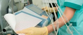
Surely many times in your life you have encountered a situation when you need to decipher a “tape”: during an exam in propaedeutics of internal diseases, when studying a medical history (and not necessarily in the department of therapy or cardiology), or even when consulting a friend of your mother who turned to you with the words “you’re a doctor.” If in all these situations you thought: “I think I studied electrocardiography in the 2nd or 3rd year of medical school, but when I pick up an electrocardiogram in my hands, I understand little. We need to do something about this,” then this article is definitely for you. If you are already well versed in electrocardiography, then in any case, the links to various sources provided in this article may be useful to you.
Where to start studying ECG?
Physiology
To understand ECG, you first need basic knowledge of physiology. If you are a 1st or 2nd year student at a medical university (or just want to repeat physiology), ECG physiology is best and most understandably described in the books “Fundamentals of Medical Physiology” by N. N. Alipov and “Medical Physiology” by Arthur Guyton and John Hall. If you are a student of 3 or more senior years and are already familiar with the basics of ECG physiology, then, most likely, it does not make much sense to return to an in-depth study of physiology and a summary repetition of electrophysiology in the initial chapters of books on ECG, which will be given below, will be enough for you .
The main thing that needs to be understood from physiology is what leads are, and most importantly, how a graphical image of an ECG is generally formed, because, in essence, an ECG is a projection of the integral vector of electrical excitation onto a lead. Knowing this, it will be much easier to understand how the waves and intervals are formed on the electrocardiogram.
ECG analysis and interpretation
The main thing that is required from the clinician is the ability to analyze any changes in the electrocardiogram and give a clinical interpretation of these changes. In fact, the most common difficulty that doctors encounter when deciphering an ECG is the absence of at least some kind of diagram in their head (even a basic one, without any subtleties), and this gives rise to fear and uncertainty when looking at the ECG. After all, how can you be confident in the conclusion if you don’t know what needed to be analyzed in the first place? So, the first guarantee of your success when deciphering an ECG is to have a well-developed scheme in your head. Even if it is the simplest one, perhaps you will periodically change some points, but there must be a scheme. Which scheme exactly should you choose? There is no exact answer here. All diagrams were invented for one purpose - so that you do not forget to analyze all the main points on the electrocardiogram. In essence, they are similar to each other, so first, remember at least one, and with practice, if necessary, you will be able to adjust this scheme to suit yourself. One of the simple but useful schemes that we can recommend to you is the ECG analysis scheme from the book by V.V. Murashko, A.V. Strutynsky:
ECG analysis scheme
I. Analysis of heart rhythm and conductivity: 1. Assessment of the regularity of heart contractions; 2. Heart rate assessment; 3. Determination of the source of rhythm; 4. Conduction assessment (duration P, PQ, QRS, QT); II. Definition of EOS; III. Atrial P wave analysis; IV. Analysis of the ventricular QRS complex; V. ST segment; VI. T wave; VII. U waves
Despite the fact that the diagram is simple, there is almost everything that needs to be analyzed on an ECG: rhythm and conductivity, the electrical axis of the heart, and then an analysis of the main waves, segments and intervals in order. After analyzing the changes in the ECG, it is necessary to make their clinical interpretation, which, of course, can be even more difficult than the analysis itself, but for the most frequent and important situations in medicine, basic knowledge is enough to make a conclusion. For interpretation, not only knowledge, but also experience in decoding ECG is important. The more ECGs you see, the better your understanding of the clinical significance of the changes you identify.
Which books to start studying ECG with?
There is no exact answer here. Rather, it is a matter of taste. In our opinion, for those new to ECG, John Hampton's ECG series is the best option. The series consists of three books:
The first is “ECG Basics.” A very simple, small but useful book. You can read it literally in a few evenings. Why do we consider this guide to be the most suitable for a beginner? It's all about its simplicity. As mentioned above, the most common problem for a beginner is the lack of even the simplest diagram in their head, as well as uncertainty when analyzing the ECG. This book will allow you to overcome this “threshold of uncertainty”, look at the ECG and know what to look for on it. And this is a very important step, laying the foundation for further study of the ECG.
The second is “Atlas of ECG. 150 clinical situations" is a logical continuation of the first book. Once you learn the basics of ECG analysis, it takes practice, practice, and more practice. Of course, this atlas is only one of countless other ECG atlases (where to look for others will be discussed below), and for practice you can use any other atlas, website, blog or public page.
The third is “ECG in a doctor’s practice.” Here, individual issues of clinical interpretation of the ECG are discussed in more depth. It is also worth noting that all books are interconnected in text and one part refers to another, which can be quite useful.
Another good option for starting to study ECG is the well-known book by V.V. Murashko and A.V. Strutynsky “ECG”, which remains the bible for many students of medical universities. And yet, we put this book in second place for beginners, since some of the domestic approaches described in it are already outdated. But, of course, today this manual remains one of the best options for beginners to study ECG.
Each requires a different approach, and therefore you, of course, can choose other books both to begin and further study ECG. The purpose of this article is not to describe absolutely all books and sources on ECG; we offer, in our opinion, the most optimal options for beginners. Links to articles discussing many other books on ECG will be provided below.
Invention of electrocardiography
In 1906, the famous Dutch scientist Willem Einthoven first recorded a clear electrical signal from the heart from the surface of the human body using a device he designed.
Photograph of an assembled electrocardiograph, demonstrating a method in which electrodes placed on the patient's arms and one leg are jars filled with a solution of table salt. Photo taken from the link:
Back in 1893, V. Einthoven proposed calling this signal an electrocardiogram (abbreviated ECG), and the device - an electrocardiograph. Later, he also developed a system for applying electrodes to the patient’s limbs (ECG lead system), introduced designations for the main fragments of the electrocardiographic signal (complex) and showed the correspondence of ECG fragments to various heart diseases.
From this moment on, the active introduction of electrocardiography in medicine began as a diagnostic method for the state of the cardiovascular system. In 1911, at the suggestion of V. Einthoven, the English company CSIC developed a desktop model of the device. In the forties of the last century, it became clear that for a detailed study of the ECG, the lead system according to V. Einthoven was not enough. In 1942, American cardiologist E. Goldberger obtained three more leads from the same electrodes placed on the limbs, which were called Goldberger enhanced. In 1946, the American cardiologist F. Wilson proposed ECG chest leads. This is how the modern system of 12 generally accepted leads was formed, which is widely used today.
Further study of the ECG
Once you have mastered the basics of the ECG and have learned to confidently look at the electrocardiogram, knowing what you should analyze on it and how to interpret the main changes you find, you need 2 things - first of all, practice, as well as in-depth study of individual ECG topics.
ECG practice
The main thing you need to understand about ECG practice is that it must be regular. ECG analysis and interpretation is a skill that needs to be practiced. It may be difficult at first and there will be many small questions about various details. This is normal, the main thing is to try to sort out unclear questions and look into books and reference books on ECG more often.
After some time of viewing ECGs, you will feel something like a “point of no return”, when you will be able to look at the ECG comprehensively, confidently excluding or confirming the main pathologies. However, there will still be complex, controversial cases that will force you to read more in-depth books and articles on ECG and ask your colleagues for advice.
Where can I find an ECG for regular practice?
There are various sources: atlases, public pages, websites, videos, programs for smartphones.
Subscribe to VKontakte public pages:
- MEDIC: ECG. In this group, every day in the morning an ECG is posted with possible answers in the form of a test, and in the evening an answer is given with an analysis of the main changes in the ECG. You can also find various sources and articles on ECG here. Link: https://vk.com/medic_ecg
- ECG tests. Another great public page with ECG tests every day. Its admin also translated many different books on ECG from English, created several reference programs on ECG for computers and phones, all of this can be found in the group. Link: https://vk.com/club122935445
Websites:
1. https://lifeinthefastlane.com/ecg-library/ - This site has many different ECG examples. It is also one of the best sites that can be used as a reference when you don't understand something, and even to some extent as a study guide.
2. Wikipedia on ECG: https://en.ecgpedia.org/index.php?title=Main_Page
3. https://www.ecg-quiz.com/
4. https://www.skillstat.com/tools/ecg-simulator - online ECG simulator (see picture) Try your hand at determining the rhythm on the monitor at speed! You can also download the ECG simulator to your computer: https://cloud.mail.ru/public/6aLN/9xaABKDea
Even more useful public pages and websites on ECG can be found in this article on “MEDIC: ECG” - https://vk.com/@medic_ecg-ssylki-na-resursy-po-ekg
Physical principles of electrocardiography
To understand the essence of the electrocardiogram, let us first recall the structure of the heart, familiar to us from school biology lessons.
Diagram of the structure of the heart. Taken from the article: https://bursaa.narod.ru/kardio.html
From the above diagram it can be seen that the heart consists of two pulsating two-chamber pumps that circulate venous and arterial blood through two circulation circles. Blood pumping is ensured by periodic changes in the volumes of the atria and ventricles (chambers). The change in chamber volumes occurs due to wave-like contraction and relaxation (relaxation) of the muscle tissue surrounding the atria and ventricles. The contraction of muscle tissue is caused by the excitation of the endings of nerve fibers that literally entangle the entire heart.
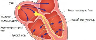
In the absence of diseases of the cardiovascular system, the SA node of nerve fibers of the heart (figure below) generates excitation impulses (from 60 to 80 impulses per minute), which, spreading along the nerve fibers to the muscles, cause their contraction.
Diagram of the distribution of nerve fibers throughout the muscle tissues of the heart. Taken from the article: https://bursaa.narod.ru/kardio.html
Due to the fact that the lengths of the nerve fibers are different, the excitation impulses of the muscle tissue are delayed differently relative to the SA node signal. As a result, a very complex movement of tissues surrounding the chambers of the heart occurs, expelling blood first from the atria into the ventricles, and then from the ventricles into the circulatory system. The cycle of the heart from excitation of the SA node to the end of excitation of all nerve endings is called systole. The cycle from the end of systole to the next excitation of the SA node is called diastole.
Due to the fact that excitation impulses are impulses of electrical potentials, they are projected onto the surface of the body. Consequently, between two points separated by a sufficient distance on the surface of the body, potential differences can be measured using a special device (differential millivoltmeter). The measured potential difference changes over time in accordance with the propagation of excitation waves along the nerve fibers during systole. The graph in mV coordinates, sec of the measured potential difference is an electrocardiogram.
It is obvious that the projections of excitation waves of the systolic cycle onto different parts of the body surface differ from each other, therefore ECGs taken from different points will also differ. In this regard, a diagnostically reliable ECG is one that is taken from certain points according to the rules established by V. Einthoven, E. Goldberger and F. Wilson.
Atlases, applications and videos on ECG
Atlases
1. Clinical electrocardiography - Franklin Zimmerman. 200 electrocardiograms with excellent analysis and explanations.
2. Podrids Real-World ECGs. One of the most famous atlases on ECG interpretation (6 volumes).
Books for a more in-depth study of ECG:
1. Marriott's Practical Electrocardiography is one of the most comprehensive guides to ECG;
2. Guide to electrocardiography - Orlov V.N.
You will find even more literature (books, atlases on ECG) in the article on the public page “MEDIC: ECG” https://vk.com/@medic_ecg-literatura-po-ekg
ECG video
There are good video courses that can help with understanding the basics of ECG: - ECG course from Strong Medicine: https://vk.com/videos-150727887?section=album_3 - “Everyone can do an ECG”: https://vk.com /videos-150727887?section=album_1 — “ECG is interesting!” from internist.ru: https://vk.com/videos-150727887?section=album_2
There are also various ECG applications for a smartphone (reference books, atlases, tests, or all together in one program):
— Of the Russian-language programs, in our opinion, the best (and this is not advertising) are the programs from the public “ECG Tests.” True, they are paid (link to the public page - see above); — ECG: Interpretation and Tests (Ecg). There is a small reference book in the appendix that might be useful to someone. For free. — ECG 100 Clinical Cases. Free application; — ECG Master. A good application with ECG tests, you answer them as a test. The downside is that if you want to open explanations for the ECG, you need to pay 140 rubles. Other ECG applications for smartphones in the article on the public page “MEDIC: ECG” https://vk.com/@medic_ecg-obzor-prilozhenii-po-ekg-na-smartfon
Presentations on ECG
Many different useful presentations on ECG at the link: https://vk.com/topic-150727887_41585478
Financial conditions
All lessons in the section “Textbook - simulator”, “Examples of cardiograms” are available freely. Exercises for the first 15 lessons in the “Textbook - Simulator” are also available freely (only authorization is required).
Starting from section 16 (“sinus tachycardia”), exercises are available for a fee. The cost of access to all sections is 10 rubles per day (for 24 hours). You can choose any number of days of access to paid sections. To transfer money, go to the "Payment" section.
General principle of diagnostics in medicine
Diagnosis of diseases in medicine is carried out according to the principle: from symptom to syndrome, from syndrome to diagnosis. Suppose we are in a forest in cloudy weather, and we need to determine the direction to the South. We look at the pines and see where their crowns are concentrated. The direction of crown concentration is a symptom of the direction to the South. However, to the south there may be a higher forest, shading the one where we are. Therefore, the crowns may thicken in a different direction, for example to the South-West. A symptom is one of the signs of an object (in our case, the direction to the South). It displays the object ambiguously due to not all known factors. Next we see an anthill. Its location relative to the tree is another evidence of the direction to the South. This is another symptom. For various reasons, the anthill may also not be exactly in the South. The sun came out from behind the clouds. Using it, knowing the time of day, you can approximately determine the desired direction. Another symptom. By comparing all three symptoms, you can more accurately determine the path to the South. This is already a syndrome. However, in order to go exactly in the right direction, a compass is required. The direction of its arrow is the diagnosis. A compass is a tool for determining direction. If it is not there, then the path is paved approximately as a result of the syndrome identified by several symptoms.
In medicine, symptoms are first identified - these are the patient’s complaints, for example, chest pain on the left. This symptom is a sign of various diseases. To find the cause of the patient's complaint, it is necessary to establish other symptoms. For example, does the patient have shortness of breath when climbing stairs? The presence of shortness of breath directs the doctor to the syndrome - disorders of the cardiovascular system. In other words, a number of symptoms (left chest pain and shortness of breath) suggest a syndrome (disorders of the cardiovascular system).
The path to diagnosis requires additional instrumental studies, the results of which can both refute and clarify the alleged syndrome until a final description of the cause of the patient’s complaint is made - a diagnosis that reveals pathological changes in the organ under study.
Cardiograph device
Most devices have a similar structural principle.
Schematic representation of the electrocardiogram recording chain
- Electrodes that need to be applied to the body and attachments to them (suction cups, belts, etc.).
- A switch that activates a pair of electrodes at the input to the amplifier.
- An amplifier that increases the signals from the electrodes so that they can be recorded, but ignores background noise, for example, from the movement of the subject.
- Recorder with the function of recording a cardiogram curve on film/paper.
- Filters that suppress network interference.
Before recording a cardiogram, it is necessary to calibrate the device - record millivolts
The technique of taking an ECG before starting recording involves calibrating the device - recording the calibration millivolt. This is the direct responsibility of nurses conducting functional diagnostics. Before recording data from the patient, a voltage of 1 millivolt is applied, and the device should display a deviation of 10 mm on the tape. If there is no calibration on the tape, then the correct reading of a given cardiogram can be questioned.
Heart functions and their disorders
As has been shown, from a technical point of view, the heart is a complex biological electromechanical device that contains: a self-oscillator (SA node), information transmission lines (nerve fibers), excitatory mechanisms (nerve endings) and an actuator (muscle tissue or the blood pump itself ). Consequently, the performance of the heart is characterized by the following functions:
- automaticity;
- conductivity;
- contractility.
Automaticity determines the possibility of self-generation of heart contractions without the influence of external factors.
Conductivity is the ability to conduct excitation impulses from the SA node to muscle tissue.
Contractility characterizes the ability of the muscle tissue of the heart to perform work when receiving an excitation impulse.
There is one more function that does not follow from the considered electromechanical model of the heart. This is a function of excitability. Excitability is defined as the ability (sensitivity) of the heart to perform the systolic cycle under the influence of external impulses. With constant excitability, conditions could arise for superposition of systolic cycles (current from the influence of the SA node impulse and a random external one). To eliminate such collisions, a mechanism is provided in the heart to reduce the excitability threshold at the time of the developed systolic cycle until the predicted beginning of the next one. By the time of the expected next impulse of the SA node, the excitability threshold is restored.
All known heart diseases cause disturbances in one or more of the four functions discussed. Violations of these functions (with the exception of contractility) cause ECG changes. Therefore, ECG diagnostics makes it possible to identify heart diseases that are not related to a violation of contractility function only. Taking into account the fact that most diseases that impair contractility affect the state of other functions, electrocardiography is an effective diagnostic tool for the state of the cardiovascular system.
Indications for the procedure
The examination is carried out only with a referral from a cardiologist. A visit to the doctor can be either planned or in case of complaints about heart function.
Frequent symptoms of impaired heart function include conditions such as:
- Compressive pain in the chest, as well as in the region of the heart, which radiates to the shoulder blade;
- Rapid pulse, which causes dizziness and increased heart rate;
- Shortness of breath with minor exertion;
- Periodic fainting;
- Dry barking cough;
- Swelling of the lower extremities.
Particular attention is paid to patients who have suffered a stroke, have congenital heart abnormalities or rheumatism.
Specialists conduct an examination and give a referral to see a doctor if they find:
- extraneous noise in the work of the heart muscle;
- with low or high blood pressure;
- heart rhythm disturbances;
The following categories of citizens are subject to mandatory examinations, namely:
- Persons with congenital or acquired heart disease;
- Athletes who have a serious load on their heart function;
- Patients undergoing examination before surgery;
- Women who are pregnant.

