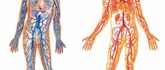
Vascular tone is the tension of blood vessels, supported by smooth muscles, an important factor in the blood supply to internal organs, one of the tools for general blood circulation. Vascular tone is influenced by the endothelium, the inner layer of blood vessels. Very thin (one cell thick), but plays a significant role in regulating vascular and cardiac tone, as well as in angiogenesis (formation of new vessels) and immune defense.
The fact that the endothelium is formed by a single layer of flat mesenchymal cells should seem to mean its insignificance, “microscopicity”. In fact, it is the largest organ in humans, present in all tissues. It affects blood clotting, kidney filtration, brain nutrition, and many other processes.
Theory
Tests
Help the site
Disable advertising
All vessels, with the exception of capillaries, have smooth muscle cells (SMCs) , due to which the lumen of the vessel , therefore the resistance to blood flow and the intensity of blood flow changes in a given region.
Local regulatory mechanisms:
- all vessels with SMCs are characterized by the original - basal tone created by the automaticity of smooth muscles;
- under the influence of various factors, basal tone can increase, while the vessels narrow and less blood enters the region;
- when vascular tone decreases, they dilate and blood flow into the region increases.
A decrease in tone leads to vasodilation, while an increase leads to vasoconstriction.
What normalizes vascular regulation in VSD
Hardening, moderate regular physical activity, and massage help to gradually improve the regulation of blood vessels.
Hardening with cold water should begin with the feet and face. The first 3-4 times - 30 seconds each, then gradually increasing the time of douches to 5 minutes and involving a larger area of the body. To enhance the effect, do contrasting douses, 2-3 changes of cold and hot water, always ending with cold. It is better to start tempering with water in the summer. Hardening is especially effective as a prevention of vascular regulation disorders in children with a tendency to VSD. Before starting hardening procedures, be sure to consult your pediatrician.
A restorative massage improves vascular regulation in all organs, as it activates reflex mechanisms through the skin and muscles. The most effective courses are 7-10 sessions every three months.
Sufficient physical activity would be daily gymnastics lasting 15-20 minutes, aimed at improving flexibility and muscle strength, as well as walking for 30 minutes. The need to expand and contract in response to adequate impulses gradually reinforces the coordinated response of the vessels.
But initially, in the human body with uncoordinated autonomic regulation, there are changes at the cellular level that disrupt energy synthesis. The amino acids glycine, cystine and glutamic acid help return metabolic processes in the cell to normal. They are directly involved in cellular respiration and activate it when there is insufficient activity. Eltacin® contains all three amino acids that are regularly needed by people with VSD.
Vascular tone
Tone is the tension created by the asynchronous contraction of the SMC of the middle layer of the vascular wall, which has automaticity.
Tone components:
- basal tone,
- humoral,
- central (neurogenic).
Mechanisms for regulating vascular tone:
- Local mechanisms that ensure blood flow through individual organs and tissues, that is, controlling the amount of blood flow in individual regions.
- The central mechanisms regulating systemic blood circulation are the constancy of blood pressure, blood flow, blood volume, etc.
Local regulatory mechanisms
The principle of local regulation is to ensure the independence of blood flow in organs from changes in systemic hemodynamics, that is, the provision of blood to a given region in its interests.
Local mechanisms for regulating blood vessel tone include :
- myogenic,
- metabolic.
Myogenic mechanism
- myogenic autoregulation is characteristic of the vessels of the brain, kidneys, heart, liver, celiac region, that is, regions where it is necessary to maintain constant blood flow;
- an adequate stimulus to SMCs is their stretching;
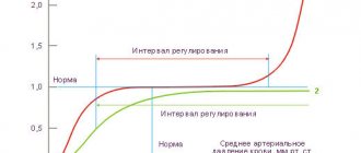
- with an increase in blood pressure (BP) -> stretching of the vascular walls -> contraction of the SMC of the vessels -> an increase in vascular tone and maintaining the same lumen -> the blood flow in the vessels does not change;
- a decrease in blood pressure causes a decrease in vascular tone due to relaxation of the SMC: in this case, despite the decrease in blood pressure, the flow of the same volume of blood into the vessels remains,
- Thus, the amount of basal tone is influenced by the level of blood pressure.
Metabolic mechanism
- metabolic products, dilating blood vessels, increase blood flow in working organs;
- as a result of insufficient supply of oxygen and nutrients to the region, metabolites accumulate in the tissues and blood flow increases due to the expansion of precapillaries.
Vascular tone decreases with a decrease in oxygen and carbon dioxide pressure, an increase in H and C3H6O3 ions and temperature - as a result, blood flow in working organs increases in proportion to their activity.
Central regulatory mechanisms
- nervous (reflex),
- humoral.
Nervous mechanisms
Vasomotor - vasomotor nerves:
- vasoconstrictors - vasoconstrictor nerves,
- Vasodilators are vasodilating nerves.
Vasoconstrictors
- All vasoconstrictors are sympathetic adrenergic nerves.
- The vasoconstrictor effect occurs when norepinephrine (NA) acts on α-adrenergic receptors.
- Impulses through sympathetic vasoconstrictors constantly arrive to the vessels from the neurons of the lateral horns of the thoracolumbar segments of the SC with a frequency of 1-3 impulses/s, maintaining resting tone.
- At a frequency of more than 3 pulses/s (from 3 to 15) - increased tone.
Vasodilators
- Parasympathetic cholinergic nerves:
- chorda tympani - drum string - dilates the vessels of the submandibular salivary gland;
- n. lingualis - lingual nerve - dilates the vessels of the tongue;
- n. glossopharyngeus - glossopharyngeal - dilates the vessels of the tonsils, posterior third of the tongue, parotid salivary gland;
- n. pelvicus - pelvic - dilates the vessels of the area of the same name.
- Sympathetic nerves:
- cholinergic, innervating vessels of skeletal muscles;
- adrenergic - a vasoconstrictor effect occurs when NA acts on β-adrenergic receptors in the vessels of the heart, brain and lungs.
- Dorsal root sensory nerves - dilate skin vessels according to the axon reflex (mediator - ACh).
Axon reflex:
- dilation of skin blood vessels is observed when insects bite, under the influence of mustard plasters, rubbing, scratching the skin;
- blood vessels that do not have special vasodilators dilate due to a decrease in the tone of vasoconstrictors (for example: in the abdominal organs).
Impulses along the vasomotor nerves to the vessels constantly come from the vasomotor center (VMC) .
The main localization of the vasomotor center is in the medulla oblongata (Ovsyannikov, 1871).
How does the endothelium affect vascular tone?
Among the products of endothelial activity, nitric oxide (NO) attracts attention. It regulates vascular tone. The production of nitric oxide increases with increasing pressure at a given vascular site. Blood pressure may rise due to increased physical activity or under the influence of certain hormones (for example, acetylcholine).
Increased pressure on the vessel wall activates a special set of enzymes called endothelial nitric oxide synthase (eNOS). These enzymes spur increased production of NO. Nitric oxide molecules are freely released through cell membranes and penetrate into smooth muscle. Under the influence of NO, muscle tissue relaxes - the walls of blood vessels, without encountering resistance from smooth muscles, expand, and the pressure inside these vessels drops.
A decrease in pressure weakens eNOS activity in the endothelium. Less nitric oxide is released - the vascular muscles tense again, maintaining the pressure at the working level.
Vasomotor center (VDC)
SC centers (lateral horns of gray matter) -> bulbar centers: vasoconstrictor, vasodilator -> hypothalamic centers (anterior ( depressor zone ) and posterior ( pressor zone ) parts of the hypothalamus) -> cortical representation of the SDC.
After transection of the brainstem above the quadrigeminal artery, blood pressure does not decrease , but when the brain is transected between the medulla oblongata and the spinal cord, it drops from 120 mm Hg. Art. up to 70-80.
SDC consists of 2 departments:
- pressor department,
- depressor department.
Both of these departments do not have clear boundaries. They are located at the bottom of the 4th ventricle among the neural structures of the reticular formation and mutually overlap each other.
Pressor and depressor neurons of the SDC are in a reciprocal relationship.
There are more pressor neurons than depressor neurons. The state of the SDC is judged by pressor neurons .
The SDC also includes other parts of the central nervous system.
At rest, the hypothalamus does not actively participate in the regulation of blood pressure.
The influence of the cortex on the regulation of blood pressure is conditioned reflex - an increase in blood pressure before the start, with excitement.
Conclusion: a multi-level system for regulating the functions of the cardiovascular system ensures adequate adaptation to the conditions of the external and internal environment.
The tone of the SDC depends on the nerve impulses constantly coming to it from the receptors of various reflexogenic zones.
Vascular reflexes
Vascular reflexes are divided into:
- own and
- conjugated.
Own reflexes
They are carried out from mechanoreceptors located in the heart and blood vessels ( baroreceptors ).
These receptors stabilize blood pressure.
There are own reflexes:
- pressor - increasing low blood pressure,
- depressor - lowering high blood pressure.
Reflexogenic zones (areas of maximum accumulation of receptors):
- aortic arch,
- carotid sinus (bifurcation of the common carotid artery into external and internal).
Depressor reflex: with an increase in blood pressure -> the baroreceptors of the aortic arch and carotid sinus are irritated -> excitation along the sensory nerves - aortic (depressor) and sinus (Hering's nerve) -> medulla oblongata -> the vagal center is excited and the vasomotor center is inhibited -> heart rate decreases - > blood vessels dilate -> blood pressure decreases (normalizes).
When blood pressure drops, the opposite is true, that is, the pressor reflex .
Own reflexes:
- are also carried out from chemoreceptors located in the aortic and carotid bodies;
- they are excited when CO2 and H ions increase in the blood and when O2 decreases;
- impulses arriving from chemoreceptors in the medulla oblongata increase the tone of the central nervous system, which leads to an increase in pressure.
Chemoreceptors are not in the vessel wall, but in the aortic and carotid bodies or glomeruli under the adventitia of the vessel and are penetrated by a network of capillaries.
From chemoreceptors -> SDC of the medulla oblongata -> SDC is excited -> vasoconstriction -> increase in blood pressure -> rapid blood renewal.
Conjugate reflexes
Carried out from receptors located outside the heart and blood vessels:
- they disrupt the stability of blood pressure, causing pressor reactions;
- distinguish conjugate reflexes: exteroceptive - from skin receptors,
- interoceptive - from internal organs.
Major vascular diseases of the legs
Vascular diseases of the lower extremities can strike at any age, sometimes with the development of complications.
Main vascular diseases of the legs:
- Atherosclerosis
- Saphenous vein thrombosis
- Phlebeurysm
Symptoms of venous disease of the legs can appear at a very early stage and if they are detected in time, the process can not only be stopped, but also completely get rid of the developing vascular disease of the lower extremities.
For atherosclerosis
arteries are affected as a result of cholesterol deposits with the formation of plaques on the walls of blood vessels, which can lead to complete blockage. As a result, ischemia develops - insufficient oxygen supply to cells and tissues.
Most often the popliteal, femoral and tibial arteries are involved in the pathological process.
The development of the disease occurs gradually and in the first stages there may be nothing to show for it. Subsequently, the patient develops pain in the lower extremities, especially with prolonged walking, and a feeling of numbness in the feet. A decrease in temperature is felt on the legs, pallor and cyanosis are visible, and cramps occur. Over time, a person develops intermittent claudication and trophic changes occur in the form of delamination of nails and the formation of ulcers on the fingers and heels.
Symptoms of atherosclerosis:
- muscle pain in the legs, in the later stages even the feet hurt, and the pain cannot be relieved with painkillers;
- limited mobility of the affected leg;
- pain along the affected artery - first during physical activity, then at rest;
- intermittent claudication - after walking some distance, a person is forced to stand up and give his legs a rest, and then he moves on until the next attack of weakness;
- tingling, numbness in the affected area;
- loss or slow growth of hair on the legs;
- paleness when raising the legs and sharp redness when lowering them;
- rapid freezing of feet;
- sores on the skin;
- purple fingers;
- absence of pulsation in the affected artery;
- areas of darkening on the skin are signs of the onset of gangrene in advanced stages;
- coldness of the skin of the leg;
- reduction in the mass and volume of the muscles of the thighs and legs.
At the beginning of the disease, as a rule, one limb suffers, then the process becomes symmetrical - this is a signal that the arteries are affected on both sides. An objective examination reveals the absence of pulsation in the popliteal fossa, thigh and ankle. Men get sick more often.
Causes
: poor nutrition, stress, smoking, diabetes.
We recommend
“TOP 15 harmful foods for adults and children, the most dangerous food additives” Read more
Saphenous vein thrombosis
- this is the formation of blood clots (thrombi) inside the lumen of the vein. Most often, venous thrombosis develops in the vessels of the lower extremities, but it can also occur in any other parts of the body, such as the veins of the upper extremities, neck, chest, etc.
There are three main causes of venous thrombosis
:
- Hypercoagulation
- increased activity of the blood coagulation system (hereditary predisposition, genetic defects, autoimmune diseases).
- Violation of the vascular wall
- Normally, the inner surface of the blood vessel (endothelium) is smooth. Pathological changes make the endothelium rough. Platelets, moving freely along with the blood flow, cling to the rough surface of the affected endothelium and a blood clot forms in this place.
- Blood flow disturbance
(stasis - stagnation of blood) blood flow is one of the main mechanisms that prevent the formation of blood clots, since the speed of blood flow does not allow platelets to accumulate in a certain place. Consequently, a decrease in blood flow speed promotes the formation of blood clots.
Provoking factors for the development of venous thrombosis
:
- Abnormal vein structure.
- Phlebeurysm.
- Excess body weight. Obesity of various stages leads to venous stagnation in the lower extremities, provoking thrombus formation.
- Prolonged bed rest.
- Oncological diseases.
- Pregnancy.
- Infections.
- Trauma and surgery.
- Taking medications that increase blood clotting.
- Smoking. When smoking, more than 300 different carcinogenic and toxic substances are released, which enter the systemic bloodstream through the alveoli of the lungs and cause damage to the vascular wall.
If a person has varicose veins of the lower extremities, a small trigger (“trigger”) is enough - blood thickening, infections of various natures, stress factors and physical activity, injuries, surgical interventions - for a blood clot to form in the venous system.
Venous complications are very dangerous, so it is important to know the first signs of superficial and deep vein thrombosis.
Clinical signs of superficial vein thrombosis:
- burning, throbbing pain along the thrombosed veins, limiting limb movements;
- a stripe of redness (hyperemia) in the projection of the affected vein;
- local increase in temperature;
- increased sensitivity (hyperesthesia) of the skin;
- in some cases, an increase in body temperature up to 38 ° C, malaise, and chills are noted.
Clinical signs of deep vein thrombosis:
- severe swelling below the site of blockage of the vessel;
- change in skin color to bluish-purple due to disruption of blood flow, often throughout the entire limb;
- bursting pain that intensifies with exercise;
- local temperature increase;
- general weakness, malaise.
Phlebeurysm
– expansion and lengthening of veins with thinning of the venous wall and the formation of nodes, as a result of pathology of the venous walls and valve failure. Normally, a healthy valve works correctly and blood flows in one direction; with varicose veins, the valve is deformed and blood flow is disrupted.
This is the most common peripheral vascular disease. According to epidemiological data, various forms of this disease occur in 26-28% of women and 10-20% of men of working age.
Risk factors for developing varicose veins:
- Pregnancy is traditionally considered one of the main risk factors for the development of varicose veins, explaining the more frequent occurrence of varicose veins in women. It is generally accepted that the main provoking factors are an increase in BCC (circulating blood volume) and compression of the pregnant uterus by the retroperitoneal veins.
- Obesity is a proven risk factor for varicose veins. At the same time, an increase in body mass index to 27 kg/m2 and above leads to an increase in the incidence of the disease by 33%. A high degree of food processing and a decrease in raw vegetables and fruits in the diet has led to a constant deficiency of plant fibers necessary for the remodeling of the venous wall and chronic constipation, which causes a permanent increase in intra-abdominal pressure.
- Lifestyle has a significant impact on the development and course of varicose veins. In particular, the adverse effects of prolonged static loads with heavy lifting or immobile standing and sitting have been proven.
- Dyshormonal conditions are involved in the pathogenesis of varicose veins. Their role has been progressively increasing in recent years, which is due to the widespread use of hormonal contraception, the popularization of hormone replacement therapy during the premenopausal period and in the treatment of osteoporosis. It has been proven that estrogens and progesterone, as well as their derivatives, reduce the tone of the venous wall due to the gradual destruction of collagen and elastic fibers.
- Varicose veins are a disease that is partial to the fair sex. On average, women suffer from varicose veins in the legs three times more often than men.
Here are the symptoms that should make you wary - these are the symptoms of varicose veins
:
- Pain - the variety of pain with varicose veins is also very rich (hot throbbing pain, night cramps and itching in the muscles, pain when walking, pain along the venous trunks, general pain and aching in the legs).
- Swelling in the legs.
- Feeling of fullness and heaviness in the legs.
- Skin changes - first the skin becomes dry, pigmentation appears - the skin of the legs darkens, becomes covered with brownish “spots”. Later, these symptoms may be joined by various dermatitis, eczema and so-called trophic disorders in the form of poorly healing wounds up to the formation of ulcers.
- Twisted, elongated varicose veins protruding above the surface of the skin of the legs and feet, with saccular, cylindrical or mixed extensions.
- Spider veins (telangiectasia). Yes, the group of patients with varicose veins also includes people with dilated small veins, since the reasons for their appearance are the same.
Targeted nutrition tips
that will increase your energy level by 10 out of 10
From TOP nutritionists of the MIIN
Get tips
The earliest stages of the development of varicose veins are very difficult to identify and diagnose, because at the earliest stages of the disease its main symptom – varicose veins – is absent. And the first and earliest symptoms of varicose veins, such as a feeling of heaviness in the legs, moderate pain in the legs, increased fatigue, also occur in the initial stages of arterial diseases, flat feet, and lumbar osteochondrosis.
The very first symptoms of varicose veins in the legs, by which you yourself can suspect the onset of varicose veins, are an increase in the pattern of veins on the skin of the legs. This defect, as we have already noted, is most often noticed by young women: they notice wreaths on their legs or thighs that were not there before. There may be no other complaints at the onset of varicose veins.
The beginning of the development and progression of varicose veins is already manifested by varicose nodes protruding above the surface of the skin. This is the main symptom of beginning varicose veins. Varicose veins can most often be found on the inner surface of the legs or thighs. When walking for a long time or standing for a long time, such symptoms of varicose veins appear as fatigue, heaviness in the legs, a feeling of fullness (often in the calves). These symptoms are especially pronounced during prolonged periods of sitting or standing. Aching or acute pain also appears in the areas of varicose veins (dilated veins), cramps in the calf muscles, especially in the evening, and sometimes at night.
The first signs of trouble in the veins are manifested by swelling of the legs at the end of the day. Swelling usually occurs in the evening, especially after standing for a long time. After an overnight rest, the swelling completely disappears. These symptoms of varicose veins go away quickly at first if you rest, especially if you lie down. However, the disappearance or significant reduction of these symptoms when walking and after a night's rest are very characteristic symptoms of varicose veins.
But varicose veins, if not seriously addressed, do not recede, but progress. Over time, symptoms of varicose veins appear such as convoluted dark blue intradermal veins protruding above the surface of the skin of the legs and feet. All this is accompanied by bursting pain in the legs and calves, a feeling of heat in the legs, night cramps in the calf muscles, and swelling. Gradually, these symptoms are accompanied by skin changes. First, the skin becomes dry, it darkens, and pigment appears on it - brown spots. A little later, so-called trophic disorders begin to appear: poorly healing wounds, eczema and even ulcers.
Humoral regulation
- Hormones formed in the endocrine glands: adrenaline, norepinephrine, vasopressin, etc. - constrict blood vessels.
- Vasoactive agents (local hormones) formed in tissues - acetylcholine, bradykinin, histamine, prostaglandins, etc. - dilate blood vessels.
- Dual-acting substances - catecholamines:
- alpha - narrowing
- betta - extension.
The hormones adrenaline and norepinephrine narrow the arteries and arterioles of the skin, skeletal muscles, and abdominal organs. Coronary vessels, vessels of the brain, and lungs expand at the same time, since it all depends on which adrenergic receptors perceive the hormone. When NA interacts with α-adrenergic receptors, the vessels narrow, and when interacting with β-adrenergic receptors , they dilate. β-adrenergic receptors predominate in the vessels of the heart, lungs, and brain.
Vasopressin mainly constricts arterioles and veins.
Angiotensin II is formed from plasma α-globulins under the influence of renin (JA cells of the renal cortex) and also constricts blood vessels.
Vascular tone:
- basal tone - the tone of the SMC and the influence of sympathetic vasoconstrictors;
- resting tone - tone of the SMC and the influence of sympathetic nerves with a frequency of 1-3 impulses/s;
- increased tone - impulses along sympathetic vasoconstrictors with a frequency of 3-15 impulses/s.
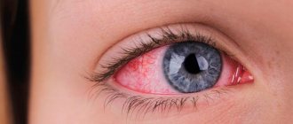
How to distinguish dystonia from heart and cerebral vascular disease
If you find symptoms that resemble VSD, you should not delay treatment, much less ignore this problem. But first of all, you need to make sure that you really have vegetative-vascular dystonia.
To clarify the diagnosis, your doctor will refer you for examination to a cardiologist and neurologist. Hardware research methods will help to exclude angina pectoris, arrhythmia, damage to the heart valves, changes in the blood vessels of the brain, and tumors.
With vegetative-vascular dystonia, symptoms periodically appear and disappear, and are associated with physical or emotional discomfort. The rest of the time the person feels healthy.