Classification
Erythrocytosis is diagnosed when the following indicators are exceeded: in men – 5.5x10⁹/ml, in women and children under 13 years of age – 4.7x10⁹/ml.
In newborns, the number of red blood cells should exceed 5.6x10⁹/ml. The following types of erythrocytosis are distinguished: 1. Based on the presence or absence of a connection with pathological processes:
- Physiological
. The change in the indicator is not associated with diseases. Occurs as part of adaptation to hypoxia caused by environmental conditions. - Pathological
. Accompanies some diseases and develops as a result of disturbances in homeostasis caused by various traumatic and non-traumatic causes.
2. According to the changes underlying erythrocytosis:
- Absolute
. It becomes a consequence of increased erythropoiesis against the background of hypoxia, increased production of erythropoietin due to kidney disease, endocrine diseases, and hormonally active tumors. - Relative
. Formed after a decrease in plasma volume as a result of profuse sweating, vomiting, and diarrhea.
3. By time of occurrence:
- Primary
. It is hereditary in nature, provoked by gene mutations that cause changes in the structure of hemoglobin or the enzyme that is responsible for transporting oxygen into red blood cells and its subsequent “return” to tissues. - Secondary (symptomatic)
. It develops throughout life and is potentiated by diseases and pathological conditions.
Symptomatic erythrocytosis is the most common type of this condition. Identified in men and women, it can be absolute or relative. Absolute symptomatic erythrocytosis is found more often than relative erythrocytosis and is provoked by the following factors:
- Kidney diseases
: ischemia of various etiologies, neoplasms, condition after kidney transplantation. - Neurohumoral regulation disorders
: excessive stimulation of the autonomic nervous system. - Endocrine disorders
: increased levels of thyroid hormones, glucocorticoids, catecholamines, adrenocorticotropic hormone. - Blood diseases
: hemoblastosis with increased production of red blood cells. - Hemic hypoxia
: poisoning with substances that reduce the oxygen capacity of the blood. - Circulatory hypoxia
: insufficient blood supply to organs and tissues due to impaired cardiac function. - Respiratory hypoxia
: a decrease in the volume of ventilation of the lungs against the background of bronchopulmonary pathologies. - Exogenous hypoxia
: normobaric hypoxia with a lack of oxygen in the air, hypobaric hypoxia with decompression sickness.
Among the relative symptomatic erythrocytoses, hemoconcentration ones predominate - arising as a result of a decrease in the amount of fluid in the vascular bed. Less common in women and men are redistributive erythrocytosis - conditions in which there is a sharp release of red blood cells from the depot.
Erythrocytosis
Why does erythrocytosis occur?
Physiological reasons
A high number of red blood cells is detected in residents of high mountain areas. Even in the absence of somatic diseases, the body suffers from hypoxia; due to a decrease in the amount of oxygen in the external environment, more blood cells are produced compensatoryly to transport it. In native-born residents of the highlands, physiological erythrocytosis is observed throughout their lives; in visitors, it develops during a long stay in the area.
Another example is erythrocytosis in infants after birth. In the womb, the fetal blood was saturated with oxygen from the mother's blood. There is less oxygen in a woman's blood than in the air, so the unborn child needed a lot of red blood cells to carry it. With the beginning of breathing atmospheric air, the supply of oxygen increases, the number of blood cells gradually decreases.
Respiratory hypoxia
Erythrocytosis is detected in pathologies accompanied by a decrease in the volume of pulmonary ventilation:
- Bronchial asthma.
Can be allergic, non-allergic (for example, aspirin), mixed. An attack of suffocation is accompanied by a feeling of tightness in the chest, a reduction in the duration of inspiration, an increase in the duration of exhalation, wheezing, forced positioning, and swelling of the neck veins. - Chronical bronchitis.
It can be obstructive or non-obstructive. Manifests itself as a cough with mucopurulent sputum. The amount of sputum increases during an exacerbation. Symptoms of obstruction are expiratory shortness of breath, persistent cough, wheezing, and swelling of the jugular veins. - COPD
Characterized by a progressive course of bronchitis or emphysematous type. In the first case, the symptoms of obstructive bronchitis predominate, in the second - expiratory shortness of breath. - Restrictive violations
. Chronic pneumonia, pneumosclerosis, pneumothorax, exudative pleurisy, widespread adhesions in the pleural cavity, and other conditions lead to restriction of the movements of the lung tissue.
Along with nonspecific diseases, various forms of pulmonary tuberculosis and pneumoconiosis play a significant role in the development of respiratory hypoxia with symptomatic erythrocytosis: silicosis, silicosis. In addition, this type of hypoxia is observed in smokers. Caused by the constant intake of tobacco smoke, the occurrence of smoker's bronchitis (a type of COPD). Men are more often affected.
Circulatory hypoxia
The phenomena of hypoxia are most clearly expressed in blue heart defects, accompanied by venous discharge or mixing of blood with hypovolemia of the pulmonary circulation, hypoxemia. Secondary erythrocytosis and the development of collaterals to a certain extent make it possible to compensate for hemodynamic disturbances. Due to the threat of irreversible dystrophic changes in the myocardium, children are recommended to undergo surgery at an early age. Erythrocytosis is characterized by the following defects:
- Fallot's disease: triad, tetrad, pentad;
- tricuspid valve atresia;
- transposition of the great vessels;
- anomalies of the confluence of the pulmonary veins;
- common arterial trunk.
Hemic hypoxia
A decrease in the oxygen capacity of the blood and compensatory erythrocytosis are potentiated by some exogenous intoxications. Carbon monoxide poisoning occurs at home or at work. Accompanied by a pressing headache, nausea, dizziness, unsteadiness of gait. Sometimes convulsions and coma are observed. Subsequently, retrograde amnesia, cardiac conduction disturbances, pneumonia, and bronchitis are possible.
Nitrate poisoning is caused by drinking water, agricultural products grown with nitrogen fertilizers, taking large doses of certain pharmaceuticals, and inhaling toxic fumes. Manifested by gastrointestinal disorders, jaundice, shortness of breath, hypotension, and disturbances of consciousness.
Exogenous hypoxia
At normal atmospheric pressure, but limited air flow, normobaric hypoxia occurs. Erythrocytosis is observed in victims of natural disasters, industrial accidents (for example, staying in a confined space without access to air after a mine collapse). The cause of hypobaric hypoxia is a decrease in barometric pressure when pilots spend a long time at high altitude. There is an extremely rapid change in environmental conditions with the development of decompression sickness.
Kidney diseases
Insufficient blood supply to the kidneys and the presence of space-occupying formations in the organ stimulate increased release of erythropoietin and activation of the red germ of hematopoiesis. Erythrocytosis is accompanied by:
- Pyelonephritis.
Acute inflammation is characterized by increased body temperature, weakness, dull pain in the lumbar region, and change in the color of urine. In a chronic process, the symptoms are smoothed out, the general condition is not disturbed. - Glomerulonephritis.
The acute form occurs with hyperthermia, signs of intoxication, hematuria, hypertension, edema, more pronounced on the face. Chronic glomerulonephritis typically has a relapsing course with the periodic appearance of acute inflammation. - Hydronephrosis.
Occurs due to obstruction of urine flow. It manifests itself as discomfort, dull pain in the lower back, which is later joined by progressive fatigue, transient hypertension, and hematuria. - Renal artery stenosis.
Increasing arterial hypertension and ischemic nephropathy are noted. The first syndrome includes cerebral manifestations, the second – low back pain, sometimes hematuria. The development of secondary hyperaldosteronism is possible. - Kidney cyst.
Erythrocytosis develops with compression of blood vessels and stimulation of the juxtaglomerular apparatus, accompanied by an increase in blood pressure. Other possible symptoms are pain and urodynamic disturbances.
The list of neoplasms that provoke erythrocytosis includes nephroblastomas, hypernephromas, and neoplasias that secrete erythropoietin. Sometimes the rate increases in patients who have undergone a kidney transplant.
Neurohumoral regulation disorders
Increased activity of the sympathetic nervous system, causing erythrocytosis, accompanies chronic heart failure and neurogenic essential arterial hypertension. A similar change in the level of red blood cells against the background of neurohumoral disorders is observed with severe sleep apnea or frequent panic attacks.
Endocrine pathologies
Some hormones increase the level of oxygen utilization, so their overproduction is accompanied by secondary erythrocytosis:
- Thyroid hormones
. Hyperthyroidism is observed in diffuse toxic goiter, autoimmune thyroiditis, and toxic thyroid adenoma. - Adrenocorticotropic hormone
. Hypercorticism is determined by pituitary microadenoma, Itsenko-Cushing syndrome caused by adenomas and adenocarcinomas of the adrenal glands, ACTH-ectopic syndrome. - Catecholamines and glucocorticoids
. Increased levels of hormones are found, respectively, in tumors of the medulla and cortex of the adrenal glands.
In addition, erythrocytosis is diagnosed in people taking androgenic steroids as doping to stimulate muscle growth and improve athletic performance.
Polycythemia
Erythremia (polycythemia or Vaquez's disease) is a chronic leukemia accompanied by increased proliferation of all hematopoiesis, primarily erythrocyte. Typical signs of the disease are plethora (telangiectasia, red or cherry skin tone, scleral hyperemia), unbearable skin itching. Many patients have erythromelalgia. As it progresses, bone pain, migraines, hypertension, and myalgia are observed.
Hereditary familial erythrocytosis
It is a group of genetically determined diseases with changes in the structure of hemoglobin, metabolic disorders in red blood cells, and excessive release of erythropoietin by the kidneys. The severity and time of onset of symptoms are determined by the form of hereditary familial erythrocytosis. Possible tachycardia, varicose veins, hemorrhoids, hepatosplenomegaly, shortness of breath, increased bleeding.
Fluid loss
Hemoconcentration with the formation of erythrocytosis occurs with significant fluid losses. In a healthy person, a transient disturbance in a laboratory indicator can be determined during prolonged thirst, after profuse sweating when working in conditions of high ambient temperature. The latter option is more common among men engaged in heavy physical labor. Erythrocytosis is also found in the following conditions:
- Intestinal infections:
cholera, dysentery, salmonellosis. Fluid is lost through repeated vomiting and profuse diarrhea with watery stools. - Extensive burns:
more than 15% of the body surface for burns of II and more severe degrees. The cause of hemoconcentration is the release of burn exudate. - Ketoacidosis:
ketoacidotic coma in patients with diabetes mellitus. The fluid is excreted from the body through the kidneys along with glucose.
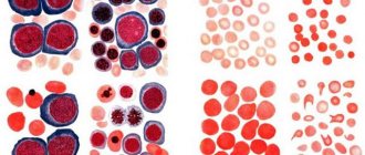
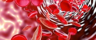
Red blood cell count
Increased hemoglobin
Diarrhea
Vomit
Thyrotoxicosis
20325 04 February
IMPORTANT!
The information in this section cannot be used for self-diagnosis and self-treatment.
In case of pain or other exacerbation of the disease, diagnostic tests should be prescribed only by the attending physician. To make a diagnosis and properly prescribe treatment, you should contact your doctor. Increased hemoglobin, or erythrocytosis: causes of occurrence, in which diseases it occurs, diagnosis and treatment methods.
Definition
Erythrocytosis is an increase in the content of red cells per unit volume of blood, accompanied by an increase in hemoglobin levels. The main symptoms of these changes are headaches, muscle pain, dizziness, nosebleeds, fatigue; more specific symptoms depend on the relevant disease.
Types of erythrocytosis
Erythrocytosis can be primary or secondary.
Primary erythrocytosis
is considered as an independent disease of the hematopoietic system and is of a genetic nature. Medically it is known as congenital polycythemia, or Vaquez's disease. This pathology provokes an increase in bone marrow volume and increased production of red blood cells and hemoglobin.
Secondary erythrocytosis is not considered a separate disease, but only a symptom of acute or chronic diseases and conditions. Relative erythrocytosis is a consequence of dehydration caused by excessive diarrhea or vomiting.
Also, an increase in hemoglobin levels can be the result of an overdose of medications, smoking, alcohol abuse and exposure to chemicals (nitrites).
Absolute erythrocytosis is a consequence of increased erythropoiesis, the process of formation of red blood cells in the bone marrow. This form of pathology is always associated with diseases of internal organs or systems.
Possible causes of increased hemoglobin
I. Hereditary:
- Changes in the structure of the Jak2 V617F gene, responsible for the production of red blood cells.
- Inability of blood to bind and transport oxygen to tissues.
- Reduced oxygen supply to the kidney tissues (this leads to the fact that they begin to intensively produce the hormone responsible for the formation of red blood cells (erythropoietin)).
- Deficiency of enzymes responsible for the production of red blood cells and their function of transporting oxygen to tissues.
II. Purchased:
- Kidney diseases (hydronephrosis, polycystic kidney disease, cancer and renal artery stenosis).
- Lung diseases (chronic obstructive bronchitis, bronchial asthma, diseases affecting lung tissue, sometimes of unknown cause).
- Heart diseases (congenital and acquired heart defects).
- Liver diseases (liver tumors).
- Brain diseases (in particular, cerebellar tumors).
- Diseases of the female reproductive system (ovarian cancer).
- Diseases of the endocrine system that affect the adrenal glands and contribute to increased blood pressure, in which the main drugs used for hypertension (Cushing's disease, pheochromocytoma) are usually ineffective, as well as diseases of the thyroid gland.
- Carbon monoxide poisoning.
- Staying at high altitudes.
- Obstructive apnea syndrome, characterized by temporary cessation of breathing during sleep.
Which doctors should you contact if your hemoglobin is increased?
If you detect an increase in the number of red blood cells, hematocrit, or hemoglobin, you must first contact a hematologist as soon as possible.
To clarify the diagnosis, a sternal puncture or bone marrow trepanobiopsy may be required.
Diagnostics
Diagnosis of lung diseases is carried out by pulmonologists. Patients with suspected kidney pathology are examined by nephrologists. Other diseases require the participation of hematologists, oncologists, and other specialists. Erythrocytosis is confirmed by a general blood test. Other indicators determined during the study are hemoglobin and hematocrit levels, the number of reticulocytes, leukocytes, and platelets.
Leukocytosis with a shift of the formula to the left indicates the presence of an inflammatory process. Reticulocytosis is detected in broncho-obstructive diseases, restrictive lesions of lung tissue, cyanotic heart defects, and polycythemia. Vaquez disease is also characterized by increased levels of platelets and leukocytes, and microcytosis is possible.
To clarify the diagnosis, the patient is prescribed an extensive laboratory examination. The level of erythropoietin is determined and the blood gas composition is examined. If endocrine diseases are suspected, hormonally active tumors are analyzed for adrenal hormones, thyroid hormones, and ACTH. Patients with polycythemia undergo trephine biopsy followed by histological examination. The list of instrumental techniques depends on the nature of the pathology:
- Lung diseases
. The basic study is chest x-ray. The images reveal signs of bronchitis, pneumonia, pneumosclerosis, tuberculosis, and other diseases. The method can be supplemented by MRI, bronchoscopy, spirometry, pneumotachography. - Heart defects
. The list of diagnostic procedures includes electrocardiography, phonocardiography, Holter monitoring, echocardiography, and chest radiography. Patients with pulmonary hypertension are advised to undergo angiography and cardiac probing. - Kidney diseases
. Patients are prescribed ultrasound, MRI, CT scan of the kidneys. Pyelography, excretory urography, nephroscintigraphy are performed. To identify secondary disorders from other organs, fundus examination, ECG, and ultrasound of the pleural cavity are performed. To clarify the nature of the pathological process, a biopsy with morphological examination is performed. - Endocrine disorders
. Depending on the existing symptoms, ultrasound or MRI of the adrenal glands or thyroid gland, radiography of the sella turcica, and contrast MRI of the brain may be recommended.
Erythremia (polycythemia vera, erythrocytosis, Vaquez disease)
In the early stages of the disease, there are usually no symptoms. However, as changes in the blood supply system increase, patients complain of a vague feeling of fullness in the head, headaches, and dizziness. Other symptoms appear depending on which body systems are affected. Paradoxically, hemorrhage can be a complication of erythremia.
The advanced stage is characterized by more pronounced clinical symptoms. The most common and characteristic symptom is headaches, sometimes in the nature of painful migraines with blurred vision.
Many patients complain of pain in the heart, sometimes like angina pectoris, pain in the bones, in the epigastric region, weight loss, blurred vision and hearing, unstable mood, and tearfulness. A common symptom of erythremia is skin itching. There may be paroxysmal pain in the tips of the fingers and toes. The pain is accompanied by redness of the skin.
Upon examination, attention is drawn to the special red-cyanotic color of the skin with a predominance of dark cherry tone. Redness of the mucous membranes (conjunctiva, tongue, soft palate) is also noted. Due to frequent thrombosis of the extremities, darkening of the skin of the legs and sometimes trophic ulcers are observed. Many patients complain of bleeding gums, bleeding after tooth extraction, and bruises on the skin. In 80% of patients, there is an enlargement of the spleen: in the advanced stage it is moderately enlarged, in the terminal stage severe splenomegaly is often observed. Usually the liver is enlarged. Often in patients with erythremia, increased blood pressure is detected. As a result of malnutrition of the mucous membrane and vascular thrombosis, ulcers of the duodenum and stomach can occur. Vascular thrombosis plays an important role in the clinical picture of the disease. Thrombosis of the cerebral and coronary arteries, as well as the vessels of the lower extremities, is usually observed. Along with thrombosis, patients with erythremia are prone to the development of hemorrhages.
In the terminal stage, the clinical picture is determined by the outcome of the disease - liver cirrhosis, coronary thrombosis, a softening focus in the brain due to thrombosis of cerebral vessels and hemorrhages, myelofibrosis accompanied by anemia, chronic myeloid leukemia and acute leukemia.
The prognosis depends on the patient's age at diagnosis, treatment, and complications. Mortality is high in the absence of treatment and in cases where erythremia is combined with leukemia and other cancers.
Treatment
Conservative therapy
Smokers are advised to give up the bad habit. The diet and physical activity regimen are selected taking into account the characteristics of the disease. Symptomatic treatment consists of bloodletting. To eliminate erythrocytosis, a course is prescribed, including 3-4 procedures with the simultaneous removal of 400-500 ml of blood.
When determining the need for manipulation, the compensatory role of erythrocytosis in patients with heart defects is taken into account; oxygen therapy is used according to indications, and antiplatelet agents are used. For renal pathologies, bloodletting is carried out until the hematocrit normalizes. In recent years, the procedure is performed less frequently than before, since studies indicate stimulation of the bone marrow and subsequent activation of hematopoiesis after bloodletting.
Etiopathogenetic therapy for hereditary erythrocytosis has not been developed. Antiplatelet agents, anticoagulants, and sometimes cytotoxic agents are recommended as symptomatic measures. Secondary erythrocytosis is eliminated by treating the underlying pathology:
- Kidney diseases
. For pyelonephritis, antibacterial and detoxification therapy, immunostimulants, and immunomodulators are indicated. Patients with glomerulonephritis are prescribed immunosuppressive therapy using cytostatics, glucocorticoids, NSAIDs, anticoagulants and antiplatelet agents. - Bronchopulmonary pathologies
. Bronchodilators and mucolytics are effective. During periods of exacerbation of inflammatory diseases, antibacterial agents are recommended. Patients with COPD and bronchial asthma need to use inhalers, nebulizers, and spacers. Anti-tuberculosis medications are effective for tuberculosis. - Endocrine disorders
. For diabetes mellitus, it is necessary to correct glucose levels by administering insulin or hypoglycemic agents. Hyperthyroidism requires thyreostatics. Itsenko-Cushing syndrome involves treatment with drugs that suppress steroidogenesis. For pituitary microadenomas, radiation therapy and dopamine agonists are effective.
Surgery
For secondary erythrocytosis, the following operations can be performed:
- Respiratory hypoxia
: drainage of the pleural cavity, thoracostomy, thoracoplasty, pleurectomy, lung resection. - Circulatory hypoxia
: aortic and pulmonary artery transplantation, Norwood operation, Fontan operation, heart transplantation. - Kidney pathologies
: bougienage, installation of stents, lithotripsy and removal of stones to restore urine passage, plastic hydronephrosis, excision of a kidney cyst, removal of a kidney tumor, kidney resection, nephrectomy. - Endocrine diseases
: resection of the thyroid lobe, hemithyroidectomy, removal of the thyroid gland, adrenalectomy, excision of a pituitary tumor.