Autoimmune thyroid diseases occur predominantly in children and women. Against the background of such an illness, the immune system incorrectly reacts to the cells of its own body and begins to actively fight it. This condition can be dangerous during pregnancy. What to do if AT to TPO is very elevated? What does this mean, what are the dangers and what treatment measures need to be taken? When can pathology be suspected and who is usually most susceptible to it? The answers to all these questions will be given below.
Medical description of AT to TPO
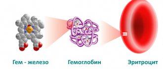
Anti-TPO Ab is an immune system protein. Determining the presence of this component in the blood reflects how aggressive the protective functions are towards the body’s own cells. Antibodies are known to serve as the basis for the human immune system. Thanks to them, dangerous harmful cells entering the body from the external environment can be recognized and destroyed. True, they often suddenly begin to fight with their own cells, because they mistake them for their enemy. Symptoms and causes of Graves' disease will also be described.
If the level of antibodies to thyroid peroxidase (AT to TPO) is significantly increased, this means only one thing - the human immune system is not reacting correctly to its own cells. In such a situation, the development of pathology is guaranteed, which entails danger in disrupting the functioning of various organs and systems, which in turn can lead to the development of serious diseases. The reasons for the increased production of antibodies, as a rule, are disorders in the thyroid gland, due to which thyroid peroxidase penetrates into the blood from this organ.
So, AT to TPO is greatly increased, what does this mean? Let's figure it out.
Thyroid peroxidase is necessary for the body to produce iodine synthesis, which in turn is required for the production of the hormones T3 and T4. As the level of antibodies increases, iodine synthesis is greatly reduced, which means this directly affects the production of hormones by the thyroid gland. With an insufficient amount of such enzymes, pathologies of the gastrointestinal tract, cardiovascular, nervous and even respiratory systems develop.
If AT to TPO is elevated, the consequences can be very serious.
Free triiodothyronine (free T3)
Thyroid hormone stimulates the exchange and absorption of oxygen by tissues (more active than T4).
Produced by follicular cells of the thyroid gland under the control of TSH (thyroid-stimulating hormone). In peripheral tissues it is formed during deiodination of T4. Free T3 is the active part of total T3, accounting for 0.2 - 0.5%.
T3 is more active than T4, but is found in the blood in lower concentrations. Increases heat production and oxygen consumption by all body tissues, with the exception of brain tissue, spleen and testicles. Stimulates the synthesis of vitamin A in the liver. Reduces the concentration of cholesterol and triglycerides in the blood, accelerates protein metabolism. Increases calcium excretion in urine, activates bone turnover, but to a greater extent, bone resorption. It has a positive chrono- and inotropic effect on the heart. Stimulates reticular formation and cortical processes in the central nervous system.
By 11–15 years, the concentration of free T3 reaches adult levels. In men and women over 65 years of age, there is a decrease in free T3 in serum and plasma. During pregnancy, T3 decreases from the first to the third trimester. One week after delivery, serum free T3 levels return to normal. Women have lower concentrations of free T3 than men by an average of 5 - 10%. Free T3 is characterized by seasonal fluctuations: the maximum level of free T3 occurs from September to February, the minimum in the summer.
Limits of determination:
1.5 pmol/l - 46.1 pmol/l.
Norms of AT protein content to TPO in the human body
In healthy people under the age of fifty, the level of such hormones in the blood should be below 5.6 mIU/ml. For those over fifty, this figure can usually increase. This value of the volume of the protein in question is quite stable and does not depend at all on the gender of the patient. It should be said that about seven percent of the world's population typically experiences an increase in antibodies to TPO. AT to TPO is greatly increased, what does this mean? More on this later.
It would not be superfluous to note that the deviation of this indicator itself is most often observed among women. Establishing the level of antibodies to thyroid peroxidase is of particular importance during pregnancy. A noticeable increase in indicators indicates greater risks associated with bearing a fetus or the birth of a child with possible congenital abnormalities. In women who are carrying a fetus, the normal level of antibodies should be no more than 2.6 mIU/ml.
When should you be tested to determine the level of antibodies to thyroid peroxidase?
A blood test for these antibodies is not considered mandatory for all categories of patients. Such research may be needed in the following situations:
- during pregnancy;
- with an enlarged thyroid gland;
- in case of suspected hypothyroidism;
- risk of autoimmune diseases;
- suspicion of thyrotoxicosis.
This analysis, of course, is most important during pregnancy. Based on its results, medical specialists are able to predict the risk of increased antibodies to thyroid peroxidase and thyroiditis in women in the postpartum period. If the volume of the hormone AT to TPO is increased, the risk of acquiring pathology increases twice as compared to normal tests.
In addition, this test may be needed before drug therapy with certain medications that have negative effects when antibody levels are high. It should also be noted that in some patients the volume of AT may be increased even in the absence of any pathologies. Also, the level of the hormone increases against the background of other autoimmune diseases that are not related to the functioning of the thyroid gland.
How to take it correctly
Blood for analysis is taken from a vein. Depending on the symptoms, the doctor identifies a group of hormones that need to be tested. It is advisable to completely limit physical and psycho-emotional stress for 12 hours, not to drink alcohol, medications or food containing iodine.
Particular attention is required in preparation for donating blood samples for representatives of the fair half - it should be carried out during a certain period of the menstrual cycle, which is designated by the doctor. The donation procedure itself, as a rule, is scheduled for the morning hours, on an empty stomach.
AT to TPO is increased - reasons
A high level of antibodies exceeding the norm is usually observed in the following diseases:
- various viral diseases;
- chronic renal failure;
- thyroiditis;
- Graves' disease;
- thyroid injuries;
- autoimmune diseases of a hereditary nature;
- diabetes;
- rheumatism.
Also, elevated antibodies to TPO occur if, shortly before the tests, the patient underwent radiation therapy in the head and neck area. It should be said that the analysis for these antibodies is not used as part of the measure to monitor the therapy being carried out. The examination is necessary only to determine whether pathology is present or not.
The danger of increasing antibody levels
An increased level of antibodies to thyroid peroxidase is justifiably considered an extremely serious deviation, indicating incorrect functioning of the immune system. As a result of such a failure, there is a risk of developing a lack of thyroid hormones, which are very important for the health of our body. They regulate the functioning of various organs and tissues, and against the background of their deficiency, there is a threat of serious diseases.
An increased level of antibodies can lead to the following number of diseases:
- The appearance of hyperthyroidism. Symptoms of this pathology are expressed in sudden weight loss, fatigue, irritability, rapid pulse, hair loss, goiter, shortness of breath, menstrual irregularities and poor sleep.
- Development of hypothyroidism. The main complaints of patients with this disease are intolerance to low temperatures, disturbances in the stomach and intestines, poor condition of hair and nails, and excess weight.
If AT to TPO is elevated and signs are detected during pregnancy, then there is a significant risk of miscarriage or the birth of a baby with all sorts of pathologies. Women who have an increased level of antibodies to thyroid peroxidase can often face a problem such as hormonal imbalance. Its appearance can lead to major problems with the health of the female genital organs.
Indications for Anti TPO analysis
The Anti-TPO definition is not included in the list of mandatory diagnostic tests during clinical examination; the decision on when to take the test is made by an endocrinologist if a patient suspects thyroid pathologies.
Primary autoimmune thyroiditis is diagnosed in patients with the development of hypothyroidism with its characteristic symptoms. In the early stages, patients complain of decreased concentration, weakness, apathy, and memory impairment. As the disease progresses, more informative signs appear:
- pathological weight loss;
- swelling of the face and lower extremities;
- cold intolerance;
- thinning hair, brittle nails;
- menstrual irregularities in women or absence of menstruation;
- infertility;
- cholelithiasis.
With long-term hypothyroidism, the patient’s face acquires a characteristic appearance for the disease (myxedematous) - swelling, weak facial expressions, an aloof look, yellowish skin. During diagnostic testing, high autoantibodies to TPO are detected in the blood test of such patients.
The clinical picture of toxic goiter has more pronounced symptoms:
- goiter;
- tachycardia;
- strong weight loss with a healthy appetite;
- goggle-eyed;
- emotional disorders: tearfulness, irritability;
- heart failure;
- weakness of the muscles of the limbs.
The first pronounced symptoms of Graves' disease appear several months before diagnosis - heart failure and eye changes.
Treatment for elevated levels of antibodies to thyroid peroxidase
The symptoms and causes of Graves' disease are of interest to many.
Treatment of deviations in the volume of AT TPO, as a rule, consists of eliminating the autoimmune diseases that lead to this pathology. In order to determine an accurate diagnosis, doctors need to study the patient's medical history, carry out various additional diagnostic procedures, and also carry out a detailed blood test.
If elevated antibodies to TPO are detected, then treatment of underlying pathologies is required:
- Postpartum thyroiditis. Often this disease goes away with virtually no symptoms. Young women encounter it, as a rule, in the first years after the birth of a baby. The most common complaints of patients are fatigue, increased irritability, rapid heartbeat, trembling in the arms and legs. As with the autoimmune type of the disease, symptomatic treatment is required. A table of antibodies to thyroid peroxidase is presented below.
- Graves' disease. This pathology manifests itself in weakness, tremors of the limbs, the formation of toxic goiter, high blood pressure, increased sweating and is accompanied by arrhythmia. True, such a disease can be treated quite successfully, especially in its first stages. The drugs most often prescribed as medicinal methods are Propicil and Thiamazol. These drugs block thyroid functions. In addition, patients may be given radiotherapy to the thyroid gland.
- Symptoms of autoimmune thyroiditis. This pathology has symptoms such as decreased concentration and performance, tremors, sudden weight gain, dry skin and hair, sweating, and arrhythmia. General therapy of the disease is reduced to alleviating the main symptoms. At the moment there are no special drugs to treat this disease. It is important to note that the detection of antibodies in the blood is not considered a clear reason for determining an accurate diagnosis. It is not uncommon for such a deviation to be observed among absolutely healthy people. If the patient’s TSH level is normal, this indicates that there is no disease.
Elevated antibodies to thyroid peroxidase are often found.
Thus, regular monitoring of thyroid function is recommended for all women. In addition, patients may be prescribed replacement therapy. In cases of failure of the normal activity of the heart muscle, it will be necessary to use appropriate medications. Vitamin therapy and adherence to standard recommendations for a healthy lifestyle are also considered mandatory. Against the background of dysfunction of the thyroid gland, hormonal therapy may well become a lifelong measure.
We looked at antibodies to thyroid peroxidase; what they are is now clear.
Thyroiditis in children
The thyroid gland is located on the front surface of the neck and, more than other endocrine organs, is exposed to the influence of the external environment, and the unity of the lymphatic ring of the pharyngeal tonsils and the thyroid gland creates conditions for the occurrence of an inflammatory process in it against the background of chronic diseases of the nasopharynx.
The child's body is predisposed to the development of autoimmune processes, since its lymphatic system is more active than that of adults.
Most often, existing classifications of inflammatory diseases of the thyroid gland are based on external symptoms. In particular, our classification includes: a) acute thyroiditis (purulent and non-purulent); b) subacute thyroiditis; c) chronic thyroiditis (fibrous - Riedel and lymphomatous - Hashimoto); d) thyroid tuberculosis; e) syphilis of the thyroid gland; f) parasitic diseases; g) actinomycosis of the thyroid gland. Obviously, the concept of “inflammatory diseases of the thyroid gland” includes pathological conditions that have different pathogenetic essences, in particular, thyroiditis often has an autoimmune rather than inflammatory genesis. This approach to the classification of this pathology still causes controversy among specialists.
Inflammatory diseases of the thyroid gland are relatively rare (1–2% of all thyroid diseases). Thyroiditis is an inflammation of a previously normal thyroid gland (strumitis is an inflammatory process in a goitrous gland). Thyroiditis is distinguished between nonspecific (acute, subacute and chronic) and specific - tuberculous, syphilitic, actinomycotic, parasitic.
Acute thyroiditis
The cause of the disease can be any acute or chronic infection (flu, tonsillitis, acute respiratory infections, chronic tonsillitis, scarlet fever, mumps, tuberculosis, syphilis, etc.).
Inflammation can develop either directly during infection or as a complication. The cause of its occurrence can also be various intoxications with iodine, lead, as well as traumatic damage to the capsule. In some cases, the etiology of thyroiditis cannot be established.
Clinical manifestations of acute thyroiditis are variable. More often the process begins unnoticed, gradually. There is pain in the neck, sometimes pain when swallowing, slight hoarseness, weakness, and malaise. The temperature reaction is often absent; less often, an increase in temperature may be observed. Often during this period the disease remains unattended, and the doctor’s efforts are aimed at examining the nasopharynx.
After a few days (sometimes after 2–3 months), the temperature rises, chills, weakness, headache, sweating, tachycardia, and in some cases nausea and vomiting appear. The thyroid gland increases in size, becomes dense and sharply painful. Movement of the head, coughing, and swallowing increase pain, which can radiate to the head, ears, and jaw. Swelling and dilation of the veins of the neck, pastiness and hyperemia of the face are noted, the cervical lymph nodes increase in size, moderate leukocytosis with a shift to the left, and accelerated ESR are detected in the blood.
At the onset of the disease, there are signs of increased functional activity of the thyroid gland.
In the future, signs of hypothyroidism are sometimes detected, which are also transient in most cases.
The acute period lasts an average of 3–6 weeks, after which the temperature normalizes, pain disappears, and the size of the thyroid gland decreases. Due to the appearance of the inflammatory process in other parts of the gland, relapses of the disease may occur, so recovery from thyroiditis (with restoration of thyroid function) can be said after 4–6 months from the onset of the disease.
Along with cases of thyroiditis that spontaneously end in recovery, severe, fulminant forms may occur, which are accompanied by necrosis and purulent melting of the parenchyma. These forms are often caused by streptococci, staphylococci, and pneumococci. In these cases, the disease develops acutely and is characterized by a general severe condition, a significant increase in temperature, chills, tachycardia, severe pain in the anterior surface of the neck, and often a feeling of suffocation. There is significant leukocytosis (15,000–20,000), neutrophilia, a shift in the leukocyte formula to the left, and accelerated ESR.
Suppuration can be diffuse and localized in the form of an abscess. A breakthrough of pus occurs through the skin, as well as the esophagus, larynx or trachea (aspiration pneumonia), and mediastinum (mediastenitis).
Purulent thyroiditis, more often than other forms, ends with the development of fibrosis of the thyroid gland, which leads to a decrease in its functional activity and the development of hypothyroidism.
Treatment of acute thyroiditis should include dietary nutrition (liquid, semi-liquid food in small portions). The patient is prescribed bed rest during the entire acute period, the position in bed is semi-sitting.
Antibiotic therapy is mandatory, in large doses during the first 7–10 days. The average course of antibiotic treatment is 3–4 weeks.
At the same time, desensitizing agents are prescribed in age-specific doses throughout the entire acute period. According to indications, oxygen therapy, cardiac, painkillers, and sleeping pills are used. For hypofunction of the thyroid gland, treatment is carried out with L-thyroxine (50–100 mcg). It is advisable to prescribe vitamin therapy (A, C, B1, B2, B12).
Warming compresses, diathermy, and Solux are not indicated for everyone.
Treatment of acute thyroiditis should begin as early as possible, the doses of drugs should be large enough, and treatment should not be stopped immediately after the signs of inflammation are eliminated.
For purulent thyroiditis, surgical treatment (opening of the abscess and drainage) is indicated.
Subacute thyroiditis
Subacute thyroiditis, known as giant cell granulomatous thyroiditis, is a rare disease. Recently, it has been assumed that it is viral in nature. Often the disease develops after suffering from influenza, measles, mumps and other infections. In a significant proportion of patients, autoantibodies to thyroid tissues are detected in the blood serum, which suggests the presence of autoimmune mechanisms in the pathogenesis of this disease. Subacute thyroiditis is characterized by the formation of giant cells and granulomas resembling a tuberculous tubercle, as well as lymphoid-plasmacytic infiltration of the stroma, which is characteristic of an autoimmune process.
Clinically, subacute thyroiditis is characterized by pain in the thyroid gland, radiating to the parotid region, neck, and back of the head. The thyroid gland increases in size and is painful on palpation. Symptoms of intoxication are noted: weakness, lethargy, fever.
The disease is often accompanied by dysfunction of the thyroid gland, with the appearance of symptoms of thyrotoxicosis: weakness, sweating, irritability, tachycardia. Subsequently, the phase of hyperthyroidism is replaced by symptoms of hypothyroidism, due to depletion of the synthesis of thyroid hormones. The levels of triiodothyronine (T3) and thyroxine (T4) in the blood decrease, and the content of thyroid-stimulating hormone increases.
At the peak of the disease, accelerated ESR, dysproteinemia (increased α2- and γ-globulin fractions) are detected, and antibodies to thyroglobulin in low titers are detected in the blood.
In the treatment of these forms of thyroiditis, the use of glucocorticoids is of particular importance. Salicylic or pyrazolone drugs are also used. Antibiotics are prescribed for the period of hormonal therapy and have no independent value.
Prescribing methizol in the hyperthyroid phase is not recommended, since thyrotoxicosis is caused by the destruction of thyrocytes and the release of previously synthesized hormones into the blood. It is more advisable to use β-blockers.
Considering that hypothyroidism in subacute thyroiditis is reversible, taking thyroid hormones is not required. Normalization of the echostructure of the thyroid gland in patients occurs with a delay compared to the normalization of clinical and laboratory parameters.
Chronic fibrous thyroiditis
Chronic fibrous thyroiditis (Riedel's goiter) is rare.
Microscopically, the thyroid gland reveals a chronic productive inflammatory nonspecific process with a pronounced proliferation of dense fibrous connective tissue with a certain amount of diffuse lymphoid cell infiltrates and very often with the presence of eosinophils, which is one of the characteristic signs of this disease. The architecture of the thyroid gland is disturbed.
The disease begins unnoticed, a protrusion appears on the neck, which is not accompanied by pain. When the trachea and esophagus are involved in the process, breathing and swallowing disorders, hoarseness, and dry cough are noted.
The thyroid gland is often diffusely enlarged. The consistency is very dense, woody, the surface is smooth. The skin over the gland is not changed. Regional lymph nodes are not enlarged. With a long course of the disease, adhesion of the goiter to the surrounding tissues is noted, which may suggest the presence of a malignant tumor of the thyroid gland. The general condition, as a rule, is not disturbed, the function of the thyroid gland does not change.
An X-ray examination reveals signs of displacement or compression of the trachea and esophagus.
The generally accepted method is surgical treatment of this disease, which consists of resection of the isthmus and nearby areas of the lobes to relieve the symptoms of tracheal compression. After the operation, the process may reverse.
Chronic autoimmune thyroiditis
Among inflammatory diseases of the thyroid gland in children, a large proportion belongs to chronic autoimmune thyroiditis (CHAT). This is due to the deterioration of the environmental situation, as well as the unity of the lymphatic ring of the pharyngeal tonsils and the thyroid gland, which creates conditions for the occurrence of an inflammatory process in it against the background of chronic tonsillitis.
Autoimmune damage to the thyroid gland was first described by Hashimoto in 1912. The author called this disease Struma lymphomatosa.
With the accumulation of clinical data related to autoimmune damage to the thyroid gland, clinical classifications have been developed, the most successful of which is the classification of R. Volpe (1984), in the rubric of which ChAT is divided into several different forms, among which lymphocytic thyroiditis of children and adolescents is specifically distinguished.
In the pathogenesis of CHAT, the main role is played by disorders of immunological control in hereditarily predisposed children. It is known that ChAT according to the HLA system is associated with the DR 3 and DR 5 loci, like many other autoimmune diseases, and the association with the DR 5 locus causes the hypertrophic, and with the DR 3 locus, the atrophic form of ChAT.
The hereditary genesis of the disease is confirmed by data on frequent cases of the disease in close relatives. Other autoimmune diseases of endocrine and somatic origin are often recorded in the pedigree of patients with CAT, which may indicate an innate predisposition to autoimmune reactions.
A genetically determined defect in immunocompetent cells leads to a breakdown of natural tolerance and activation of cellular and humoral immunity. This process can be caused by a viral infection, medications, or, less commonly, injury. However, there is still no convincing data on the etiology of CHAT.
The process of producing antibodies to thyroglobulin, the microsomal fraction of the thyroid gland, the thyroid-stimulating hormone receptor of the pituitary gland, the release of lymphotoxins, tumor necrosis factor, etc. is launched, which ultimately causes destructive changes in thyrocytes and a decrease in thyroid function. The outcome of ChAT is the development of hypothyroidism. This condition leads to an increased release of thyroid-stimulating hormone from the pituitary gland, which provokes the growth of surviving thyrocytes followed by their infiltration by lymphocytes, macrophages, and plasma cells.
Histological examination of the thyroid gland reveals diffuse or focal infiltration of lymphocytes and plasma cells. The presence of large epithelioid Hürthle-Ashkenazi cells is characteristic. Depending on the phase and duration of the process, stromal fibrosis can be detected. The disease occurs in hypertrophic and atrophic variants.
In children, CHAT often develops in the pre- and puberty period, less often in preschool and primary school age. In the vast majority of children, autoimmune thyroiditis is accompanied by the development of goiter; in 5–10% of patients, an atrophic form of the disease is noted.
ChAT may be one of the components of autoimmune polyendocrine syndrome (APS). For the first time, the combination of autoimmune thyroiditis with primary adrenal insufficiency was described by E. V. Schmidt. Currently, two types of APS are distinguished.
The first type of APS involves adrenal insufficiency, hypoparathyroidism, autoimmune thyroiditis, type 1 diabetes mellitus, candidiasis, nail dystrophy, hypoplasia of tooth enamel, vitiligo, alopecia, chronic active hepatitis, autoimmune gastritis, hypogonadism, malabsorption syndrome. This syndrome is caused by a mutation in the autoimmune regulator gene. The second type of APS includes type 1 diabetes mellitus, adrenal insufficiency, autoimmune thyroiditis, and hypogonadism.
ChAT can be combined with rheumatoid arthritis, pernicious anemia, and systemic lupus erythematosus.
The disease is 4–5 times more common in girls. A family predisposition for thyroid pathology is detected in 65% of children, and 3 times more often on the maternal side.
In most cases, CHAT develops gradually. Clinical diagnosis of CHAT in children is based on the syndrome of enlarged thyroid gland.
Currently, one should use a classification of goiter sizes based on palpation data (WHO, 1994). According to WHO criteria, the following sizes of the thyroid gland are distinguished:
- 0 degree - no goiter;
- I degree - the size of the lobes of the thyroid gland is larger than the distal phalanx of the thumb of the patient being examined, but is not visible to the eye;
- II degree - the goiter is palpable and visible to the eye.
In case of CAT in children, the size of the goiter most often corresponds to the II degree of increase in the lobes of the thyroid gland. On palpation, the thyroid gland is compacted, less often - elastic in consistency, heterogeneous, with an uneven surface, mobile when swallowing.
The chronic course of the disease ultimately leads to hypothyroidism. But unlike adults, 1/3 of patients with ChAT debut with symptoms of increased thyroid function. The hyperthyroid phase of ChAT - this phenomenon is called hashitoxicosis, in contrast to similar manifestations in diffuse toxic goiter - develops as a result of the destruction of thyroid tissue and the release of a large amount of deposited hormones into the blood. In these cases, against the background of an enlarged thyroid gland, tachycardia, changes in blood pressure with an increase in pulse pressure, tremor of the eyelids and outstretched fingers, and increased bowel movements are observed. As a rule, exophthalmos, one of the typical symptoms of diffuse toxic goiter, is not recorded at this phase of ChAT.
The hyperthyroid phase in its natural course lasts no more than six months. In the future, spontaneous remission may occur. In 19% of patients, symptoms of hypofunction of the thyroid gland are detected, which manifest themselves in decreased performance at school, a tendency to constipation, chilliness, bradycardia, and bradypsychism syndrome.
When studying thyroid hormones, despite various clinical dysfunctions of the thyroid gland, the levels of T4 and T3 remain within the normal range. With hypofunction of the thyroid gland, the levels of thyroid-stimulating hormone of the pituitary gland are 2–2.5 times higher than its average values.
In tests of children who are diagnosed with thyrotoxicosis upon admission to the hospital, high levels of thyroid hormones are detected, thyroid-stimulating hormone levels are at the lower limit of normal or are below normal values.
In case of CAT, ultrasound examination reveals a diffuse or focal decrease in the echodensity of thyroid tissue. Hypoechoic areas without clear contours are observed in most patients. In some patients, hyperechoic “strands”—foci of fibrosis—are visualized. In cases where the echographic picture resembles a nodular formation with unclear contours, a dynamic examination of such a gland is necessary.
Thyroid antibodies to thyroglobulin and thyroid peroxidase are detected in 80–95% of patients with CAT in various diagnostic titers.
Fine needle biopsy is the most accurate diagnostic method. The morphological sign of ChAT is typical lymphoid infiltration, which always predominates over the epithelial component. In addition to lymphocytes, the presence of plasma cells and macrophages, large Hürthle-Ashkenazi cells is characteristic. The main indication for a puncture biopsy is suspicion of thyroid nodules or cancer suspicion.
So, the disease does not have a specific clinical picture, just as none of the research methods by itself can reliably make a diagnosis of ChAT. A comprehensive assessment of the pathological process in the thyroid gland involves studying the echostructure, thyroid status, and thyroid antibodies.
In differential diagnosis, certain difficulties are presented by the hyperthyroid phase of ChAT and the presence of diffuse toxic goiter, since ultrasound data, thyroid antibodies, and the morphological picture of punctate do not always allow an accurate diagnosis.
To diagnose CHAT, one should rely on the following indicators.
- Typically, thyrotoxicosis develops at the onset of the disease, followed by restoration of thyroid function after 4–6 months or the gradual development of hypothyroidism.
- Symptoms of thyrotoxicosis do not reach the intensity that is observed with diffuse toxic goiter.
- Thyroid-stimulating antibodies are markers of diffuse toxic goiter, and their detection in patients with CAT indicates a combination of two autoimmune diseases.
- The presence of ChAT may be indicated by the rapid development of euthyroidism during treatment of thyrotoxicosis. The course dose of methizol in children with ChAT in our observations averaged 600 mg, which is lower than for Graves' disease.
Effective therapy for ChAT as an immunopathological disease has not been developed. In case of pain in the thyroid gland during palpation and spontaneous pain in it, non-steroidal anti-inflammatory drugs (ortofen, indomethacin) should be prescribed in an age-specific dosage.
There is no convincing data on the effectiveness of immunocorrective drugs. In the hyperthyroid phase of mild CHAT, it is advisable to start treatment with β-blockers and sedatives. For more severe thyrotoxicosis, treatment is carried out with methizol - 10–15 mg per day.
To treat hypothyroidism, L-thyroxine is prescribed at a dose of 50–100 mcg per day under the control of thyroid-stimulating hormone and T4.
Surgical intervention for ChAT is carried out when the goiter is large and in the case of a combination of ChAT with a tumor process in the thyroid tissue.
V. V. Smirnov , Doctor of Medical Sciences, Professor I. S. Mavricheva , Candidate of Medical Sciences, Russian State Medical University, Moscow
Additional medical and public assistance
The importance of the thyroid gland should never be underestimated. And if malfunctions occur in its working functions, you need to immediately contact a doctor. As a rule, this refers to situations where there is a significantly increased titer of antibodies to an enzyme such as peroxidase. Treatment of this type of disorder is carried out through the use of medications. The doctor usually prescribes hormone replacement therapy on an individual basis.
As part of the development of autoimmune thyroiditis, the occurrence of hypothyroidism cannot usually be excluded. It is necessary to use medications until it becomes clear which one is most suitable.
For ordinary patients, just like pregnant women, doctors prescribe thyroid medications, for example, L-thyroxine. Patients are required to donate blood regularly. This is done so that the doctor can better examine the overall clinical picture and determine whether the treatment is successful.
Medicines
Against the background of such treatment, therapy is carried out using the following means:
- non-steroidal anti-inflammatory drugs;
- glucocorticoids, for example, Prednisolone.
For some patients, surgery is necessary, and the indications for surgery may be as follows;
- Graves' disease;
- nodular toxic goiter;
- iodine-induced thyrotoxicosis.
In order to generally strengthen the entire body, patients are recommended to take vitamins and adaptogens. Subsequently, doctors prescribe medications that you will have to take throughout your life.
Traditional medicine will also be useful in treatment when the level of antibodies to thyroid peroxidase begins to increase. As a rule, for three to four months the patient drinks tea, for example, from celandine, chamomile or licorice root, and at the end of the period it will be advisable for him to switch to other remedies.
If AT to TPO is elevated, treatment should be comprehensive and timely.
You can prepare herbal preparations yourself. An example is persimmon tincture, which will help normalize hormone levels. The procedure is as follows:
- squeeze the juice out of the fruit;
- mix two hundred milligrams of the resulting substance with a couple of drops of alcohol;
- leave the product for two days;
- drink one tablespoon of the resulting infusion before meals three times a day.
Free thyroxine (free T4)
The most important stimulator of protein synthesis.
Produced by follicular cells of the thyroid gland under the control of TSH (thyroid-stimulating hormone). It is the predecessor of T3. By increasing the basal metabolic rate, it increases heat production and oxygen consumption by all tissues of the body, with the exception of the tissues of the brain, spleen and testicles. Increases the body's need for vitamins. Stimulates the synthesis of vitamin A in the liver. Reduces the concentration of cholesterol and triglycerides in the blood, accelerates protein metabolism. Increases calcium excretion in urine, activates bone turnover, but to a greater extent, bone resorption. It has a positive chrono- and inotropic effect on the heart. Stimulates reticular formation and cortical processes in the central nervous system.
During the day, the maximum concentration of thyroxine is determined from 8 to 12 hours, the minimum - from 23 to 3 hours. During the year, maximum T4 values are observed between September and February, and minimum values are observed in the summer. Women have lower thyroxine concentrations than men. During pregnancy, the concentration of thyroxine increases, reaching maximum values in the third trimester. Levels of the hormone in men and women remain relatively constant throughout life, decreasing only after 40 years.
The concentration of free thyroxine, as a rule, remains within the normal range in severe diseases not related to the thyroid gland (the concentration of total T4 may be reduced!).
An increase in T4 levels is facilitated by high serum bilirubin concentrations, obesity, and the use of a tourniquet when drawing blood.
Limits of determination:
5.1 pmol/l - 77.2 pmol/l.
special instructions
But no matter how good and useful traditional medicine is, it is necessary to take into account that against the background of severely advanced forms of the disease, when AT to TPO is greatly increased (we explained what this means above), no herbs and herbal mixtures can correct the situation. Therefore, to prevent the patient’s condition from worsening further, it is necessary to engage in regular prevention. In addition, it is important to strictly observe and fulfill all medical instructions. Any signs that demonstrate or hint at problems in the proper functioning of the thyroid gland should be a signal and an incentive to urgently undergo the necessary examination to determine the causes of the disorders.
conclusions
If a person has been tested for antibodies to thyroid peroxidase, and the required norm is exceeded, under no circumstances should you immediately panic. Small deviations in values are quite possible even among healthy people. If you still have some minor deviations, you can bring your tests back to normal without using additional medications. To do this, you will simply need to reconsider your diet and give up all sorts of bad habits, after first getting rid of excess weight. Most doctors advise completely avoiding regularly wearing necklaces and chains around the neck, as some metals can negatively affect the functioning of the thyroid gland.