On average, 7-10% of Russians have chronic heart failure (CHF), 4.5% suffer from clinical symptoms of heart failure that reduce quality of life and ability to work, and among people over 65 years of age, half of patients with cardiovascular diseases have symptoms of CHF, and These numbers are increasing every year.
Heart failure is a syndrome caused by abnormal structure and decreased function of the heart. It can be the outcome of diseases such as arterial hypertension, coronary heart disease, heart defects, arrhythmias, kidney and thyroid diseases, cardiomyopathies, hereditary or as a result of infections, intoxications, metabolic and other disorders.
Classification
There is a classification of CHF according to the stages of development of the disease (N.D. Strazhesko, V.Kh. Vasilenko):
Stage I (initial) - signs of deficiency appear only during physical activity, there are no symptoms at rest.
Stage II is divided into two periods:
- period A – clinically pronounced disorder of the right or left side of the heart, congestion in the pulmonary or systemic circulation, shortness of breath and symptoms occur with little physical effort;
- period B – stagnation in both circles of blood circulation (manifested by shortness of breath, swelling), performance is sharply reduced.
Stage III (final) - changes in the structure of organs and tissues, due to impaired blood supply and trophism, shortness of breath at rest.
The severity of the disease is determined by the functional class (FC), which shows how limited the patient’s physical activity is.
- I FC - no restrictions on physical activity. Normal physical activity does not cause excessive shortness of breath, fatigue, or palpitations;
- FC II - minor limitation in physical activity. Feel comfortable at rest, but normal physical activity causes shortness of breath, fatigue, or palpitations;
- III FC is a clear limitation of physical activity. Feeling comfortable at rest but doing less physical activity than usual causes excessive shortness of breath, fatigue, or palpitations;
- IV FC - inability to perform any physical activity without discomfort. Symptoms may be present at rest. With any physical activity, discomfort increases.
The disease should not be allowed to develop; timely diagnosis will help prevent the occurrence of pathology. Our cardiology center of the Federal Scientific and Clinical Center of the Federal Medical and Biological Agency invites you to undergo a comprehensive heart examination. Timely identification of the cause of heart failure and its treatment will help maintain your quality of life.
Cardiovascular consequences of COVID-19: pathogenesis, diagnosis and treatment
Summary. The article presents a review of the scientific literature containing data on the pathogenesis, diagnosis and treatment of cardiovascular consequences of a new coronavirus infection. Cardiac involvement occurs in 7-28% of hospitalized patients with COVID-19. Myocardial damage when exposed to coronavirus infection can be realized through two pathological mechanisms: direct myocardial damage due to the interaction of SARS-CoV-2 with myocardial angiotensin-converting enzyme 2 receptors, as well as indirect myocardial damage, which can be caused by cytokines and other pro-inflammatory factors, impaired microcirculation, hypoxic changes in cardiomyocytes. Frequent arrhythmic complications of COVID-19 are atrial fibrillation and ventricular extrasystole. Despite numerous publications on cardiac damage in the acute phase of this disease, data on disorders that persist after recovery are insufficient, and there are no clinical recommendations for the management of such patients. Based on the presented clinical case, the mechanisms of combination therapy with bisoprolol and amlodipine for frequent ventricular extrasystoles that occurred after COVID-19 are described.
The COVID-19 pandemic, caused by the SARS-CoV-2 coronavirus, has demonstrated a wide range of its manifestations, including cardiovascular ones. The incidence of heart damage in this case ranges from 7% to 28% of hospitalized patients, which depends on the severity of the disease in the analyzed population [1-5]. Depending on the nature of myocardial injury, including type 1 or type 2 myocardial infarction, myocarditis, vasculitis, drug exposure, or other mechanisms, recovered patients may have long-term subclinical or overt cardiovascular dysfunction. The consequences of severe acute respiratory syndrome can include heart failure, arrhythmias, chest pain, and shortness of breath [6]. Thus, in a study by Yvonne MJ Goërtz et al., based on a survey of 2113 patients who had suffered COVID-19, it was shown that after 79 days from the onset of the disease, 44% of people still had complaints of tightness in the chest, and 32% had complaints of palpitations [7 ].
It is sometimes difficult to differentiate changes caused by infection and exacerbation of chronic cardiovascular disease (CVD) that preceded it. It is known that the presence of comorbid CVD, for example, arterial hypertension, increases the risk of severe COVID-19 and affects its prognosis [8]. Currently, there is no systematic data describing the dynamics of the condition in patients who have suffered cardiovascular complications after COVID-19. The UK's National Institute for Health and Care Excellence (NICE) has defined post-Covid syndrome as signs and symptoms that develop during or after COVID-19, last for more than 12 weeks, and are not explained by an alternative diagnosis. Among the cardiovascular manifestations of post-Covid syndrome, the most frequently recorded are cardialgia, including sensations of tightness in the chest, and rapid heartbeat [9].
Issues of clinical assessment of cardiovascular manifestations of post-Covid syndrome and the tactics of its treatment inevitably arise before general practitioners, therapists, and cardiologists. The prescription of medications should be reconsidered taking into account the characteristics of the pathogenesis of the disease.
Mechanisms of cardiovascular damage in COVID-19
Myocardial damage when exposed to coronavirus infection can occur through two pathological mechanisms [10]. First, there is direct myocardial damage due to the interaction of SARS-CoV-2 with myocardial angiotensin-converting enzyme 2 (ACE2) receptors. Secondly, indirect myocardial damage can be caused by cytokines and other pro-inflammatory factors, microcirculation disorders, and hypoxic changes in cardiomyocytes [11, 12]. In addition to the above, one cannot fail to mention the medicinal effects of drugs used for COVID-19, which prolong the QT interval and may have a proarrhythmic predisposition [13].
Studies have shown that calcium ions play an important role in the penetration of SARS-CoV-2 into a cell. It is assumed that the virus requires two Ca2+ ions to fuse with the cell at the entry stage (Fig. 1). Some calcium channel blockers, such as nimodipine and memantine, reduce the penetration of the virus into the cell [14].
SARS-CoV-2 causes a number of pathophysiological processes, including hypersympathicotonia, hypercoagulability, systemic inflammation, and endothelial dysfunction [15, 16]. Hyperactivation of the sympathetic nervous system inevitably develops in patients with COVID-19 due to many factors: hyperactivation of the immune system, release of a huge amount of cytokines, interleukin-6, tumor necrosis factor alpha, hypoxia, which, in turn, contributes to myocardial damage and a more severe course of the disease . Frequent complications of COVID-19 include atrial fibrillation and ventricular extrasystole [17]. Magnetic resonance imaging of the heart of patients recovered from COVID-19 reveals myocardial edema, fibrosis, and dysfunction of the left and right ventricles [18, 19], which may be the morphological substrate of the cardiovascular manifestations of coronavirus infection.
Treatment of cardiovascular complications associated with COVID-19
Treatment of acute inflammation and cardiac damage in COVID-19 has been widely studied and described in the literature. Thus, a published scientific statement from the American Heart Association recommends an initial protocol for the treatment of cardiogenic shock in patients with fulminant myocarditis, including the administration of inotropes and/or vasopressors and mechanical ventilation [20]. Treatment of long-term manifestations of cardiac damage caused by COVID-19 has not been sufficiently studied and mainly consists of symptomatic therapy [21].
Clinical case
Patient D., 43 years old, contacted a cardiologist for the first time on April 15, 2021 with complaints of stabbing, aching, sometimes burning pain in the heart and interscapular region, not associated with any provoking factors, lasting from several minutes to 1- 2 hours, self-limiting; feeling of rapid heartbeat, lability of blood pressure (BP), interruptions in the heart. Similar symptoms have appeared since late October 2021 and have intensified over the last 2 weeks. Previously, there were no complaints from the cardiovascular system, she considered herself practically healthy, noted a high tolerance to physical activity, and no pathologies were detected during annual medical examinations.
In September 2021, she suffered from a coronavirus infection COVID-19 (laboratory confirmed), complicated by bilateral polysegmental pneumonia. From the protocol of spiral computed tomography of the chest (September 2021): “The area of damage to the parenchyma of the right lung is 20%, the left lung is 5%. Conclusion: signs of bilateral polysegmental pneumonia, high probability of viral pneumonia COVID-19). The degree of lung damage is CT-1.” She was treated on an outpatient basis, taking olsemitavir 75 mg 2 times a day, nasal interferon alpha-2b, then levofloxacin 500 mg + ceftriaxone 2 g/day for 10 days, acetylcysteine, vitamin D.
Since October 2021, she has noted the appearance of numerous symptoms: a feeling of rapid heartbeat, cardialgia, a decrease in blood pressure to 100/70 mm Hg. Art. and increase to 150/90 mm Hg. Art. (I took captopril when my blood pressure increased); frequent cephalgia (relieved by taking analgesics), a feeling of dizziness not associated with changes in blood pressure or other provoking factors; daily repeated “hot flashes”, a feeling of heat in the face, increased sweating; “flying” arthralgia (elbow, wrist, knee joints, cervicothoracic spine); myalgia (mainly in the muscles of the neck, shoulder girdle, upper and lower extremities); swelling of the feet and legs appeared occasionally (without clear provoking factors); emotional lability, decreased mood, anxiety, absent-mindedness, fatigue, decreased concentration. I consulted a therapist several times and underwent examinations prescribed by a gynecologist, surgeon, and neurologist. Electrocardiography (repeatedly in dynamics), laboratory tests (blood tests, including troponin, C-reactive protein, urine tests, assessment of thyroid hormones and sex hormones), instrumental studies (ultrasound examination of the pelvic organs, abdominal cavity and retroperitoneal space, thyroid gland, vessels of the brachiocephalic trunk and lower extremities; magnetic resonance imaging of the spine). No significant deviations were identified from the survey data. Sedative therapy, vitamin therapy (D, group B), non-steroidal anti-inflammatory drugs, physiotherapy, massage, and courses of metabolic drugs were prescribed. The intensity of most of the above-described complaints gradually decreased over the next 4-5 months; complaints of episodes of rapid heartbeat, interruptions in heart function, and blood pressure lability persisted.
Upon examination, the condition is satisfactory. Consciousness is clear and oriented. The skin is clean, there is no swelling. BMI 36 kg/m2. In the lungs, percussion above all fields, there is a clear pulmonary sound, vesicular breathing, wheezing is not heard. The frequency of respiratory movements at rest is 18 per minute. On percussion, the left border is shifted to the midclavicular line. Heart sounds are muffled and rhythmic. Heart rate (HR) – 90 per minute. Pulse – 80 per minute. Blood pressure – 150/90 mm Hg. Art. The tongue is clean and moist. The abdomen is symmetrical, participates in the act of breathing, is soft and painless on palpation. The liver is not enlarged. Urination is free and painless.
Blood and urine tests are unremarkable. Blood for troponins – 0.001 ng/ml. Blood test for IgG to SARS-CoV-2 (ELISA) is positive, positivity rate (PF) is 15.9.
Electrocardiography: sinus rhythm, heart rate – 83 beats/min. in 1 minute. Normal position of the electrical axis of the heart (Fig. 2). Echocardiography: chamber sizes are not enlarged, left ventricular myocardial mass index is 92 g/m2, hyperdynamic syndrome. Daily ECG monitoring (Fig. 3): the main rhythm is sinus. HRav. – 82/min, maximum heart rate – 145 beats/min, minimum heart rate – 51 beats/min. Circadian index – 1.5. Duration of tachycardia with heart rate - 90-100/min - 3 hours 17 minutes, with heart rate - 100-120 beats/min - 55 minutes, with heart rate - 120-150 120-150 beats/min - 4 minutes (all episodes of tachycardia during the day). 2892 single ventricular extrasystoles were registered (mainly during the daytime), 7 single supraventricular extrasystoles. The maximum QTc duration is 0.42 seconds (at night). Daily blood pressure monitoring: labile systolic arterial hypertension of the 1st degree was registered during the daytime. The degree of night-time decrease in systolic blood pressure is 12% (dipper), night-time decrease in diastolic blood pressure is 13% (dipper).
The diagnosis was made: “Hypertension stage I, degree II, risk 3. Condition after coronavirus infection in September 2021. Post-Covid syndrome. Complications: ventricular extrasystole.”
The patient was prescribed treatment: a rational diet with salt restriction, selective β-blockers and dihydropyridine calcium channel blockers with titration and subsequent transfer to a fixed combination - Concor AM, metabolic drugs, acupuncture, physiotherapy, massage of the cervical-collar area. During the treatment, positive dynamics were observed, blood pressure was within normal limits – 130/80 mm Hg. Art. According to daily ECG monitoring in dynamics: average heart rate – 64 beats/min, single ventricular extrasystoles – 68 per day.
Discussion
Treatment of cardiovascular manifestations after COVID-19 infection is challenging. Currently, clinical recommendations for the treatment of post-Covid syndrome have not been developed. Most available reports on observations of patients with arrhythmia contain information about the development of complications in the acute phase of coronavirus infection [22], so the significance of COVID-19 in the pathophysiology of arrhythmia in recovered patients is unknown.
In the presented clinical case, a patient who had not previously been observed for CVD, immediately after suffering from COVID-19, began to complain of frequent interruptions in heart function and arterial hypertension developed for the first time. The examination revealed frequent ventricular extrasystole. The proarrhythmic effect of some drugs used to treat coronavirus infection is known. In the analyzed case, a course of antibiotic therapy was used to treat bilateral pneumonia, including cephalosporins and fluoroquinolones. The toxic effect of fluoroquinolones on the heart is manifested in the appearance of arrhythmias against the background of prolongation of the QT interval, which also occurs with the use of macrolides and hydroxychloroquine. The mechanism of this disorder was described in the work of J. Kang et al. [23]. Fluoroquinolones have been proven to induce mutations in a gene called human ether-a-go-go-related, or HERG, which controls one of the subunits of potassium channels involved in the transport of ionized potassium ions into the cardiomyocyte. The mutation leads to impaired penetration of potassium into the cell and an increase in the repolarization period, prolongation of the QT interval on the electrocardiogram, and clinically can lead to ventricular arrhythmia. Azithromycin has also been described to influence potassium and magnesium channels, changing the ion content in eukaryotic myocardial cells [24].
Despite the fact that at the time of examination the patient’s QT interval was not prolonged, it was necessary to prescribe drugs that, while having an antiarrhythmic effect, do not increase the repolarization period. It is known that beta blockers in combination with magnesium preparations are effective treatments for ventricular arrhythmias in patients with acquired forms of long QT interval syndrome [25]. In addition, in the described case, the multifactorial influence of the previous infection on the pathogenesis of the observed rhythm disturbances cannot be excluded. Thus, in a study by ST Lau et al. It has been reported that patients recovering from infection with SARS viruses experienced palpitations in the form of tachycardia at rest or during mild exercise. Possible causes, according to the authors, were impaired pulmonary and cardiac function, thyroid dysfunction, anemia, autonomic dysfunction and anxiety [26].
The use of beta-blockers in patients with arterial hypertension and ventricular extrasystole is due to their following effects: a decrease in cardiac output as a result of a weakening of the contractility of the left ventricular myocardium and a decrease in heart rate, inhibition of renin secretion, an increase in the release of vasodilating substances, a decrease in total peripheral vascular resistance and an effect on blood vessels -motor centers of the medulla oblongata. This group of drugs has a positive effect on the renin-angiotensin-aldosterone system along with the ability to suppress the activity of the sympathoadrenal system [27], which determines their effectiveness in cardiovascular manifestations of post-Covid syndrome. Thus, according to the observations of P. Gao, in critically ill patients hospitalized for COVID-19, ventricular tachyarrhythmias were associated with a 3-fold increase in the risk of mortality, while taking beta-blockers reduced it by 80% [28]. It is important to use cardioselective drugs that do not have a negative effect on the smooth muscles of the bronchi. For example, one of the most highly selective beta-blockers is bisoprolol, whose selectivity for β1-adrenergic receptors is more than 3 times greater than that of metoprolol.
Considering possible metabolic disorders in cardiomyocytes due to hypoxia caused by COVID-19, it was advisable to prescribe cardiotropic drugs and drugs with an antianginal effect. In this regard, it was worth paying attention to the group of dihydropyridine calcium channel blockers, of which amlodipine is a prominent representative. It has a slight effect on myocardial contractility and does not affect the function of the sinus node and atrioventricular conduction, which, along with its antihypertensive and antianginal effect, has a beneficial effect on the condition of the myocardium after injury [29]. In the Chi Peng study, which included 4569 patients hospitalized for COVID-19, the mortality rate in patients taking calcium channel blockers was 3 times lower compared to those who did not take them [30].
A common manifestation of post-Covid syndrome are disorders caused by the direct effect of coronavirus on the sympathetic nervous system [31], the symptoms of which were observed in our patient. During treatment, we observed a decrease in complaints of headache, dizziness, irritability, and emotional lability. The feasibility of this therapy is confirmed by the study of I. L. Zapesochnaya et al. [32], who described the positive effect of the combined administration of the cardioselective beta-blocker bisoprolol and the dihydropyridine calcium channel blocker amlodipine on the parameters of cerebral blood flow, manifested by an improvement in Doppler ultrasonography.
Thus, in patients with cardiac manifestations of post-Covid syndrome, the optimal solution is the use of a fixed combination of amlodipine and bisoprolol, which seems to be the most effective and safe today due to the characteristics of the drugs that make it up. By influencing different mechanisms of virus penetration into cells, this combination of drugs leads to a decrease in the infectivity of SARS-CoV-2, has a positive effect on disease outcomes and reduces cardiovascular manifestations of post-Covid syndrome.
Conclusion
Treatment of cardiovascular manifestations of the new coronavirus infection COVID-19 should be based on the pathophysiological mechanisms of the identified disorders. The use of a combination of selective beta-blockers and dihydropyridine calcium channel blockers for ventricular arrhythmias and arterial hypertension significantly expands the possibilities of treating post-Covid syndrome.
CONFLICT OF INTEREST. The authors of the article have confirmed that there is no conflict of interest to disclose.
CONFLICT OF INTERESTS. Not declared.
Literature/References
- Akhmerov A., Marbán E. COVID-19 and the heart // Circ. Res. 2020; 126: 1443-1455.
- Liu P.P., Blet A., Smyth D. et all. The science underlying COVID-19: implications for the cardiovascular system // Circulation. 2020; 142: 68-78.
- Wang D., Hu B., Hu C. Clinical characteristics of 138 hospitalized patients with 2021 novel coronavirus-infected pneumonia in Wuhan, China // JAMA. 2020; 323(11):1061-1069.
- Guo T., Fan Y., Chen M. et all. Cardiovascular implications of fatal outcomes of patients with coronavirus disease 2021 (COVID-19) // JAMA Cardiol. 2020; 5 (7): 811-818.
- Shi S., Qin M., Shen B. et all. Association of cardiac injury with mortality in hospitalized patients with COVID-19 in Wuhan, China // JAMA Cardiol. 2020; 5 (7): 802-810.
- Richter D., Guasti L., Koehler F. et all. Late phase of COVID-19 pandemic in General Cardiology. A position paper of the ESC Council for Cardiology Practice // ESC Heart Fail. 2021 Jun 25.
- Goërtz YMJ, Van Herck M., Delbressine JM et all. Persistent symptoms 3 months after a SARS-CoV-2 infection: the post-COVID-19 syndrome? // ERJ Open Res. 2021 Oct 26; 6(4):00542-2020.
- Mitrani RD, Dabas N., Goldberger JJ COVID-19 cardiac injury: Implications for long-term surveillance and outcomes in survivors // Heart Rhythm. 2020; 17 (11): 1984-1990.
- COVID-19 rapid guideline: managing the long-term effects of COVID-19. London: National Institute for Health and Care Excellence (UK); 2021 Dec 18.
- Zheng Y., Ma Y., Zhang J. et all. COVID-19 and the cardiovascular system // Nat Rev Cardiol. 2020; 17: 259-260.
- Huang C., Wang Y., Li X. Clinical features of patients infected with 2021 novel coronavirus in Wuhan, China // Lancet. 2020; 6736: 1-10.
- Kochi AN, Tagliari AP, Forleo GB et all. Cardiac and arrhythmic complications in patients with COVID-19 // J Cardiovasc Electrophysiol. 2020; 31 (5): 1003-1008.
- Chorin E., Wadhwani L., Magnani S. et all. QT interval prolongation and torsade de pointes in patients with COVID-19 treated with hydroxychloroquine/azithromycin // Heart Rhythm. 2020; 17 (9): 1425-1433.
- Danta CC Calcium Channel Blockers: A Possible Potential Therapeutic Strategy for the Treatment of Alzheimer's Dementia Patients with SARS-CoV-2 Infection. // ACS Chem Neurosci. 2021 Aug 5; 11(15): 2145-2148.
- Porzionato A., Emmi A., Barbon S. et all. Sympathetic activation: a potential link between comorbidities and COVID-19 // FEBS J. 2020; 287(17): 3681-3688.
- Talasaz A.H., Kakavand H., Van Tassell B. et all. Cardiovascular Complications of COVID-19: Pharmacotherapy Perspective // Cardiovasc Drugs Ther. 2021; 35 (2): 249-259.
- Suthahar N., Meijers WC, Sillje HHW et all. From inflammation to fibrosis-molecular and cellular mechanisms of myocardial tissue remodeling and perspectives on differential treatment opportunities // Curr Heart Fail Rep. 2017; 14: 235-250.
- Huang L., Zhao P., Tang D. et all. Cardiac Involvement in Patients Recovered From COVID-2019 Identified Using Magnetic Resonance Imaging // JACC Cardiovasc Imaging. 2020; 13 (11): 2330-2339.
- Puntmann V.O., Carerj M.L., Wieters I. et all. Outcomes of Cardiovascular Magnetic Resonance Imaging in Patients Recently Recovered From Coronavirus Disease 2021 (COVID-19) // JAMA Cardiol. 2020; 5 (11): 1265-1273.
- Kociol RD, Cooper LT, Fang JC Recognition and initial management of fulminant myocarditis: a scientific statement from the American Heart Association // Circulation. 2020; 141: e69-e92.
- Siripanthong B., Nazarian S., Muser D., et al. Recognizing COVID-19-related myocarditis: The possible pathophysiology and proposed guideline for diagnosis and management // Heart Rhythm. 2020; 17 (9): 1463-1471.
- Lakkireddy DR, Chung MK, Gopinathannair R. et all. Guidance for cardiac electrophysiology during the COVID-19 pandemic from the Heart Rhythm Society COVID-19 Task Force; Electrophysiology Section of the American College of Cardiology; and the Electrocardiography and Arrhythmias Committee of the Council on Clinical Cardiology, American Heart Association // Heart Rhythm. 2020; 17(9):e233-e241.
- Kang J., Wang L., Chen XL. et al. Interactions of a series of fluoroquinolone antibacterial drugs with the human cardiac K+ channel HERG // Mol. Pharmacol. 2001; 59: 122-126.
- Ray WA, Murray KT, Hall K. et al. Azithromycin and the risk of cardiovascular death // N. Engl. J. Med. 2012; 366(20): 1881-1890.
- Ostroumova O. D. Prolongation of the QT interval // Russian Medical Journal. Cardiology. 2001; 18: 750. // RMJ. Kardiologiya. 2001; 18:750.]
- Lau ST, Yu WC, Mok NS et all. Tachycardia among subjects recovering from severe acute respiratory syndrome (SARS) // Int J Cardiol. 2005; 100: 167-169.
- Nedogoda S.V. Fixed combination of bisoprolol and amlodipine: new possibilities of antihypertensive therapy // Farmateka. 2013; 6 (259): 90-97. // Farmateka. 2013; 6 (259): 90-97.]
- Gao P., Wu W., Tian R. et al. Association between tachyarrhythmia and mortality in a cohort of critically ill patients with coronavirus disease 2021 (COVID-19) // Ann Transl Med. 2021; 9(10): 883.
- Golgsmith SR Effect of amlodipine and felodipine on sympathetic activity and baroreflex function in normal humans // Am J Hypertens 1995; 8 (9): 902-908.
- Peng C, Wang H, Guo YF et al. Calcium channel blockers improve prognosis of patients with coronavirus disease 2021 and hypertension // Chin Med J (Engl). 2021; Jun 16; 134(13): 1602-1609.
- Novikova L. B., Akopyan A. P., Sharapova K. M. et al. Neurological and mental disorders associated with COVID-19 // Arterial hypertension. 2020; 26 (3): 317-326. // Arterial'naya gipertenziya. 2020; 26 (3): 317-326.]
- Zapesochnaya I. L., Avtandilov A. G. Dynamics of cerebral blood flow parameters during combination therapy with amlodipine and bisoprolol // Clinical Medicine. 2016; 94 (9): 908-914. // Klinicheskaya medicsina. 2016; 94 (9): 908-914.]
L. V. Melnikova*, 1, Doctor of Medical Sciences, Professor T. V. Lokhina**, Doctor of Medical Sciences N. V. Berenshtein**, Candidate of Medical Sciences M. G. Ivanchukova**
* Federal State Budgetary Educational Institution of Further Professional Education RMANPE of the Ministry of Health of the Russian Federation, Moscow, Russia ** PIUV - branch of the Federal State Budgetary Educational Institution of Advanced Professional Education RMANPO of the Ministry of Health of the Russian Federation, Penza, Russia
1Contact information
Cardiovascular consequences of COVID-19: pathogenesis, diagnosis and treatment/ L.V. Melnikova, T.V. Lokhina, N.V. Berenshtein, M.G. Ivanchukova For citation: Melnikova L.V., Lokhina T. V., Berenshtein N.V., Ivanchukova M.G. Cardiovascular consequences of COVID-19: pathogenesis, diagnosis and treatment // Attending Physician. 2021; 7 (24): 8-13. DOI: 10.51793/OS.2021.24.7.002 Tags: coronavirus infection, heart damage, arrhythmia
Causes of heart failure
The causes of heart failure may lie in concomitant diseases:
- arterial hypertension;
- cardiac ischemia;
- myocardial infarction;
- cardiomyopathy;
- heart defects;
- diabetes.
Experts identify a number of factors that can affect the development of the disease:
- arrhythmia;
- hypertensive crises;
- pneumonia;
- psycho-emotional or physical stress;
- ARVI;
- long-term use of certain medications;
- significant weight gain;
- alcoholism.
Eliminating risk factors will help avoid and prevent the onset of heart failure.
Regular comprehensive research: why it is useful
Prevention of heart and vascular diseases does not always protect against the influence of concomitant ailments and negative external factors. Treatment of any disease is best started in the early stages. In such situations, it will not cause unwanted side effects and will bring maximum positive effect.
Pathological conditions associated with malfunctions of the heart and blood vessels can be identified through examination. You need to undergo it in a medical facility at least once a year. The following will help identify the first symptoms of disease:
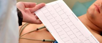
- electrocardiography, i.e. determining the correct rhythm of heart contractions;
- Doppler ultrasound, in which ultrasound makes it possible to analyze the functioning of the heart and its valves;
- daily monitoring of pressure and ECG, allowing to identify the occurrence of arrhythmia and increased pressure during exercise and at rest throughout the day.
People at risk should be especially careful about their health. They need to visit medical specialists every five to six months and undergo a comprehensive examination.
Monitoring changes in the body and a healthy lifestyle will help avoid the appearance and development of heart and vascular diseases. A person will be able to lead a full life, maintain and improve its quality.
Symptoms of heart failure
The symptoms are associated with the inability of the heart to provide adequate blood circulation and the development against this background of congestion in the pulmonary and systemic circulation (in the vessels of the lungs and the vessels of other organs and systems). Stagnation of blood in the lungs interferes with its normal saturation with oxygen and is manifested by shortness of breath. Edema - stagnation in the systemic circulation disrupts the functioning of almost all organs. Patients feel:
- increased fatigue;
- shortness of breath;
- swelling of the legs and feet;
- pain or discomfort in the abdominal cavity due to liver enlargement.
Symptoms of heart failure develop gradually and can sometimes go unnoticed for a long time, so it is necessary to undergo regular medical examinations. At the cardiology center of the Federal Medical and Clinical Center of the Federal Medical and Biological Agency, you can undergo a comprehensive heart examination in order to promptly recognize this syndrome and the diseases that accompany it.
Treatment of functional disorders of the cardiovascular system
Healing from this disease implies complex and individual therapy, which is carried out only under the close supervision of a cardiologist, neurologist and always a psychotherapist. Treatment of a functional disorder of the cardiovascular system depends on the manifestation of symptoms in the patient. It is recommended to avoid stressful situations, proper rest and good nutrition are recommended. Massage, water treatments and reflexology are also prescribed. It is possible to prescribe medications: lemon balm, St. John's wort, valerian, motherwort, glycine, antidepressants, glutamic acid.
Diagnostics
When collecting anamnesis, the doctor pays special attention to the presence of complaints of shortness of breath and fatigue. Collects information about the existence of other diseases. If heart failure is suspected, the patient is referred for instrumental studies and laboratory tests.
The Cardiology Center of the Federal Medical and Clinical Center of the Federal Medical and Biological Agency carries out a full range of diagnostic measures:
- ECG in 12 leads;
- ECHO-KG;
- Holter monitoring;
- ECG with dosed physical activity;
- transesophageal ECHO-CG;
- ABPM (24-hour blood pressure monitoring);
- Ultrasound of the abdominal organs;
- general blood analysis;
- biochemical blood test (lipid spectrum, blood glucose, liver and kidney function indicators);
- thyroid hormones;
- coagulogram (a test to determine blood clotting);
- general urine analysis;
- spirometry (respiratory function test);
Symptoms of functional disorders of the cardiovascular system
Symptoms of FNS include increased sweating, pallor or slight redness of the skin on the face, periodic fainting, headache, paroxysmal or constantly increased beating of the heart muscle (tachycardia), tachypnea, heaviness and pressure in the chest, impaired blood pressure, shortness of breath. The patient may also experience rapid fatigue, decreased attention and memory, be overly hot-tempered, irritable, suffer from insomnia and experience a constant state of anxiety. There may be various jumps in body temperature (from 35 to 37-38 degrees), nausea, vomiting, belching, diarrhea, frequent urination, anorgasmia with normal sexual desire, constipation. All this signals the Federal Tax Service.
Treatment of heart failure
When treating heart failure, the main focus is on eliminating the cause of the disease. Depending on the nature of the disease, its course and the general condition of the patient, the doctor chooses a treatment method.
On the basis of our cardiology center, the Federal Scientific and Clinical Center of the Federal Medical and Biological Agency, there are therapeutic and surgical departments. You may be required to undergo examination and selection of drug therapy in our hospital. If drug treatment is not effective enough, the cardiologist may recommend surgery. Specialists of the Cardiac Surgery Department of the Federal Scientific Center for Medical and Biological Agency successfully use advanced methods of treating heart failure. This could be an operation to correct valve disease, coronary heart disease, or arrhythmias. Our clinic performs unique minimally invasive surgeries.
Causes of functional disorders of the cardiovascular system
Nowadays, there are many reasons that can lead to the acquisition of the Federal Tax Service. The cause of this disease can be the harmful effects of various factors on the human nervous system. Our body is one whole, and the fact that negative influences on the nervous system lead to changes in the functioning of the entire organism has long been no secret.
The reason for the FTS may be factors such as:
- Impact of chronic diseases
- Improper lifestyle (disturbance in daily routine, poor sleep, etc.)
- Depression
- Severe stress
- Heredity