“Jellyfish head” is one of the symptoms of portal hypertension developing in various pathologies, which is manifested by the appearance of visible, convoluted and dilated veins on the anterior surface of the abdominal wall, diverging in different directions from the navel. The appearance of the blood vessels visible through the skin resembles the head of the character from the myths of Ancient Greece, Medusa the Gorgon, who had snakes on her head instead of hair.
The appearance of the symptom in question may indicate the development of various pathologies, which are accompanied by an increase in pressure in the portal vein and its branches. This vessel collects blood from all organs of the abdominal cavity, and disruption of its outflow provokes the appearance of multiple additional collateral vessels. They branch in different directions, expand and become visible on the skin of the abdomen. Due to increased pressure in the portal vein, the patient develops ascites, in which the skin on the anterior abdominal wall becomes thinner due to overstretching. As a result, the network of dilated vessels becomes even more noticeable.
Causes
Many severe heart diseases lead to portal hypertension, including myocarditis and cardiomyopathies.
“Jellyfish head” can be detected in patients with various pathologies of the liver and other organs:
- liver pathologies: alcoholic, acute or chronic hepatitis, hepatosis, primary or secondary biliary cirrhosis, sarcoidosis, carbohydrate liver degeneration, amyloidosis, polyps and liver tumors, metastases from other organs;
- pancreatic tumors;
- cholelithiasis;
- schistosomiasis;
- pathologies of the cardiovascular system: heart defects (congenital and acquired), restrictive cardiomyopathy, phlebothrombosis of the liver veins in Budd-Chiari syndrome, constrictive pericarditis;
- chronic poisoning with hepatotropic poisons;
- complications of surgical interventions, injuries and burns;
- taking chemotherapy drugs;
- bleeding from the stomach and intestines;
- infectious diseases of the digestive system;
- taking high doses of diuretics and tranquilizers;
- intrauterine thrombosis of the liver veins.
OVERBIG ZAVROYUVANNYA
Sculo-stravochoidal varicose veins (PVVs) are the most clinically important portosystemic collaterals, so their ruptures lead to varicose bleeding, the most common lethal form of cirrhosis - the first This episode is associated with a mortality rate of 30–50%. Varicose veins and varicose bleeding are aggravated by cirrhosis, which is directly caused by portal hypertension. Patients with cirrhosis and gastroesophageal varix show HPVT readings of less than 10–12 mmHg. Art.
Schlunkovo-stravochodnye varicose veins are detected in about 50% of patients with cirrhosis. Their presence correlates with the importance of liver disease (Table 2): while in class A for Child-Pugh, only 40% of patients have varicose veins, then in class C veins, 85% of patients have varicose veins. In patients with primary biliary cirrosis, varicose veins and varicose bleeding may develop at an early stage of illness, leading to the diagnosis of cirrhosis. It was also found that 16% of patients with hepatitis C and pontic fibrosis have varicose veins.
In patients without varicose veins, the remaining varicose veins develop with a frequency of 8% per river; the strongest predictor for the development of varicose veins in patients who are not visible during the initial endoscopic examination is an HPVT reading >10 mm Hg. Art. In patients with small varices, large varices develop with a frequency of 8% per population. Decompensated cirrhosis (Child B/C), alcoholic cirrhosis and the presence of “worm signs” during the first endoscopy are the head signs associated with the transition from small varixes to large ones (Merli M. et al., 2003). Japanese authors, in their classification, give a substantive meaning to the bluish bark of the VRV.
Variceal bleeding occurs with a frequency of 5–15% per river, and the most important predictor of bleeding is the size of the variceal vein, with the highest risk of first bleeding (15% per river) in patients with large variceal veins (The North Italian Endoscopic Club for the Study and Treatment of Es. ophageal Varices , 1988). Other predictors of bleeding are decompensated cirrhosis (Child B/C), bluish coloration of the veins, and the appearance of red signs during endoscopy. While bleeding from varicose veins usually disappears on its own in up to 40% of patients, regardless of treatment in the hospital for the remaining 10 years, stench is associated with a mortality rate of at least 20% between 6 years iv. Patients with HPVT >20 mm Hg. Art. (over a period of 24 years after varicose bleeding) it was found that there is a high risk of early recurrence of bleeding (re-bleeding in the first life after surgery) or distant spots of bleeding (83% versus 29%) and I see a single mortality rate (64% per against 20%) was equal to patients with lower pressure. Later, relapses of bleeding occur in about 60% of serious patients, mostly in 1-2 cases after the initial bleeding.
The portal pressure, the size of the vein and the thickness of its wall are related to each other by Laplace’s law - the stress of the vein wall (T) is directly proportional to the pressure in the middle of the vessel (P) and its radius (R) and is proportional to the thickness of the vessel Inki (W): T = P × R/W.
Apparently, dilatation of the node from the thinning of its wall is the main factor that causes stress on the wall and rupture of the varix. Under pressure, however, a vessel with a larger diameter will rupture, but a vessel with a smaller diameter will not. Around the diameter of the vessel, another source of stretching of the varicose wall is the pressure in the middle of the VRV, which is directly connected with the HPVT. Thus, a decrease in HPVT is likely to reflect a change in the stretching of the wall of the varicose vein and, apparently, a change in the risk of rupture. In fact, varicose veins do not bleed when HPVT decreases to <12 mmHg. Art. It has also been shown that the risk of re-bleeding significantly changes when GPVT is reduced by more than 20% of the cob level. Diseases in which HPVT decreases to <12 mmHg. However, at least 20% of the cob level (“HVPG responders”) may reduce the risk of recurrent variceal bleeding, as well as a lower risk of ascites, spontaneous bacterial peritonitis, and death. Table 2. Classification of the importance of cirrhosis according to Child-Pugh
| Items* | |||
| 1 | 2 | 3 | |
| Encephalopathy | No | Riven 1–2 (induced) | Riven 3–4 (chronic) |
| Ascites | No | Minor/moderate (responds to diuretics) | Tension (resistant to diuretics) |
| Bilirubin (mg/dl) | <2 | 2–3 | >3 |
| Albumin (g/dl) | >3,5 | 2,3–3,5 | <2,8 |
| Prothrombin hour (PT), daytime (s) or international normalized ratio (INR) | <4 <1,7 | 4–6 1,7–2,3 | >6 >2,3 |
*5–6 points: Child A. 7–9 points: Child B. 10–15 points: Child C.
Symptoms
An increase in pressure in the portal vein is accompanied not only by the appearance of the “head of jellyfish” symptom. Usually the first signs of this condition are digestive disorders:
- feeling of fullness in the stomach when eating small portions of food;
- constipation alternating with diarrhea;
- stomach ache;
- loss of appetite;
- bloating.
When the hepatic ducts are compressed, the patient develops obstructive jaundice:
- yellowness of the skin and mucous membranes;
- darkening of urine;
- discoloration of stool;
- skin itching;
- pain in the right hypochondrium.
Portal hypertension causes asthenia in the patient and is accompanied by intoxication:
- frequent feeling of weakness;
- dizziness;
- instability of blood pressure;
- weight loss;
- pain in muscles and joints;
- muscle weakness;
- fever;
- hair loss and brittle nails;
- sleep disorders.
Changes in blood circulation lead to multiple organ failure:
- increase in the size of the liver and spleen;
- tendency to nosebleeds;
- dyspnea;
- rhythm disturbances;
- swelling in the ankles;
- cough;
- ascites;
- disorders of the nervous system.
A prolonged increase in pressure in the portal vein leads to the development of varicose veins of the esophagus, stomach and rectum. Because of this, the patient may experience bleeding, which can cause posthemorrhagic anemia.
If untreated, portal hypertension is complicated by encephalopathy and the patient has complaints of drowsiness, memory impairment, decreased concentration and excessive irritability.
Prevention
Preventive measures to avoid the appearance of the “jellyfish head” symptom:
- Fight the causes that provoke the development of portal hypertension syndrome,
- Take blood and urine tests regularly,
- Get tested in a timely manner if you are at risk,
- Eat properly and balanced
- Take vitamins
- Do not smoke or drink alcohol,
- Lead a healthy lifestyle.
And the most important thing to remember is that there are no harmless diseases that affect vital organs. Only timely detected pathologies can be completely eliminated with the help of adequate therapy that gives effective results.
Diagnostics
Ultrasound will help detect liver disease, which has led to portal hypertension and varicose veins of the anterior abdominal wall.
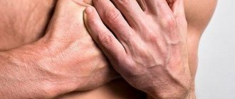
“The head of a jellyfish” is detected when examining the anterior abdominal wall - dilated, tortuous veins protruding above the surface of the skin are visible on the abdomen. In addition, the patient has swelling in the ankles and signs of ascites (increased abdominal volume). When palpating the liver, its increase is determined. The organ becomes lumpy and dense.
To identify the cause of the appearance of the “jellyfish head”, the patient is prescribed the following studies:
- blood tests: clinical, biochemistry, serum immunoglobulins, coagulogram;
- Ultrasound of the abdominal organs;
- Dopplerometry of liver vessels;
- radiographic techniques: angiography of mesenteric vessels, celiacography, portography, cavography, splenoportography;
- CT;
- MRI;
- liver scintigraphy;
- esophagoscopy;
- fibrogastroduodenoscopy;
- sigmoidoscopy.
In some cases, the examination of the patient is supplemented by a liver biopsy followed by histological analysis.
How does the head of a jellyfish appear?
The photo shows the intricate pattern formed by the dilated vessels. It's called "jellyfish head". Sometimes pineal venous formations are observed, which protrude into large tubercles on the abdomen. The protruding vessels resemble a ball of snakes; they are concentrated in the navel area and diverge from it in different directions.
Sometimes a varicose node forms right in the area of the umbilical hollow. Externally, the stomach of a patient with cirrhosis with dilation of the saphenous veins resembles a ball of snakes. Doctors gave the symptom a sonorous name due to its resemblance to the head of the Gorgon, a character in the myths of Ancient Greece. This houria had thin living snakes growing on its head instead of hair.
Treatment
To eliminate the “head of the jellyfish”, the underlying disease and the portal hypertension that provokes the appearance of this symptom must be treated.
The following medications are used to reduce pressure in the portal vein:
- vasoconstrictors (vasopressin, somatostatin) - used to narrow arterioles and reduce pressure in the portal vein;
- organic nitrates (nitroglycerin, isosorbide, etc.) – help reduce blood volume in the portal vein by increasing its volume in small vessels;
- ACE inhibitors (fosinopril, lisinopril, enalapril, captopril, etc.) – are used to eliminate arterial hypertension and improve blood flow in peripheral vessels;
- beta-blockers (propranolol, metroprolol, etc.) – reduce the number of heart contractions, lower blood pressure and increase tissue resistance to lack of oxygen;
- diuretics (furosemide, etc.) – eliminate swelling and promote the elimination of toxic substances along with urine;
- lactulose – helps eliminate toxic substances from the intestines;
- hepatoprotectors;
- B vitamins.
With portal hypertension, the patient must follow a diet that involves limiting the consumption of salt and protein foods.
If conservative therapy is ineffective, the patient is prescribed surgical treatment. In case of bleeding from varicose veins of the esophagus or stomach, endoscopic operations can be performed to ligate or sclerosis the affected vessels. If these minimally invasive techniques are ineffective, then the varicose veins are stitched through the mucous membrane.
When portal hypertension is complicated by gastrointestinal bleeding, splenomegaly and accumulation of fluid in the abdominal cavity, it is recommended to perform operations aimed at creating a bypass anastomosis, which will ensure the outflow of blood from the portal vein into the inferior vena cava or renal vein. Depending on the clinical case, the following types of bypass surgery are performed:
- mesentericocaval - portocaval anastomosis is created between the inferior vena cava and the superior mesenteric vein;
- portocaval - a shunt is performed between the portal and cava veins;
- selective splenorenal - distal vascular anastomosis is applied between the splenic and left renal veins;
- transjugular intrahepatic portosystemic - a stent is implanted through the jugular vein, which creates an anastomosis between the portal and hepatic veins;
- reduction of splenic arterial blood flow - a spiral is introduced endovascularly (through the femoral artery) into the splenic artery, which reduces the volume of arterial blood flow and prevents venous outflow into the portal vein.
In case of decompensated or complicated portal hypertension, laparocentesis or drainage of the abdominal cavity is performed. In some advanced cases, the patient may be recommended liver transplantation and splenectomy.
Complications of liver cirrhosis
They are the result of carelessness or lack of awareness of the seriousness of the consequences. Severe complications cannot be cured and cause death. These include:
Ascites is an accumulation of liquid substance in the peritoneum. The abdomen increases, which is noticeable even in a lying position. There is weight gain and swelling of the legs. Swelling does not go away after rest and makes movement difficult.
Bacterial peritonitis occurs when fluid in the abdominal cavity becomes infected. Body temperature rises - up to 40C, severe pain and malaise appear. In the absence of emergency assistance, death occurs.
Hepatic encephalopathy is a neurological symptom complex. Headache, dizziness, and unsteady gait are observed. Often progresses into a coma.
Acute renal failure due to dysfunction of the hepatobiliary system. In the absence of timely resuscitation, it ends in death.
Portal hypertension is accompanied by varicose veins of the gastrointestinal tract. Because of this, bleeding begins, during which the pressure drops, and vomiting with bloody elements appears. The condition often ends in death. Resuscitation does not always help save the patient.
Hepatocellular carcinoma is the formation of a cancerous tumor. Occurs when the disease lasts for a long time. Provoking factors are considered to be heredity and bad habits - smoking, alcoholism. In case of single tumors and the absence of metastases, it is removed surgically. In case of extensive degeneration, transplantation will help solve the problem.
The likelihood of complications of liver cirrhosis increases significantly with an inadequate approach or late detection of problems. To protect yourself, you need to undergo a preventive examination once a year, take general and biochemical tests, and do an ultrasound scan of the body. At the slightest suspicion, it is better to consult a doctor.
To prevent liver cirrhosis, play sports or engage in other adequate physical activities. Classes should be regular, preferably in the fresh air. This will help avoid stagnation. The psycho-emotional background is of great importance. Stress causes spasm of internal organs, which affects their functioning. It is better to avoid negative emotions as much as possible.
You should not ignore problems with bile discharge and excess weight. It is important to follow healthy eating rules. Doctors advise keeping a food diary. More movement, less negative emotions - the rule for restoring strength. An active lifestyle and giving up bad habits - drinking alcohol and smoking - will help avoid serious health misunderstandings and eliminate the causes of liver cirrhosis.
Author of the article: Yakovlev Evgeniy Anatolyevich
Narcologist, Candidate of Medical Sciences.
Which doctor should I contact?
If a “jellyfish head” appears (visible convoluted dilated veins on the abdomen), signs of indigestion, jaundice, deterioration in general condition and an increase in the abdomen in volume, you should consult a physician. After conducting a series of studies (ultrasound of the abdominal organs, Doppler ultrasound of liver vessels, CT, MRI, portography, liver scintigraphy, etc.) and identifying signs of portal hypertension, the doctor may prescribe a consultation with a specialized specialist (cardiologist, hepatologist, etc.) and treatment with a vascular surgeon
“Jellyfish head” is a symptom of portal hypertension and manifests itself in the form of dilated and tortuous veins on the anterior abdominal wall. It can occur with various diseases and conditions and always requires immediate treatment of the underlying ailment. To reduce pressure in the portal vein, conservative therapy and surgical treatment are prescribed.
About portal hypertension in the program “Live Healthy!” with Elena Malysheva:
VARICOSE VEINS
Varicose veins, the veins are smaller, the lower varicose veins, and are now found in 5–33% of patients with portal hypertension, with a bleeding frequency of about 25%, with a length of 2 lines and with a large amount of bleeding. veins of the bottom of the scutum (Sarin SK et al., 1992). Risk factors for vulvar varicose bleeding include the size of the nodules at the bottom of the vulva (large > medium > small, values >10 mm, 5–10 mm and <5 mm uniform), classification of cyrrhosis as Child (C > B > A) and the presence of worms them signs on the surface of the varix during the hour of endoscopy (Kim T. et al., 1999). The schulci varicose veins are usually divided on the basis of their relationship to the splanchnic veins, as well as their localization in the scutum. Gastroesophageal varices (GEVs) are extensions of varicose veins along the line and are divided into 2 types. The most common types of varix are type 1 (GEV 1 – GOV1), which widen the lesser curve. The stinks are respected by the continued VRV voyage and are guilty of rejoicing in the same way. Type 2 slug ERVs (GEV 2) are expanding at the bottom and tend to be long and sinuous. Isolated hose VRV (ISHV) are identified for the presence of varicose veins in the traveler and are also divided into 2 types. VRV type 1 (ІШВ 1 – IGV1) are localized near the day and are often sinuous and complex, and VRV type 2 (ІШВ 2) are located near the body, antrum and near the hilum. The presence of varicose veins at the bottom of the scutum will require excluding the diagnosis of splenic vein thrombosis.
Consequences
Since dilation of the veins of the abdominal wall is a serious symptom, the consequences of the diseases that cause it can be severe:
- Cirrhosis of the liver;
- Malignant neoplasms;
- Bleeding;
- Ascites;
- Death.
Therefore, if this symptom is detected, you must immediately consult a doctor and begin treatment. In most cases, such diseases require the patient to be in a hospital.